

The DOI matches its Crossref record; 3 of the 3 references it cites were found in a registry or an index.
8-oxoguanine and 8-oxodeoxyguanosine biomarkers of oxidative DNA damage: a review on HPLC-ECD determination
Abstract
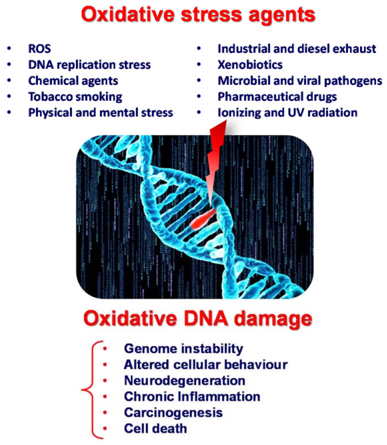
Reactive oxygen species (ROS) are continuously produced in living cells due to metabolic and biochemical reactions and due to exposure to physical, chemical and biological agents. Excessive ROS cause oxidative stress and lead to oxidative DNA damage. Within ROS-mediated DNA lesions, 8-oxoguanine (8-oxoG) and its nucleotide 8-oxo-2′-deoxyguanosine (8-oxodG)—the guanine and deoxyguanosine oxidation products, respectively, are regarded as the most significant biomarkers for oxidative DNA damage. The quantification of 8-oxoG and 8-oxodG in urine, blood, tissue and saliva is essential, being employed to determine the overall effects of oxidative stress and to assess the risk, diagnose, and evaluate the treatment of autoimmune, inflammatory, neurodegenerative and cardiovascular diseases, diabetes, cancer and other age-related diseases. High-performance liquid chromatography with electrochemical detection (HPLC–ECD) is largely employed for 8-oxoG and 8-oxodG determination in biological samples due to its high selectivity and sensitivity, down to the femtomolar range. This review seeks to provide an exhaustive analysis of the most recent reports on the HPLC–ECD determination of 8-oxoG and 8-oxodG in cellular DNA and body fluids, which is relevant for health research.
Overview
The claims and statistics of this paper haven’t been checked yet.
Run the check — free, no account needed. It reads the open-access text and checks the sources it cites.
Integrity signals
Registry
Cites
Statistics
Figures
Verification
Claims the paper makes
The claims and statistics of this paper haven’t been checked yet.
Run the check — free, no account needed. It reads the open-access text and checks the sources it cites.
Claims other papers make about this one
Each is checked against this paper’s own text: does it say what the citing paper says it does?
In the citing paper
“DNA damage, in the form of 8-hydroxy-2'-deoxyguanosine (8-OHdG), is considered a useful biomarker of oxidative DNA damage that has been used to evaluate the risk of developing inflammatory-related diseases and cancer (Chiorcea-Paquim, 2022;Graille et al., 2020).”
From Airborne microplastic exposure and urinary oxidative damage biomarkers in populations living in traffic-congested areas (2026) · cites this paper as Chiorcea-Paquim, 2022
Self-check
Percentages, sums and reported differences re-computed from the paper’s own numbers.
The self-check needs the paper’s full text.
Citations
Cites
Base-excision repair of oxidative DNA damage
David, S. S., O'Shea, V. L., & Kundu, S. (2007) · Nature · doi:10.1038/nature05978 · Cited by 1,238
Oxidative damage to mitochondrial DNA shows marked age-dependent increases in human brain
Mecocci, P., MacGarvey, U., Kaufman, A. E., Koontz, D., Shoffner, J. M., Wallace, D. C., & Beal, M. F. (1993) · Annals of Neurology · doi:10.1002/ana.410340416 · Cited by 757 · cited 1×
“The ECD analytical determination was revealed to be 1000 times more sensitive than the UV detection method [19], which made the quantification of 8-oxodG possible in HeLa cells [20], mouse liver [20], rat kidney [21,22], rat liver [23,24], rat heart [25], and rat urine [21], as well as in human leukocytes [26], blood…”
MethodSerum 8-Hydroxy-Guanine Levels Are Increased in Diabetic Patients
Shin, C. S., Moon, B. S., Park, K. S., Kim, S. Y., Park, S. J., Chung, M. H., & Lee, H. K. (2001) · Diabetes Care · doi:10.2337/diacare.24.4.733 · Cited by 133
Research works printed but not linked: 129
- Yudkina, A.V., Shilkin, E.S., Endutkin, A.V., Makarova, A.V., and Zharkov, D.O. (2019). Reading and misreading 8-oxoguanine, a paradigmatic ambiguous nucleobase. Crystals, 9. · DOI
- Loft, 2012, Urinary excretion of 8-oxo-7,8-dihydroguanine as biomarker of oxidative damage to DNA, Arch. Biochem. Biophys., 518, 142, 10.1016/j.abb.2011.12.026 · DOI
- Nakabeppu, 2014, Cellular levels of 8-oxoguanine in either DNA or the nucleotide pool play pivotal roles in carcinogenesis and survival of cancer cells, Int. J. Mol. Sci., 15, 12543, 10.3390/ijms150712543 · DOI
- Piedade, 2004, Voltammetric determination of all DNA nucleotides, Anal. Biochem., 332, 321, 10.1016/j.ab.2004.06.021 · DOI
- Kino, 2017, Generation, repair and replication of guanine oxidation products, Genes Environ., 39, 21, 10.1186/s41021-017-0081-0 · DOI
- Goyal, 1997, Electrochemical and enzymic oxidation of guanosine and 8-hydroxyguanosine and the effects of oxidation products in mice, Bioelectrochemistry Bioenerg., 43, 105, 10.1016/S0302-4598(96)05182-3 · DOI
- Brett, 1994, On the adsorption and electrochemical oxidation of DNA at glassy carbon electrodes, J. Electroanal. Chem., 366, 225, 10.1016/0022-0728(93)02994-S · DOI
- Goyal, 1982, Redox chemistry of guanine and 8-oxyguanine and a comparison of the peroxidase-catalyzed and electrochemical oxidation of 8-oxyguanine, J. Electroanal. Chem., 135, 75, 10.1016/0022-0728(82)90006-7 · DOI
- Venkateswarlu, 1998, Tautomeric equilibria in 8-oxopurines: Implications for mutagenicity, J. Comput. Aided. Mol. Des., 12, 373, 10.1023/A:1008067110965 · DOI
- Drake, 2019, Measurement of the oxidative DNA lesion 8-oxoguanine (8-oxoG) by ELISA or by high-performance liquid chromatography (HPLC) with electrochemical detection, Methods in Molecular Biology, Volume 1965, 313, 10.1007/978-1-4939-9182-2_21 · DOI
- Valavanidis, 2009, 8-Hydroxy-2′ -deoxyguanosine (8-OHdG): A critical biomarker of oxidative stress and carcinogenesis, J. Environ. Sci. Health Part C Environ. Carcinog. Ecotoxicol. Rev., 27, 120, 10.1080/10590500902885684 · DOI
- Floyd, 1986, Hydroxyl free radical adduct of deoxyguanosine: Sensitive detection and mechanisms of formation, Free Radic. Res., 1, 163
- Poulsen, 2002, HPLC-ECD, HPLC-MS/MS (Urinary Biomarkers), Critical Reviews of Oxidative Stress and Aging, Volume 1, 233, 10.1142/9789812775733_0013 · DOI
- Wang, 2016, The relationship between plasma and urinary 8-hydroxy-2-deoxyguanosine biomarkers measured by liquid chromatography tandem mass spectrometry, Environ. Sci. Pollut. Res., 23, 17496, 10.1007/s11356-016-6898-4 · DOI
- Lin, 2004, A high-throughput and sensitive methodology for the quantification of urinary 8-hydroxy-2′-deoxyguanosine: Measurement with gas chromatography-mass spectrometry after single solid-phase extraction, Biochem. J., 380, 541, 10.1042/bj20040004 · DOI
- Saito, 2000, Quantitative determination of urinary 8-hydroxydeoxyguanosine (8-OH-dG) by using ELISA, Res. Commun. Mol. Pathol. Pharmacol., 107, 39
- 2021, Nanostructured material–based electrochemical sensing of oxidative DNA damage biomarkers 8-oxoguanine and 8-oxodeoxyguanosine: A comprehensive review, Microchim. Acta, 188, 1
- Tagesson, 1991, Determination of urinary 8-hydroxydeoxyguanosine by coupled-column high-performance liquid chromatography with electrochemical detection: A noninvasive assay for in vivo oxidative DNA damage in humans, Toxicol. Mech. Methods, 1, 242, 10.3109/15376519109050855 · DOI
- Kasai, 1986, Formation of 8-hydroxyguanine moiety in cellular dna by agents producing oxygen radicals and evidence for its repair, Carcinogenesis, 7, 1849, 10.1093/carcin/7.11.1849 · DOI
- Fraga, 1990, Oxidative damage to DNA during aging: 8-Hydroxy-2′-deoxyguanosine in rat organ DNA and urine, Proc. Natl. Acad. Sci. USA, 87, 4533, 10.1073/pnas.87.12.4533 · DOI · Paper page
- Kasai, 1987, Oral administration of the renal carcinogen, potassium bromate, specifically produces 8-hydroxydeoxyguanosine in rat target organ DNA, Carcinogenesis, 8, 1959, 10.1093/carcin/8.12.1959 · DOI
- Richter, 1988, Normal oxidative damage to mitochondrial and nuclear DNA is extensive, Proc. Natl. Acad. Sci. USA, 85, 6465, 10.1073/pnas.85.17.6465 · DOI
- Fiala, 1989, Oxidative DNA and RNA Damage in the Livers of Sprague-Dawley Rats Treated with the Hepatocarcinogen 2-Nitropropane, Cancer Res., 49, 5518
- You, 2000, Increased 8-hydroxyguanine formation and endonuclease activity for its repair in ischemic-reperfused heads of rats, J. Mol. Cell. Cardiol., 32, 1053, 10.1006/jmcc.2000.1142 · DOI
- Kiyosawa, 1990, Cigarette Smoking Induces Formation of 8-Hydroxydeoxyguanosine, One of the Oxidative DNA Damages in Human Peripheral Leukocytes, Free Radic. Res. Commun., 11, 23, 10.3109/10715769009109664 · DOI
- Dandona, 1996, Oxidative damage to DNA in diabetes mellitus, Lancet, 347, 444, 10.1016/S0140-6736(96)90013-6 · DOI
- Asami, 1997, Cigarette smoking induces an increase in oxidative DNA damage, 8-hydroxydeoxyguanosine, in a central site of the human lung, Carcinogenesis, 18, 1763, 10.1093/carcin/18.9.1763 · DOI
- Musarrat, 1996, Prognostic and aetiological relevance of 8-hydroxyguanosine in human breast carcinogenesis, Eur. J. Cancer Part A, 32, 1209, 10.1016/0959-8049(96)00031-7 · DOI
- Kondo, 2000, Overexpression of the hOGG1 gene and high 8-hydroxy-2′-deoxyguanosine (8-OHdG) lyase activity in human colorectal carcinoma: Regulation mechanism of the 8-OHdG level in DNA, Clin. Cancer Res., 6, 1394
- Loft, 1993, 8-hydroxydeoxyguanosine as a urinary biomarker of oxidative DNA damage, J. Toxicol. Environ. Health, 40, 391, 10.1080/15287399309531806 · DOI
- Shigenaga, 1989, Urinary 8-hydroxy-2′-deoxyguanosine as a biological marker of in vivo oxidative DNA damage, Proc. Natl. Acad. Sci. USA, 86, 9697, 10.1073/pnas.86.24.9697 · DOI · Paper page
- Shigenaga, 1991, Assays for 8-hydroxy-2′-deoxyguanosine: A biomarker of in vivo oxidative DNA damage, In Free. Radic. Biol. Med., 10, 211, 10.1016/0891-5849(91)90078-H · DOI
- Cundy, 1988, Determination of 8-hydroxydeoxyguanosine in human urine: A possible assay for in vivo oxidative DNA damage, Basic Life Sci., 49, 479
- Kasai, 1997, Analysis of a form of oxidative DNA damage, 8-hydroxy-2′-deoxyguanosine, as a marker of cellular oxidative stress during carcinogenesis, Mutat. Res. Rev. Mutat. Res., 387, 147, 10.1016/S1383-5742(97)00035-5 · DOI
- Koide, 2010, Determination of human serum 8-hydroxy-2′-deoxyguanosine (8-OHdG) by HPLC-ECD combined with solid phase extraction (SPE), J. Chromatogr. B Anal. Technol. Biomed. Life Sci., 878, 2163, 10.1016/j.jchromb.2010.06.015 · DOI
- Loft, 1992, Oxidative DNA damage estimated by 8-hydroxydeoxyguanosine excretion in humans: Influence of smoking, gender and body mass index, Carcinogenesis, 13, 2241, 10.1093/carcin/13.12.2241 · DOI
- Loft, 2006, Prospective study of 8-oxo-7,8-dihydro-2′-deoxyguanosine excretion and the risk of lung cancer, Carcinogenesis, 27, 1245, 10.1093/carcin/bgi313 · DOI
- Bogdanov, 1999, A carbon column-based liquid chromatography electrochemical approach to routine 8-hydroxy-2′-deoxyguanosine measurements in urine and other biologic matrices: A one-year evaluation of methods, Free Radic. Biol. Med., 27, 647, 10.1016/S0891-5849(99)00113-6 · DOI
- Lengger, 2000, A high-performance liquid chromatographic method for the determination of 8-oxo-7,8-dihydro-2′-deoxyguanosine in urine from man and rat, Anal. Biochem., 287, 65, 10.1006/abio.2000.4839 · DOI
- Kasai, 2005, Simultaneous determination of 8-hydroxydeoxyguanosine, a marker of oxidative stress, and creatinine, a standardization compound, in urine, Ind. Health, 43, 333, 10.2486/indhealth.43.333 · DOI
- Svoboda, 2004, Simultaneous HPLC analysis of 8-hydroxydeoxyguanosine and 7-methylguanine in urine from humans and rodents, Anal. Biochem., 334, 239, 10.1016/j.ab.2004.08.021 · DOI
- Kasai, 2003, A new automated method to analyze urinary 8-hydroxydeoxyguanosine by a high-performance liquid chromatography-electrochemical detector system, J. Radiat. Res., 44, 185, 10.1269/jrr.44.185 · DOI
- Kasai, 2001, Life style and urinary 8-hydroxydeoxyguanosine, a marker of oxidative DNA damage: Effects of exercise, working conditions, meat intake, body mass index, and smoking, Jpn. J. Cancer Res., 92, 9, 10.1111/j.1349-7006.2001.tb01041.x · DOI
- Degan, 1991, Immunoaffinity isolation of urinary 8-hydroxy-2′-deoxygunosine and 8-hydroxyguanine and quantitation of 8-hydroxy-2′deoxyguanosine in DNA by polyclonal antibodies, Carcinogenesis, 12, 865, 10.1093/carcin/12.5.865 · DOI
- Park, 1992, Assay of excised oxidative DNA lesions: Isolation of 8-oxoguanine and its nucleoside derivatives from biological fluids with a monoclonal antibody column, Proc. Natl. Acad. Sci. USA, 89, 3375, 10.1073/pnas.89.8.3375 · DOI
- Shigenaga, 1994, Assays of oxidative DNA damage biomarkers 8-oxo-2′-deoxyguanosine and 8-oxoguanine in nuclear DNA and biological fluids by high-performance liquid chromatography with electrochemical detection, Methods Enzymol., 234, 16, 10.1016/0076-6879(94)34073-0 · DOI
- Helbock, 1998, DNA oxidation matters: The HPLC-electrochemical detection assay of 8-oxo-deoxyguanosine and 8-oxo-guanine, Proc. Natl. Acad. Sci. USA, 95, 288, 10.1073/pnas.95.1.288 · DOI
- Hofer, 2002, Optimization of the workup procedure for the analysis of 8-oxo-7,8-dihydro-2′-deoxyguanosine with electrochemical detection, Chem. Res. Toxicol., 15, 426, 10.1021/tx015573j · DOI
- Hamilton, 2001, A reliable assessment of 8-oxo-2-deoxyguanosine levels in nuclear and mitochondrial DNA using the sodium iodide method to isolate DNA, Nucleic Acids Res., 29, 2117, 10.1093/nar/29.10.2117 · DOI
- Bolin, 2004, Single extraction protocol for the analysis of 8-hydroxy-2′- deoxyguanosine (oxo8dG) and the associated activity of 8-oxoguanine DNA glycosylase, J. Neurosci. Methods, 136, 69, 10.1016/j.jneumeth.2003.12.025 · DOI
- Piedade, 2000, Electrochemical oxidation of 8-oxoguanine, Electroanalysis, 12, 969, 10.1002/1521-4109(200008)12:12<969::AID-ELAN969>3.0.CO;2-O · DOI
- Rebelo, 2004, Development of an HPLC method with electrochemical detection of femtomoles of 8-oxo-7,8-dihydroguanine and 8-oxo-7,8-dihydro-2′-deoxyguanosine in the presence of uric acid, Talanta, 63, 323, 10.1016/j.talanta.2003.10.051 · DOI
- Diculescu, 2005, Electrochemical DNA Sensors for Detection of DNA Damage, Sensors, 5, 377, 10.3390/s5060377 · DOI
- Chiorcea-Paquim, A.M., and Oliveira-Brett, A.M. (2021). Dna electrochemical biosensors for in situ probing of pharmaceutical drug oxidative dna damage. Sensors, 21. · DOI
- (2016). 8-Hydroxy-2′-deoxyguanosine as a marker of oxidative DNA damage related to occupational and environmental exposures [BAT Value Documentation, 2010]. The MAK-Collection for Occupational Health and Safety, Wiley-VCH Verlag GmbH & Co. KGaA.
- Collins, 1997, Problems in the measurement of 8-oxoguanine in human DNA—Report of a Workshop, DNA Oxidation, Aberdeen, UK, 19–21 January 1997, Carcinogenesis, 18, 1833, 10.1093/carcin/18.9.1833 · DOI
- Riis, 2002, Comparison of results from different laboratories in measuring 8-oxo-2′-deoxyguanosine in synthetic oligonucleotides, Free Radic. Res., 36, 649, 10.1080/10715760290029047 · DOI
- Collins, 2004, Are we sure we know how to measure 8-oxo-7,8-dihydroguanine in DNA from human cells?, Arch. Biochem. Biophys., 423, 57, 10.1016/j.abb.2003.12.022 · DOI
- Harris, 1994, 7, 8-dihydro-8-oxo-2′-deoxyguanosine present in DNA is not simply an artefact of isolation, Carcinogenesis, 15, 411, 10.1093/carcin/15.2.411 · DOI
- Collins, 2002, Inter-laboratory validation of procedures for measuring 8-oxo-7,8-dihydroguanine/8-oxo-7,8-dihydro-2′-deoxyguanosine in DNA, Free Radic. Res., 36, 239, 10.1080/10715760290019246 · DOI
- Cadet, 1998, Facts and artifacts in the measurement of oxidative base damage to DNA, Free Radic. Res., 29, 541, 10.1080/10715769800300581 · DOI
- Collins, 2003, Measurement of DNA oxidation in human cells by chromatographic and enzymic methods, Free Radic. Biol. Med., 34, 1089, 10.1016/S0891-5849(03)00041-8 · DOI
- Collins, 2002, Comparative analysis of baseline 8-oxo-7,8-dihydroguanine in mammalian cell DNA, by different methods in different laboratories: An approach to consensus, Carcinogenesis, 23, 2129, 10.1093/carcin/23.12.2129 · DOI
- Gedik, 2005, Establishing the background level of base oxidation in human lymphocyte DNA: Results of an interlaboratory validation study, FASEB J., 19, 82, 10.1096/fj.04-1767fje · DOI
- Barnett, 2000, Measurement of 8-oxo-deoxyguanosine in lymphocytes, cultured cells, and tissue samples by HPLC with electrochemical detection, Aging Methods and Protocols, Volume 38, 171, 10.1385/1-59259-070-5:171 · DOI
- Chepelev, N.L., Kennedy, D.A., Gagné, R., White, T., Long, A.S., Yauk, C.L., and White, P.A. (2015). HPLC measurement of the DNA oxidation biomarker, 8-oxo-7,8-dihydro-2′-deoxyguanosine, in cultured cells and animal tissues. J. Vis. Exp., 2015. · DOI
- Hofer, 1998, Reduction of oxidation during the preparation of DNA and analysis of 8- hydroxy-2′-deoxyguanosine, Chem. Res. Toxicol., 11, 882, 10.1021/tx980041x · DOI
- Gedik, 1998, Measuring oxidative damage to DNA; HPLC and the comet assay compared, Free Radic. Res., 29, 609, 10.1080/10715769800300661 · DOI
- Ravanat, 2002, Cellular background level of 8-oxo-7,8-dihydro-2′-deoxyguanosine: An isotope based method to evaluate artefactual oxidation of DNA during its extraction and subsequent work-up, Carcinogenesis, 23, 1911, 10.1093/carcin/23.11.1911 · DOI
- Lodovici, 2000, Levels of 8-hydroxydeoxyguanosine as a marker of DNA damage in human leukocytes, Free Radic. Biol. Med., 28, 13, 10.1016/S0891-5849(99)00194-X · DOI
- Kim, 2003, Effects of genetic polymorphisms in metabolic enzymes on the relationships between 8-hydroxydeoxyguanosine levels in human leukocytes and urinary 1-hydroxypyrene and 2-naphthol concentrations, J. Occup. Health, 45, 160, 10.1539/joh.45.160 · DOI
- Tarng, 2004, Protective effect of vitamin C on 8-hydroxy-2′-deoxyguanosine level in peripheral blood lymphocytes of chronic hemodialysis patients, Kidney Int., 66, 820, 10.1111/j.1523-1755.2004.00809.x · DOI
- Herbert, 2006, Dietary supplementation with different vitamin C doses: No effect on oxidative DNA damage in healthy people, Eur. J. Nutr., 45, 97, 10.1007/s00394-005-0569-4 · DOI
- Yen, 2004, Increased 8-hydroxy-2′-deoxyguanosine in leukocyte DNA in Leber’s hereditary optic neuropathy, Investig. Ophthalmol. Vis. Sci., 45, 1688, 10.1167/iovs.03-0568 · DOI
- Karahan, 2019, Oxidative DNA Damage Is Increased in Living Kidney Donors, Transplant. Proc., 51, 1049, 10.1016/j.transproceed.2019.02.011 · DOI
- Katerji, M., Filippova, M., and Duerksen-Hughes, P. (2019). Approaches and methods to measure oxidative stress in clinical samples: Research applications in the cancer field. Oxid. Med. Cell. Longev. · DOI
- Matsui, 2000, Increased formation of oxidative DNA damage, 8-hydroxy-2′- deoxyguanosine, in human breast cancer tissue and its relationship to GSTP1 and COMT genotypes, Cancer Lett., 151, 87, 10.1016/S0304-3835(99)00424-3 · DOI
- Li, 2001, Oxidative DNA damage and 8-hydroxy-2-deoxyguanosine DNA glycosylase/apurinic lyase in human breast cancer, Mol. Carcinog., 31, 214, 10.1002/mc.1056 · DOI
- Kondo, 1999, Persistent oxidative stress in human colorectal carcinoma, but not in adenoma, Free Radic. Biol. Med., 27, 401, 10.1016/S0891-5849(99)00087-8 · DOI
- Adamek, 2013, 8-hydroxy-2′-deoxyguanosine in colorectal adenocarcinoma—Is it a result of oxidative stress?, Med. Sci. Monit., 19, 690, 10.12659/MSM.883999 · DOI
- Park, 2001, Genetic changes of hOGG1 and the activity of oh8Gua glycosylase in colon cancer, Eur. J. Cancer, 37, 340, 10.1016/S0959-8049(00)00388-9 · DOI
- Autrup, 2003, Personal exposure to PM2.5 and biomarkers of DNA damage, Cancer Epidemiol. Biomark. Prev., 12, 191
- Marczynski, 2000, Association between 8-hydroxy-2′-deoxyguanosine levels in DNA of workers highly exposed to asbestos and their clinical data, occupational and non-occupational confounding factors, and cancer, Mutat. Res. Genet. Toxicol. Environ. Mutagen., 468, 203, 10.1016/S1383-5718(00)00054-1 · DOI
- Marczynski, 2000, Levels of 8-hydroxy-2′-deoxyguanosine in DNA of white blood cells from workers highly exposed to asbestos in Germany, Mutat. Res. Genet. Toxicol. Environ. Mutagen., 468, 195, 10.1016/S1383-5718(00)00053-X · DOI
- Pilger, 2000, 8-Hydroxydeoxyguanosine in leukocyte DNA and urine of quartz-exposed workers and patients with silicosis, Int. Arch. Occup. Environ. Health, 73, 305, 10.1007/s004200000117 · DOI
- Toraason, 2001, DNA strand breaks, oxidative damage, and 1-OH pyrene in roofers with coal-tar pitch dust and/or asphalt fume exposure, Int. Arch. Occup. Environ. Health, 74, 396, 10.1007/s004200100238 · DOI
- Zhang, 2003, Leukocyte 8-hydroxydeoxyguanosine and aromatic DNA adduct in coke-oven workers with polycyclic aromatic hydrocarbon exposure, Int. Arch. Occup. Environ. Health, 76, 499, 10.1007/s00420-003-0449-2 · DOI
- McDorman, 2005, Oxidative DNA damage from potassium bromate exposure in Long-Evans rats is not enhanced by a mixture of drinking water disinfection by-products, Chem. Biol. Interact., 152, 107, 10.1016/j.cbi.2005.02.003 · DOI
- Gupta, 2013, Acute in vivo treatment with 4-(methylnitrosamino)-1-(3-pyridyl)-1-butanone does not alter base excision repair activities in murine lung and liver, DNA Repair, 12, 1031, 10.1016/j.dnarep.2013.09.009 · DOI
- Mulder, 2014, In vivo treatment with aflatoxin B1 increases DNA oxidation, base excision repair activity and 8-oxoguanine DNA glycosylase 1 levels in mouse lung, Toxicology, 321, 21, 10.1016/j.tox.2014.03.004 · DOI
- Orimo, 2004, Analysis of 8-hydroxyguanine (8-OH-Gua) released from DNA by the formamidopyrimidine DNA glycosylase (Fpg) protein: A reliable method to estimate cellular oxidative stress, J. Radiat. Res., 45, 455, 10.1269/jrr.45.455 · DOI
- Graille, M., Wild, P., Sauvain, J.J., Hemmendinger, M., Canu, I.G., and Hopf, N.B. (2020). Urinary 8-OHdG as a biomarker for oxidative stress: A systematic literature review and meta-analysis. Int. J. Mol. Sci., 21. · DOI · Paper page
- ESCULA [European Standards Committee on Urinary (DNA) Lesion Analysis], Evans, M.D., Olinski, R., Loft, S., and Cooke, M.S. (2010). Toward consensus in the analysis of urinary 8-oxo-7,8-dihydro-2′-deoxyguanosine as a noninvasive biomarker of oxidative stress. FASEB J., 24, 1249–1260. · DOI
- Cooke, 2009, Interlaboratory comparison of methodologies for the measurement of urinary 8-oxo-7,8-dihydro-2′-deoxyguanosine, Biomarkers, 14, 103, 10.1080/13547500802706012 · DOI
- Cooke, 2008, Measurement and meaning of oxidatively modified DNA lesions in urine, Cancer Epidemiol. Biomark. Prev., 17, 3, 10.1158/1055-9965.EPI-07-0751 · DOI
- Li, 2013, 8-Hydroxyguanine in urine and serum as an oxidative stress marker: Effects of diabetes and aging, J. UOEH, 35, 119, 10.7888/juoeh.35.119 · DOI
- Borrego, 2013, Oxidative stress and DNA damage in human gastric carcinoma: 8-Oxo-7′8-dihydro-2′-deoxyguanosine (8-oxo-dG) as a possible tumor marker, Int. J. Mol. Sci., 14, 3467, 10.3390/ijms14023467 · DOI
- Inaba, 2011, Development of urinary 8-hydroxy-2′-deoxyguanosine (8-OHdG) measurement method combined with SPE, J. Chromatogr. Sci., 49, 303, 10.1093/chrsci/49.4.303 · DOI
- Kato, 2011, Electrochemical determination of oxidative damaged DNA with high sensitivity and stability using a nanocarbon film, Anal. Sci., 27, 703, 10.2116/analsci.27.703 · DOI
- Rebelo, 2004, Electrochemical determination of 8-oxoguanine in the presence of uric acid, Bioelectrochemistry, 63, 267, 10.1016/j.bioelechem.2003.11.007 · DOI
- Saravanabhavan, 2010, A high performance liquid chromatography-electrochemical array method for the measurement of oxidative/nitrative changes in human urine, J. Chromatogr. A, 1217, 3269, 10.1016/j.chroma.2010.01.048 · DOI
- Richie, 2002, Influence of selenium-enriched yeast supplementation on biomarkers of oxidative damage and hormone status in healthy adult males: A clinical pilot study, Cancer Epidemiol. Biomark. Prev., 11, 1459
- Miyata, 2008, Changes of urinary 8-hydroxydeoxyguanosine levels during a two-day ultramarathon race period in Japanese non-professional runners, Int. J. Sports Med., 29, 27, 10.1055/s-2007-965072 · DOI
- Li, 2007, Almond consumption reduces oxidative DNA damage and lipid peroxidation in male smokers, J. Nutr., 137, 2717, 10.1093/jn/137.12.2717 · DOI
- Pilger, 2001, Longitudinal study of urinary 8-hydroxy-2′-deoxyguanosine excretion in healthy adults, Free Radic. Res., 35, 273, 10.1080/10715760100300811 · DOI
- Kuo, 2007, Urinary 8-hydroxy-2′-deoxyguanosine (8-OHdG) and genetic polymorphisms in breast cancer patients, Mutat. Res. Genet. Toxicol. Environ. Mutagen., 631, 62, 10.1016/j.mrgentox.2007.04.009 · DOI
- Liu, 2021, The expression and significance of 8-hydroxydeoxyguanosine in breast cancer patients’ blood, urine and cancer tissue, Trends Immunother., 5, 36, 10.24294/ti.v5.i2.1.1374 · DOI
- Watanabe, S., Li, Y.S., Kawasaki, Y., Ootsuyama, Y., and Kawai, K. (2021). Health examination results and work environment factors affecting urinary 8-hydroxy-2′-deoxyguanosine levels. J. Occup. Health, 63. · DOI
- Irie, 2005, Occupational and lifestyle factors and urinary 8-hydroxydeoxyguanosine, Cancer Sci., 96, 600, 10.1111/j.1349-7006.2005.00083.x · DOI
- Hara, 2016, Intensity-specific effect of physical activity on urinary levels of 8-hydroxydeoxyguanosine in middle-aged Japanese, Cancer Sci., 107, 1653, 10.1111/cas.13070 · DOI
- Kikuchi, 2013, Lower serum levels of total cholesterol are associated with higher urinary levels of 8-hydroxydeoxyguanosine, Nutr. Metab., 10, 1, 10.1186/1743-7075-10-59 · DOI
- Li, 2021, Diurnal and day-to-day variation of urinary oxidative stress marker 8-hydroxy-2′- deoxyguanosine, J. Clin. Biochem. Nutr., 68, 18, 10.3164/jcbn.19-105 · DOI
- Shimanoe, 2018, Perceived Stress, Depressive Symptoms, and Oxidative DNA Damage, Psychosom. Med., 80, 28, 10.1097/PSY.0000000000000513 · DOI
- Kawasaki, 2020, Effects of smoking cessation on biological monitoring markers in urine, Genes Environ., 42, 26, 10.1186/s41021-020-00165-z · DOI
- Foksinski, 2004, Urinary excretion of DNA repair products correlates with metabolic rates as well as with maximum life spans of different mammalian species, Free Radic. Biol. Med., 37, 1449, 10.1016/j.freeradbiomed.2004.07.014 · DOI
- Svoboda, 2006, Urinary 8-hydroxyguanine may be a better marker of oxidative stress than 8-hydroxydeoxyguanosine in relation to the life spans of various species, Antioxidants Redox Signal., 8, 985, 10.1089/ars.2006.8.985 · DOI
- Loft, 2012, Association between 8-oxo-7,8-dihydroguanine excretion and risk of lung cancer in a prospective study, Free Radic. Biol. Med., 52, 167, 10.1016/j.freeradbiomed.2011.10.439 · DOI
- Haghdoost, 2001, Can 8-oxo-dG be used as a predictor for individual radiosensitivity?, Int. J. Radiat. Oncol. Biol. Phys., 50, 405, 10.1016/S0360-3016(00)01580-7 · DOI
- Roszkowski, 2012, Urinary 8-oxoguanine as a predictor of survival in patients undergoing radiotherapy, Cancer Epidemiol. Biomark. Prev., 21, 629, 10.1158/1055-9965.EPI-11-0981 · DOI
- Collado, 2012, Early ROS-mediated DNA damage and oxidative stress biomarkers in Monoclonal B Lymphocytosis, Cancer Lett., 317, 144, 10.1016/j.canlet.2011.11.018 · DOI
- Schins, 2005, Transition metals in personal samples of PM2.5 and oxidative stress in human volunteers, Cancer Epidemiol. Biomark. Prev., 14, 1340, 10.1158/1055-9965.EPI-04-0899 · DOI
- Kuo, 2003, Chromium (VI) induced oxidative damage to DNA: Increase of urinary 8-hydroxydeoxyguanosine concentrations (8-OHdG) among electroplating workers, Occup. Environ. Med., 60, 590, 10.1136/oem.60.8.590 · DOI
- Lardau, 2000, Absence of significant genotoxicity in lymphocytes and urine from workers exposed to moderate levels of cobalt-containing dust: A cross-sectional study, Environ. Mol. Mutagen., 36, 151, 10.1002/1098-2280(2000)36:2<151::AID-EM10>3.0.CO;2-V · DOI
- Pilger, 2002, Urinary 8-hydroxydeoxyguanosine and sister chromatid exchanges in patients with total hip replacements, J. Toxicol. Environ. Health Part A, 65, 655, 10.1080/15287390252900359 · DOI
- Nilsson, 2004, Increased urinary excretion of 8-hydroxydeoxyquanosine in engine room personnel exposed to polycyclic aromatic hydrocarbons, Occup. Environ. Med., 61, 692, 10.1136/oem.2003.007435 · DOI
- Graczyk, 2016, Increase in oxidative stress levels following welding fume inhalation: A controlled human exposure study, Part. Fibre Toxicol., 13, 31, 10.1186/s12989-016-0143-7 · DOI
- Yoshioka, 2008, Urinary excretion of an oxidative stress marker, 8-hydroxyguanine (8-OH-GUA), among nickel-cadmium battery workers, J. Occup. Health, 50, 229, 10.1539/joh.L7125 · DOI
- Kawai, 2018, Measurement of 8-hydroxyguanine as an oxidative stress biomarker in saliva by HPLC-ECD, Genes Environ., 40, 5, 10.1186/s41021-018-0095-2 · DOI
- Watanabe, 2020, Salivary 8-hydroxyguanine as a lifestyle-related oxidative stress biomarker in workers, J. Clin. Biochem. Nutr., 66, 57, 10.3164/jcbn.19-72 · DOI
Cited by
Sorted by citations
Nuñez-Selles, A. J., Nuñez-Musa, R. A., & Guillen-Marmolejos, R. A. (2025) · Frontiers in Molecular Biosciences · doi:10.3389/fmolb.2025.1611842 · Cited by 51
Navasumrit, P., Chompoobut, C., Rattanayut, P., Kuntinuguntanon, A., Waeya, S., Kanitwithayanun, J., & Ruchirawat, M. (2026) · Journal of Hazardous Materials Advances · doi:10.1016/j.hazadv.2026.101571
Metadata
- Authors
- Chiorcea-Paquim, A.-M.
- Affiliations
- University of Coimbra, Centre for Mechanical Engineering, Materials and Processes (CEMMPRE), Department of Chemistry, 3004-535 Coimbra, Portugal
- Venue
- Molecules
- Publisher
- MDPI AG
- Published
- 2022-03-01
- Volume
- 27
- Issue
- 5
- Pages
- 1620
- ISSN
- 1420-3049
- PMC
- PMC8911600
- Cited by
- 188
- References
- 3
- Language
- en
- Funders
- Fundação para a Ciência e Tecnologia (UID/EMS/00285/2020)
About these checks
Registry, retraction, Cites and statistics results are computed, not judged, with one exception: a printed reference that no index lists is read once by a model before it counts as a citation error. Claims are extracted by a model and kept only when their quote appears verbatim in the text; each cited source is then checked, with a model’s help, for whether it says what the claim relies on. Those verdicts can be wrong — read the claim’s quote and the cited source before relying on one.
Statistics and arithmetic are recomputed, not judged.
Confidence bands are shown only for fields calibrated on our annotated test set. None is calibrated yet, so no band is shown.
What counts as a claim ›How we measure confidence ›How we check this ›
Are you an author, or do you see a problem with this page? Request a review
How to cite
Cite CoChat for the checks on this page, and the paper for its own findings.
- This page
- CoChat. (2026). Citation check: 8-oxoguanine and 8-oxodeoxyguanosine biomarkers of oxidative DNA damage: a review on HPLC-ECD determination. https://app.cochat.ai/papers/10.3390/molecules27051620
- The paper
- Chiorcea-Paquim, A.-M. (2022). 8-oxoguanine and 8-oxodeoxyguanosine biomarkers of oxidative DNA damage: a review on HPLC-ECD determination. Molecules. https://doi.org/10.3390/molecules27051620
