

The DOI matches its Crossref record; the references it cites are not checked yet.
Sonication-Assisted Method for Decellularization of Human Umbilical Artery for Small-Caliber Vascular Tissue Engineering
Open accessAbstract
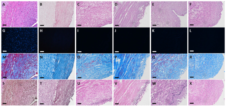
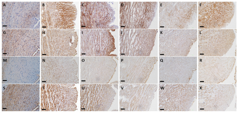
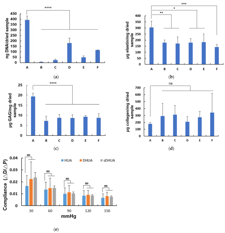
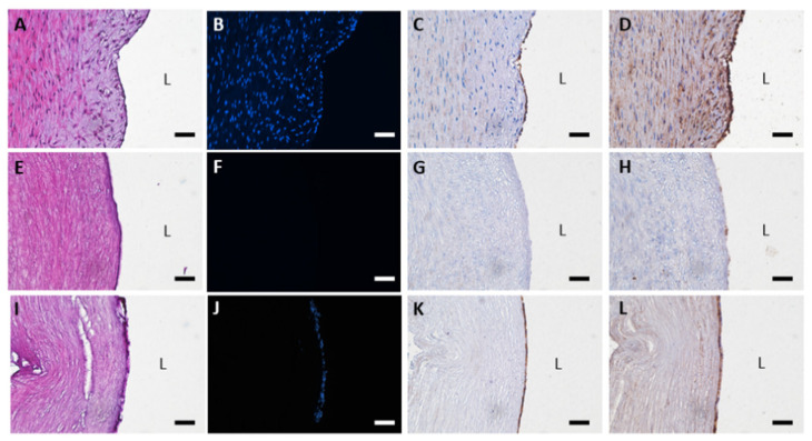
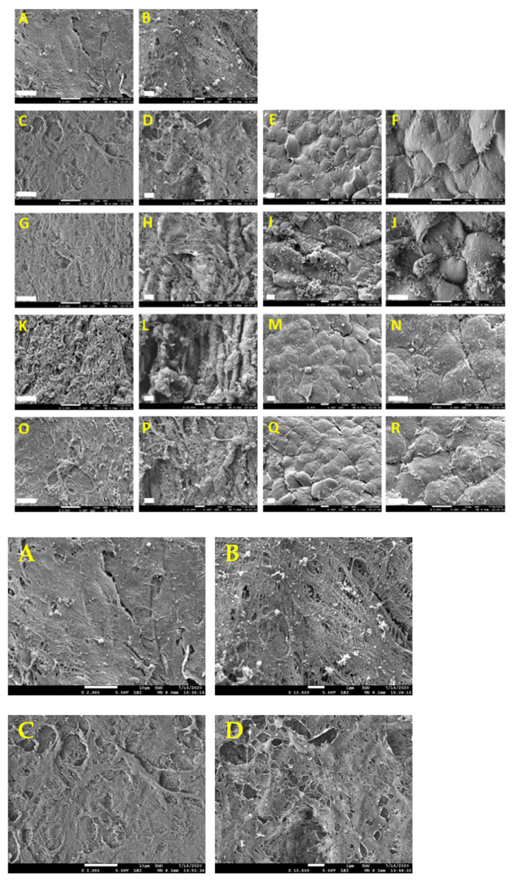
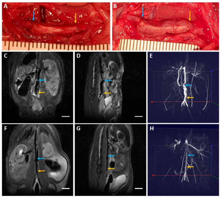
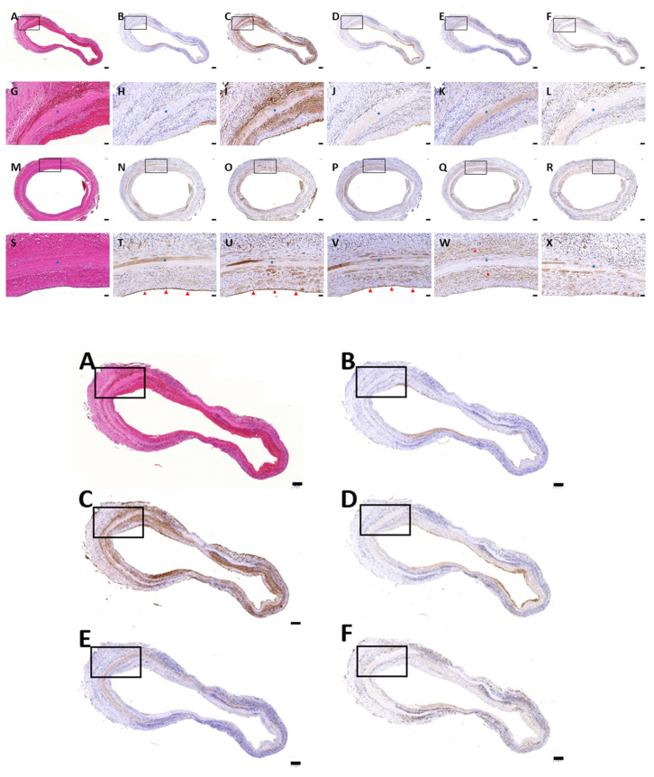
Decellularized vascular grafts are useful for the construction of biological small-diameter tissue-engineered vascular grafts (≤6 mm). Traditional chemical decellularization requires a long treatment time, which may damage the structure and alter the mechanical properties. Decellularization using sonication is expected to solve this problem. The aim of this study was to develop an effective decellularization method using ultrasound followed by washing. Different power values of sonication at 40 kHz were tested for 2, 4, and 8 h followed by a washing procedure. The efficacy of sonication of decellularized human umbilical artery (sDHUA) was evaluated via DNA content, histological staining, mechanical properties, and biocompatibility. The sDHUAs were further implanted into rats for up to 90 days and magnetic resonance angiography (MRA) was performed for the implanted grafts. The results demonstrated that treatment of human umbilical artery (HUA) by sonication at ultrasonic power of 204 W for 4 h followed by washing for 24 h in 2% SDS buffer could eliminate more than 90% of cells and retain similar mechanical properties of the HUA. Recellularization was assessed by scanning electron microscopy (SEM), which indicated that sDHUA provided niches for human umbilical vein endothelial cells (HUVECs) to reside, indicating in vitro cytocompatibility. Further implantation tests also indicated the fitness of the sonication-treated HUA as a scaffold for small-caliber tissue engineering vascular grafts.
Overview
The claims and statistics of this paper haven’t been checked yet.
Run the check — free, no account needed. It reads the open-access text and checks the sources it cites.
Integrity signals
Registry
Cites
Statistics
Figures
Verification
Claims the paper makes
The claims and statistics of this paper haven’t been checked yet.
Run the check — free, no account needed. It reads the open-access text and checks the sources it cites.
Claims other papers make about this one
Each is checked against this paper’s own text: does it say what the citing paper says it does?
The source backs part of the claim, not all of it — or backs it for a different population, condition or comparison.
In the citing paper
“These surfactants disrupt various cellular interactions, effectively eliminating cells and genetic material from tissues.”
From Bioactive scaffolds for tissue engineering: A review of decellularized extracellular matrix applications and innovations (2025) · cites this paper as [64]
Experimental — accuracy not yet measured
Self-check
Percentages, sums and reported differences re-computed from the paper’s own numbers.
The self-check needs the paper’s full text.
Citations
The citation lists haven’t been fetched yet
References 55 · Cited by 44
Research works printed but not linked: 52
- Salama, 2017, A systemic analysis for the global burden of disease study 2017, Lancet, 393, 1958
- Rashid, 2008, Tissue engineering of a hybrid bypass graft for coronary and lower limb bypass surgery, FASEB J., 22, 2084, 10.1096/fj.07-096586 · DOI
- Kannan, 2005, Current status of prosthetic bypass graft: A review, J. Biomed. Mater. Res. Part B, 74, 570, 10.1002/jbm.b.30247 · DOI
- Bennion, 1985, Patency of autogenous saphenous vein versus polytetrafluoroethylene grafts in femoropopliteal bypass for advanced ischemia of the extremity, Surg. Gynecol. Obstet., 160, 239
- Abbott, 1993, Evaluation and performance standards for arterial prostheses, J. Vasc. Surg., 17, 746, 10.1016/0741-5214(93)90120-B · DOI
- Yokota, 2008, In situ tissue regeneration using a novel tissue-engineered, small-caliber vascular graft without cell seeding, J. Thorac. Cardiovasc. Surg., 136, 900, 10.1016/j.jtcvs.2008.02.058 · DOI
- Mallis, P., Kostakis, A., Stavropoulos-Giokas, C., and Michalopoulos, E. (2020). Future perspectives in small-diameter vascular graft engineering. Bioengineering, 7. · DOI
- Crapo, 2011, An overview of tissue and whole organ decellularization processes, Biomaterials, 32, 3233, 10.1016/j.biomaterials.2011.01.057 · DOI · Paper page
- Keane, 2012, Consequences of ineffective decellularization of biologic scaffolds on the host response, Biomaterials, 33, 1771, 10.1016/j.biomaterials.2011.10.054 · DOI · Paper page
- Hawkins, 2003, Immunogenicity of decellularized cryopreserved allografts in pediatric cardiac surgery: Comparison with standard cryopreserved allografts, J. Thorac. Cardiovasc. Surg., 126, 247, 10.1016/S0022-5223(03)00116-8 · DOI
- Badylak, 2009, Extracellular matrix as a biological scaffold material: Structure and function, Acta Biomater., 5, 1, 10.1016/j.actbio.2008.09.013 · DOI · Paper page
- Sawada, 2008, Cell removal with supercritical carbon dioxide for acellular artificial tissue, J. Chem. Technol. Biotechnol. Int. Res. Process.Environ. Clean Technol., 83, 943
- Sasaki, 2009, In vivo evaluation of a novel scaffold for artificial corneas prepared by using ultrahigh hydrostatic pressure to decellularize porcine corneas, Mol. Vis., 15, 2022
- Funamoto, 2010, The use of high-hydrostatic pressure treatment to decellularize blood vessels, Biomaterials, 31, 3590, 10.1016/j.biomaterials.2010.01.073 · DOI
- Azhim, A., Yamagami, K., Muramatsu, K., Morimoto, Y., Furukawa, K.S., Tanaka, M., Fukui, Y., and Ushida, T. (2012, January 26–31). The use of sonication treatment to completely decellularize aorta tissue. Proceedings of the World Congress on Medical Physics and Biomedical Engineering, Beijing, China. · DOI
- Syazwani, 2015, Decellularization of aorta tissue using sonication treatment as potential scaffold for vascular tissue engineering, J. Med. Biol. Eng., 35, 258, 10.1007/s40846-015-0028-5 · DOI
- Say, S., Dugos, N., Roces, S., and Mondragon, J.M. (2019). Effect of sonication power on perfusion decellularization of cadaveric porcine kidney. MATEC Web of Conferences, 5–7 June 2019, EDP Sciences. · DOI
- Azhim, A., Ono, T., Fukui, Y., Morimoto, Y., Furukawa, K., and Ushida, T. (2013, January 3–7). Preparation of decellularized meniscal scaffolds using sonication treatment for tissue engineering. Proceedings of the 2013 35th Annual International Conference of the IEEE Engineering in Medicine and Biology Society (EMBC), Osaka, Japan. · DOI
- Azhim, 2015, The impact of acoustic intensity on solution parameters and decellularization using sonication treatment, J. Biomater. Tissue Eng., 5, 195, 10.1166/jbt.2015.1300 · DOI
- Niemczewski, 1999, Chemical activation of ultrasonic cavitation, Ultrason. Sonochemistry, 6, 211, 10.1016/S1350-4177(99)00008-5 · DOI
- Sun, D.-W. (2014). Emerging Technologies for Food Processing, Elsevier. [2nd ed.].
- Hung, 2013, Larynx decellularization: Combining freeze-drying and sonication as an effective method, J. Voice, 27, 289, 10.1016/j.jvoice.2013.01.018 · DOI
- Lin, C.H., Lu, J.H., Hsia, K., Lee, H., Yao, C.L., and Ma, H. (2019). The Antithrombotic Function of Sphingosine-1-Phosphate on Human Adipose-Stem-Cell-Recellularized Tissue Engineered Vascular Graft In Vitro. Int. J. Mol. Sci., 20. · DOI
- Kawecki, 2018, A review of decellurization methods caused by an urgent need for quality control of cell-free extracellular matrix’ scaffolds and their role in regenerative medicine, J. Biomed. Mater. Res. Part. B Appl. Biomater., 106, 909, 10.1002/jbm.b.33865 · DOI
- Gui, 2009, Development of decellularized human umbilical arteries as small-diameter vascular grafts, Tissue Eng. Part. A, 15, 2665, 10.1089/ten.tea.2008.0526 · DOI
- Miller, 1986, Effects of a high-amplitude 1-MHz standing ultrasonic field on the algae hydrodictyon, IEEE Trans. Ultrason. Ferroelectr. Freq. Control., 33, 165, 10.1109/T-UFFC.1986.26810 · DOI
- Xu, J., and Mosher, D. (2011). Fibronectin and other adhesive glycoproteins. The Extracellular Matrix: An Overview, Springer. · DOI
- Gui, 2010, Novel utilization of serum in tissue decellularization, Tissue Eng. Part. C Methods, 16, 173, 10.1089/ten.tec.2009.0120 · DOI · Paper page
- Woods, 2005, Effectiveness of three extraction techniques in the development of a decellularized bone–anterior cruciate ligament–bone graft, Biomaterials, 26, 7339, 10.1016/j.biomaterials.2005.05.066 · DOI
- Deeken, 2011, Method of preparing a decellularized porcine tendon using tributyl phosphate, J. Biomed. Mater. Res. Part. B Appl. Biomater., 96, 199, 10.1002/jbm.b.31753 · DOI
- Lee, 1995, The sodium lauryl sulfate model: An overview, Contact Dermat., 33, 1, 10.1111/j.1600-0536.1995.tb00438.x · DOI
- Gratzer, 2006, Matrix alteration and not residual sodium dodecyl sulfate cytotoxicity affects the cellular repopulation of a decellularized matrix, Tissue Eng., 12, 2975, 10.1089/ten.2006.12.2975 · DOI
- Azhim, A., Yamagami, K., Muramatsu, K., Morimoto, Y., and Tanaka, M. (September, January 30). The use of sonication treatment to completely decellularize blood arteries: A pilot study. Proceedings of the 2011 Annual International Conference of the IEEE Engineering in Medicine and Biology Society, Boston, MA, USA. · DOI
- Chattopadhyay, 1996, Dependence of critical micelle concentration of a zwitterionic detergent on ionic strength: Implications in receptor solubilization, Febs Lett., 391, 199, 10.1016/0014-5793(96)00733-8 · DOI
- Daniel, 2005, Development of the human umbilical vein scaffold for cardiovascular tissue engineering applications, Asaio, J., 51, 252, 10.1097/01.MAT.0000160872.41871.7E · DOI
- Rodriguez, 2012, Development of a mechanically tuneable 3D scaffold for vascular reconstruction, J. Biomed. Mater. Res. Part. A, 100, 3480, 10.1002/jbm.a.34267 · DOI
- Barnwal, 2012, Histomorphometry of umbilical cord and its vessels in pre-eclampsia as compared to normal pregnancies, Nepal, J. Obstet. Gynaecol., 7, 28, 10.3126/njog.v7i1.8832 · DOI
- Blanco, 2011, Histopathology and histomorphometry of umbilical cord blood vessels. Findings in normal and high risk pregnancies, Artery Res., 5, 50, 10.1016/j.artres.2011.02.001 · DOI
- Bańkowski, E. (1999). Collagen of the umbilical cord and its alteration in EPH-gestosis (preeclampsia). Proceedings of the Indian Academy of Sciences-Chemical Sciences, 1999, Springer. · DOI
- Ramos, 2014, Decontamination by ultrasound application in fresh fruits and vegetables, Food Control., 45, 36, 10.1016/j.foodcont.2014.04.015 · DOI
- Seidel, A., and Bickford, M. (2007). Sonochemistry. Kirk-Othmer Encyclopedia of Chemical Technology, John Wiley & Sons.
- Wang, Y. (2010). Vascular biology of the placenta. Colloquium Series on Integrated Systems Physiology: From Molecule to Function, Morgan & Claypool Life Sciences. · DOI
- Wagenseil, 2009, Vascular extracellular matrix and arterial mechanics, Physiol. Rev., 89, 957, 10.1152/physrev.00041.2008 · DOI
- Yu, 2014, On the decellularization of fresh or frozen human umbilical arteries: Implications for small-diameter tissue engineered vascular grafts, Ann. Biomed. Eng., 42, 1305, 10.1007/s10439-014-1000-1 · DOI
- Pennati, 2001, Biomechanical properties of the human umbilical cord, Biorheology, 38, 355
- Brey, 2018, Endothelialization mechanisms in vascular grafts, J. Tissue Eng. Regen. Med., 12, 2164, 10.1002/term.2747 · DOI
- Martin, 2005, In vivo behavior of decellularized vein Allograft, J. Surg. Res., 129, 17, 10.1016/j.jss.2005.06.037 · DOI
- Allaire, 1997, J.B. The immunogenicity of the extracellular matrix in arterial xenografts, Surgery, 122, 73, 10.1016/S0039-6060(97)90267-1 · DOI
- Talacua, 2015, In situ tissue engineering of functional small-diameter blood vessels by host circulating cells only, Tissue Eng. Part A, 21, 2583, 10.1089/ten.tea.2015.0066 · DOI
- Badylak, 2014, Decellularized allogeneic and xenogeneic tissue as a bioscaffold for regenerative medicine: Factors that influence the host response, Ann. Biomed. Eng., 42, 1517, 10.1007/s10439-013-0963-7 · DOI · Paper page
- Allaire, 1998, Local overexpression of TIMP-1 prevents aortic aneurysm degeneration and rupture in a rat model, J. Clin. Investig., 102, 1413, 10.1172/JCI2909 · DOI
- Ariganello, 2011, Macrophage differentiation and polarization on a decellularized pericardial biomaterial, Biomaterials, 32, 439, 10.1016/j.biomaterials.2010.09.004 · DOI
Metadata
- Authors
- Lin, C.-H., Hsia, K., Su, C.-K., Chen, C.-C., Yeh, C.-C., Ma, H., & Lu, J.-H.
- Affiliations
- National Yang Ming Chiao Tung University (TW)
- Taipei Veterans General Hospital (TW)
- Chia-Yi Christian Hospital (TW)
- Taipei Medical University Hospital (TW)
- Corresponding author
- Lu, Jen-Her
- Venue
- Polymers
- Publisher
- MDPI AG
- Published
- 2021-05-22
- Volume
- 13
- Issue
- 11
- Pages
- 1699
- ISSN
- 2073-4360 (ISSN-L)
- PMC
- PMC8196986
- OpenAlex
- W3164513037
- Cited by
- 44
- References
- 55
- Citations by year
- 2026: 9 · 2025: 10 · 2024: 13 · 2023: 8 · 2022: 3 · 2021: 1
- FWCI
- 5.87
- Type
- journal article
- Language
- en
- Open access
- gold
- doi.org
- Licence
- Locations
- Polymers (publishedVersion, cc-by)
- PubMed Central (publishedVersion, cc-by)
- PubMed (publishedVersion)
- DOAJ (DOAJ: Directory of Open Access Journals) (submittedVersion, cc-by-sa)
- Funders
- Ministry of Science and Technology, Taiwan (MoST108-2314-B-075 -053, 108-2314-B-075 -045, and MOST 109-2314-B-075 -007)
- Topics
- Health Sciences › Medicine › Surgery › Tissue Engineering and Regenerative Medicine
- Physical Sciences › Materials Science › Biomaterials › Electrospun Nanofibers in Biomedical Applications
- Physical Sciences › Engineering › Biomedical Engineering › 3D Printing in Biomedical Research
- Keywords
- decellularization, sonication, human umbilical artery, tissue-engineered vascular grafts, small-diameter vascular grafts, ultrasonic power, recellularization, human umbilical vein endothelial cells, graft biocompatibility, graft mechanical properties, graft implantation, magnetic resonance angiography
About these checks
Registry, retraction, Cites and statistics results are computed, not judged, with one exception: a printed reference that no index lists is read once by a model before it counts as a citation error. Claims are extracted by a model and kept only when their quote appears verbatim in the text; each cited source is then checked, with a model’s help, for whether it says what the claim relies on. Those verdicts can be wrong — read the claim’s quote and the cited source before relying on one.
Statistics and arithmetic are recomputed, not judged.
Confidence bands are shown only for fields calibrated on our annotated test set. None is calibrated yet, so no band is shown.
What counts as a claim ›How we measure confidence ›How we check this ›
Are you an author, or do you see a problem with this page? Request a review
How to cite
Cite CoChat for the checks on this page, and the paper for its own findings.
- This page
- CoChat. (2026). Citation check: Sonication-Assisted Method for Decellularization of Human Umbilical Artery for Small-Caliber Vascular Tissue Engineering. https://app.cochat.ai/papers/10.3390/polym13111699
- The paper
- Lin, C.-H., Hsia, K., Su, C.-K., Chen, C.-C., Yeh, C.-C., Ma, H., & Lu, J.-H. (2021). Sonication-Assisted Method for Decellularization of Human Umbilical Artery for Small-Caliber Vascular Tissue Engineering. Polymers. https://doi.org/10.3390/polym13111699
