

The DOI matches its Crossref record; 149 of the 149 references it cites were found in a registry or an index.
Non-Invasive Blood Glucose Monitoring Technology: A Review
Open accessAbstract
In recent years, with the rise of global diabetes, a growing number of subjects are suffering from pain and infections caused by the invasive nature of mainstream commercial glucose meters. Non-invasive blood glucose monitoring technology has become an international research topic and a new method which could bring relief to a vast number of patients. This paper reviews the research progress and major challenges of non-invasive blood glucose detection technology in recent years, and divides it into three categories: optics, microwave and electrochemistry, based on the detection principle. The technology covers medical, materials, optics, electromagnetic wave, chemistry, biology, computational science and other related fields. The advantages and limitations of non-invasive and invasive technologies as well as electrochemistry and optics in non-invasives are compared horizontally in this paper. In addition, the current research achievements and limitations of non-invasive electrochemical glucose sensing systems in continuous monitoring, point-of-care and clinical settings are highlighted, so as to discuss the development tendency in future research. With the rapid development of wearable technology and transdermal biosensors, non-invasive blood glucose monitoring will become more efficient, affordable, robust, and more competitive on the market.
Overview
The claims and statistics of this paper haven’t been checked yet.
Run the check — free, no account needed. It reads the open-access text and checks the sources it cites.
Integrity signals
Registry
Cites
Statistics
Figures
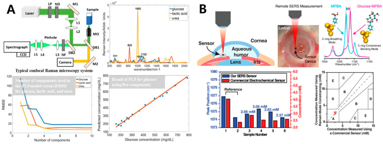
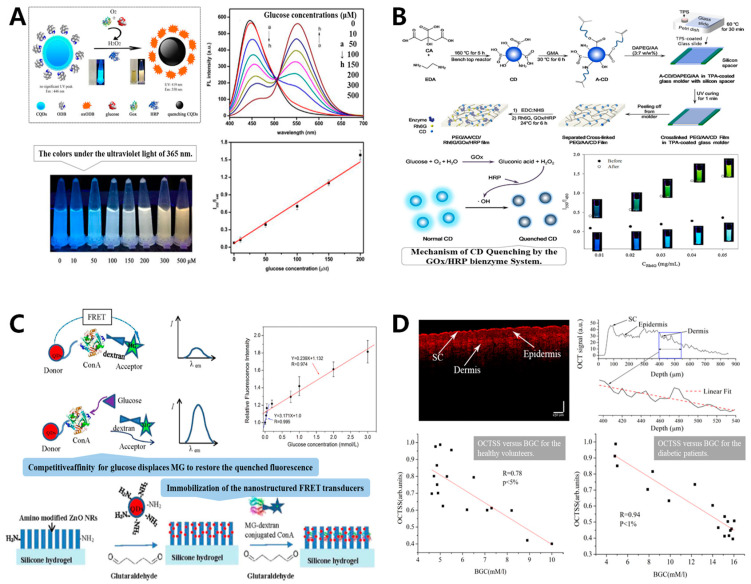
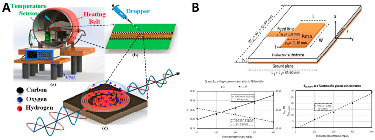
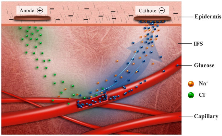
Verification
Claims the paper makes
The claims and statistics of this paper haven’t been checked yet.
Run the check — free, no account needed. It reads the open-access text and checks the sources it cites.
Claims other papers make about this one
Each is checked against this paper’s own text: does it say what the citing paper says it does?
The source backs part of the claim, not all of it — or backs it for a different population, condition or comparison.
In the citing paper
“Indeed, it is remarkable that glucose levels in biofluids, such as ISF (Figure 4B), sweat (Figure 4C), saliva, tears, and urine (Figure 4 D, E, F, respectively) are correlated with those in the blood 76.”
From Glucose biosensors in clinical practice: principles, limits and perspectives of currently used devices (2022) · cites this paper as [76]
Experimental — accuracy not yet measuredThe cited source says what the claim attributes to it.
In the citing paper
“As stated above, over the years, enzymatic amperometric glucose sensors were the first and widespread glucose sensors available 76.”
From Glucose biosensors in clinical practice: principles, limits and perspectives of currently used devices (2022) · cites this paper as [76]
The check wasn’t confident either way, so it gives no verdict — never disputed.
In the citing paper
“It is widely known that enzymatic sensors such as those based on glucose oxidase are more stable than other enzymes, easy to obtain, and cheap; on the other hand, it quickly loses its activity at pH<2 or pH>8 and can be irrevocably damaged at temperatures above 40 °C 76.”
From Glucose biosensors in clinical practice: principles, limits and perspectives of currently used devices (2022) · cites this paper as [76]
Experimental — accuracy not yet measured
Self-check
Percentages, sums and reported differences re-computed from the paper’s own numbers.
The self-check needs the paper’s full text.
Citations
Cites
Standards of Medical Care in Diabetes
Power, D. V. (2006) · Diabetes Care · doi:10.2337/diacare.29.02.06.dc05-1593 · Cited by 8,676
Standards of medical care in diabetes—2014
American Diabetes Association (2014) · Diabetes Care · doi:10.2337/dc14-s014 · Cited by 4,572
Gabriel, S., Lau, R. W., & Gabriel, C. (1996) · Physics in Medicine and Biology · doi:10.1088/0031-9155/41/11/003 · Cited by 4,175
ELECTRODE SYSTEMS FOR CONTINUOUS MONITORING IN CARDIOVASCULAR SURGERY
Clark, L. C., & Lyons, C. (1962) · Annals of the New York Academy of Sciences · doi:10.1111/j.1749-6632.1962.tb13623.x · Cited by 3,945
Electrochemical Glucose Biosensors
Wang, J. (2008) · Chemical Reviews · doi:10.1021/cr068123a · Cited by 3,498
Standards of medical care in diabetes 2010
American Diabetes Association (2010) · Diabetes Care · doi:10.2337/dc10-s011 · Cited by 3,470
Nathan, D. M., & for the DCCT/EDIC Research Group (2014) · Diabetes Care · doi:10.2337/dc13-2112 · Cited by 1,832
Wearable and flexible electronics for continuous molecular monitoring
Yang, Y., & Gao, W. (2019) · Chemical Society Reviews · doi:10.1039/c7cs00730b · Cited by 1,315
Flexible Electronics toward Wearable Sensing
Gao, W. F., Ota, H., Kiriya, D., Takei, K., & Javey, A. (2019) · Accounts of Chemical Research · doi:10.1021/acs.accounts.8b00500 · Cited by 1,301
Microneedles: A smart approach and increasing potential for transdermal drug delivery system
Waghule, T., Singhvi, G., Dubey, S. K., Pandey, M. M., Gupta, G., Singh, M., & Dua, K. (2018) · Biomedicine & Pharmacotherapy · doi:10.1016/j.biopha.2018.10.078 · Cited by 1,277
Cited by
Sorted by citations
Flexible Wearable Sensors for Cardiovascular Health Monitoring
Chen, S., Qi, J., Fan, S., Qiao, Z., Yeo, J. C., & Lim, C. T. (2021) · Advanced Healthcare Materials · doi:10.1002/adhm.202100116 · Cited by 453
Pullano, S. A., Greco, M., Bianco, M. G., Foti, D., Brunetti, A., & Fiorillo, A. S. (2022) · Theranostics · doi:10.7150/thno.64035 · Cited by 210
“Indeed, it is remarkable that glucose levels in biofluids, such as ISF (Figure 4B), sweat (Figure 4C), saliva, tears, and urine (Figure 4 D, E, F, respectively) are correlated with those in the blood [76].”
BackgroundMethodInfluentialWu, Z., Qiao, Z., Chen, S., Fan, S., Liu, Y., Qi, J., & Lim, C. T. (2024) · Communications Materials · doi:10.1038/s43246-024-00468-6 · Cited by 204
Magnesium Oxide (MgO) Nanoparticles: Synthetic Strategies and Biomedical Applications
Gatou, Μ.-A., Skylla, E., Dourou, P., Pippa, Ν., Gazouli, M., Lаgopati, N., & Pavlatou, E. A. (2024) · Crystals · doi:10.3390/cryst14030215 · Cited by 176
Thwala, L. N., Ndlovu, S. C., Mpofu, K. T., Lugongolo, M. Y., & Mthunzi-Kufa, P. T. (2023) · Nanomaterials · doi:10.3390/nano13071247 · Cited by 171
Metadata
- Authors
- Tang, L., Chang, S. J., Chen, C.-J., & Liu, J.-T.
- Affiliations
- Research Center for Materials Science and Opti-Electronic Technology, College of Materials Science and Opti-Electronic Technology, University of Chinese Academy of Sciences, Beijing 100049, China
- I-Shou University (TW)
- University of Chinese Academy of Sciences (CN)
- Corresponding author
- Chen, Ching-Jung
- Liu, Jen-Tsai
- Venue
- Sensors
- Publisher
- MDPI AG
- Published
- 2020-12-04
- Volume
- 20
- Issue
- 23
- Pages
- 6925
- ISSN
- 1424-8220 (ISSN-L)
- PMC
- PMC7731259
- OpenAlex
- W3106856419
- Cited by
- 481
- References
- 149
- Citations by year
- 2026: 66 · 2025: 100 · 2024: 101 · 2023: 111 · 2022: 71 · 2021: 32
- FWCI
- 2.65
- Type
- journal article
- Language
- en
- Open access
- gold
- doi.org
- Licence
- Locations
- Sensors (publishedVersion, cc-by)
- PubMed Central (publishedVersion, cc-by)
- PubMed (publishedVersion)
- DOAJ (DOAJ: Directory of Open Access Journals) (submittedVersion, cc-by-sa)
- Funders
- Chinese Academy of Sciences
- University of Chinese Academy of Sciences
- Topics
- Physical Sciences › Chemistry › Analytical Chemistry › Electrochemical sensors and biosensors
- Life Sciences › Biochemistry, Genetics and Molecular Biology › Biophysics › Spectroscopy Techniques in Biomedical and Chemical Research
- Physical Sciences › Engineering › Biomedical Engineering › Non-Invasive Vital Sign Monitoring
- Keywords
- non-invasive glucose monitoring, diabetes mellitus, optical glucose sensing, electrochemical glucose sensor, continuous glucose monitoring, point-of-care testing, wearable sensors
- MeSH terms
- Wearable Electronic Devices, Blood Glucose, Humans, Monitoring, Physiologic, Blood Glucose Self-Monitoring, Biosensing Techniques
About these checks
Registry, retraction, Cites and statistics results are computed, not judged, with one exception: a printed reference that no index lists is read once by a model before it counts as a citation error. Claims are extracted by a model and kept only when their quote appears verbatim in the text; each cited source is then checked, with a model’s help, for whether it says what the claim relies on. Those verdicts can be wrong — read the claim’s quote and the cited source before relying on one.
Statistics and arithmetic are recomputed, not judged.
Confidence bands are shown only for fields calibrated on our annotated test set. None is calibrated yet, so no band is shown.
What counts as a claim ›How we measure confidence ›How we check this ›
Are you an author, or do you see a problem with this page? Request a review
