

Structurally Robust and Functionally Highly Versatile—C-Type Lectin (-Related) Proteins in Snake Venoms
Open accessThe DOI matches its Crossref record; 217 of the 217 references it cites were found in a registry or an index.
Abstract
Snake venoms contain an astounding variety of different proteins. Among them are numerous C-type lectin family members, which are grouped into classical Ca2+- and sugar-binding lectins and the non-sugar-binding snake venom C-type lectin-related proteins (SV-CLRPs), also called snaclecs. Both groups share the robust C-type lectin domain (CTLD) fold but differ in a long loop, which either contributes to a sugar-binding site or is expanded into a loop-swapping heterodimerization domain between two CLRP subunits. Most C-type lectin (-related) proteins assemble in ordered supramolecular complexes with a high versatility of subunit numbers and geometric arrays. Similarly versatile is their ability to inhibit or block their target molecules as well as to agonistically stimulate or antagonistically blunt a cellular reaction triggered by their target receptor. By utilizing distinct interaction sites differentially, SV-CLRPs target a plethora of molecules, such as distinct coagulation factors and receptors of platelets and endothelial cells that are involved in hemostasis, thrombus formation, inflammation and hematogenous metastasis. Because of their robust structure and their high affinity towards their clinically relevant targets, SV-CLRPs are and will potentially be valuable prototypes to develop new diagnostic and therapeutic tools in medicine, provided that the molecular mechanisms underlying their versatility are disclosed.
Overview
The claims and statistics of this paper haven’t been checked yet.
Free: it reads the open-access text and checks the first 5 claims that cite a source we can check. Create a free account to check every claim.
Integrity signals
Registry
Cites
Statistics
Figures
Verification
Claims the paper makes
The claims and statistics of this paper haven’t been checked yet.
Free: it reads the open-access text and checks the first 5 claims that cite a source we can check. Create a free account to check every claim.
Claims other papers make about this one
Each is checked against this paper’s own text: does it say what the citing paper says it does?
The cited source says what the claim attributes to it.
In the citing paper
“C-type lectins (CTLs/snaclecs) are predominantly found in venoms of viperids. Although structurally related to classical C-type lectins, most snaclecs lack carbohydrate binding activity [19].”
From Role of Regulated Cell Death Pathways in Snakebite Envenomation: Mechanisms, Crosstalk, and Therapeutic Opportunities (2026) · cites this paper as [19]
The check wasn’t confident either way, so it gives no verdict — never disputed.
In the citing paper
“This can result in cellular swelling or the release of cellular contents, leading to severe necrosis or more regulated pathways for cellular destruction [19].”
From Role of Regulated Cell Death Pathways in Snakebite Envenomation: Mechanisms, Crosstalk, and Therapeutic Opportunities (2026) · cites this paper as [19]
Experimental — accuracy not yet measured
Self-check
Percentages, sums and reported differences re-computed from the paper’s own numbers.
The self-check needs the paper’s full text.
Citations
Cites
Mizgerd, J. P., Kubo, H., Kutkoski, G. J., Bhagwan, S. D., Scharffetter-Kochanek, K., Beaudet, A. L., & Doerschuk, C. M. (1997) · The Journal of Experimental Medicine · doi:10.1084/jem.186.8.1357 · Cited by 7,306
Stöckl, S., Göttl, C., Grifka, J., & Grässel, S. (2013) · Cellular Physiology and Biochemistry · doi:10.1159/000350089 · Cited by 4,656
Ricard-Blum, S. (2011) · Cold Spring Harbor Perspectives in Biology · doi:10.1101/cshperspect.a004978 · Cited by 2,305
Humphries, J. D., Byron, A., & Humphries, M. J. (2006) · Journal of Cell Science · doi:10.1242/jcs.03098 · Cited by 1,708 · cited 1×
“The extracellular domains of both subunits form one head domain, which harbours the binding site for the extracellular matrix ligands [136].”
BackgroundStructural Basis of Integrin Regulation and Signaling
Luo, B.-H., Carman, C. V., & Springer, T. A. (2007) · Annual Review of Immunology · doi:10.1146/annurev.immunol.25.022106.141618 · Cited by 1,660 · cited 1×
“This enables not only firm connection between the cytoskeleton and the extracellular matrix, but also signal transduction via the integrin-mediated cell-extra cellular matrix contacts [137,138].”
BackgroundNanoscale architecture of integrin-based cell adhesions
Kanchanawong, P., Shtengel, G., Pasapera, A. M., Ramko, E. B., Davidson, M. W., Hess, H. F., & Waterman, C. M. (2010) · Nature · doi:10.1038/nature09621 · Cited by 1,567 · cited 1×
“Moreover, integrin signalling also encompasses clustering of several integrin molecules and associated proteins into specific cell-adhesive cell organelles, termed adhesomes [139].”
BackgroundBarczyk, M., Carracedo, S., & Gullberg, D. (2010) · Cell and Tissue Research · doi:10.1007/s00441-009-0834-6 · Cited by 1,512
The C‐type lectin‐like domain superfamily
Zelensky, A. N., & Gready, J. E. (2005) · The FEBS Journal · doi:10.1111/j.1742-4658.2005.05031.x · Cited by 1,407 · cited 8×
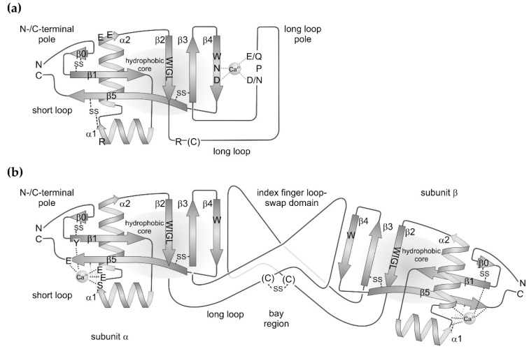
“This N-terminal expansion is stabilized by a disulfide bridge between two cysteine residues, generically named C0 and C0′ [2].”
BackgroundInfluentialProteoglycan form and function: A comprehensive nomenclature of proteoglycans
Iozzo, R. V., & Schaefer, L. (2015) · Matrix Biology · doi:10.1016/j.matbio.2015.02.003 · Cited by 1,263 · cited 1×
“Such variants are found in the hyaluronic acid-binding link domain (synonym: protein tandem repeat (PTR) domain) of lecticans, among them aggrecan, versican and other proteoglycans [12] and of the hyaluronan receptor CD44, as well as in some bacterial CTLD-containing proteins, such as in intimin and invasin [8].”
BackgroundMET signalling: principles and functions in development, organ regeneration and cancer
Trusolino, L., Bertotti, A., & Comoglio, P. M. (2010) · Nature Reviews Molecular Cell Biology · doi:10.1038/nrm3012 · Cited by 1,240
Cited by
Phylogenetically diverse diets favor more complex venoms in North American pitvipers
Holding, M. L., Strickland, J. L., Rautsaw, R. M., Hofmann, E. P., Mason, A. J., Hogan, M., Nystrom, G. S., Ellsworth, S. A., Colston, T. J., Borja, M., et al. (2021) · Proceedings of the National Academy of Sciences · doi:10.1073/pnas.2015579118 · Cited by 117
Old World Vipers—A Review about Snake Venom Proteomics of Viperinae and Their Variations
Damm, M., Hempel, B.-F., & Süßmuth, R. D. (2021) · Toxins · doi:10.3390/toxins13060427 · Cited by 94
Larréché, S., Chippaux, J., Chevillard, L., Mathé, S., Résière, D., Siguret, V., & Mégarbane, B. (2021) · International Journal of Molecular Sciences · doi:10.3390/ijms22179643 · Cited by 103
“The concave surface is probably the main ligand binding site [95].”
BackgroundTeixeira, C., Fernándes, C. M., Leiguez, E., & Chudzinski‐Tavassi, A. M. (2019) · Frontiers in Immunology · doi:10.3389/fimmu.2019.02082 · Cited by 78
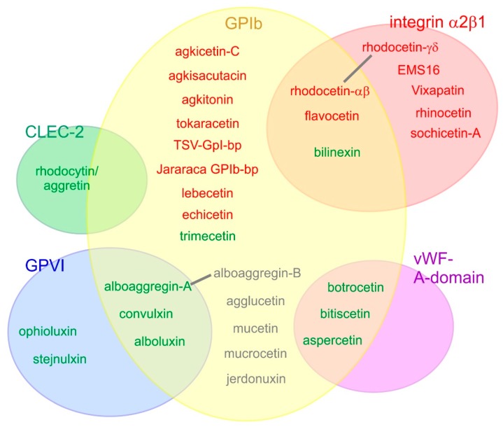
“SV-CLRPs connect with amultitude of molecules implicated in hemostasis that are present on endothelial cells, coagulation factors, and receptors of platelets, having a role in thrombus formation and inflammation (150).”
BackgroundJenkins, T. P., Ahmadi, S., Bittenbinder, M. A., Stewart, T. K., Akgun, D. E., Hale, M., Nasrabadi, N. N., Wolff, D. S., Vonk, F. J., Kool, J., et al. (2021) · PLoS neglected tropical diseases · doi:10.1371/journal.pntd.0009880 · Cited by 58
“, bitiscetin) are hemostasis disruptive and consequently prevent blood clotting by interfering with platelet aggregation and the coagulation cascade [35].”
Background
Metadata
- Authors
- Eble, J. A.
- Affiliations
- University of Münster (DE)
- Corresponding author
- Eble, Johannes A.
- ORCID
- Eble, Johannes A.
- Venue
- Toxins
- Publisher
- MDPI AG
- Published
- 2019-03-01
- Volume
- 11
- Issue
- 3
- Pages
- 136
- ISSN
- 2072-6651 (ISSN-L)
- PMC
- PMC6468738
- OpenAlex
- W2919970718
- Cited by
- 82
- References
- 217
- Citations by year
- 2026: 8 · 2025: 11 · 2024: 15 · 2023: 9 · 2022: 23 · 2021: 11 · 2020: 3 · 2019: 2
- FWCI
- 1.31
- Type
- journal article
- Language
- en
- Open access
- gold
- dx.doi.org
- Licence
- Locations
- Toxins (publishedVersion, cc-by)
- Europe PMC (PubMed Central) (publishedVersion, cc-by)
- PubMed Central (publishedVersion, cc-by)
- PubMed (publishedVersion)
- DOAJ (DOAJ: Directory of Open Access Journals) (submittedVersion, cc-by-sa)
- Funders
- Deutsche Forschungsgemeinschaft (Eb177/13-1)
- Topics
- Life Sciences › Pharmacology, Toxicology and Pharmaceutics › Toxicology › Venomous Animal Envenomation and Studies
- Life Sciences › Biochemistry, Genetics and Molecular Biology › Molecular Biology › Glycosylation and Glycoproteins Research
- Life Sciences › Immunology and Microbiology › Immunology › Toxin Mechanisms and Immunotoxins
- Keywords
- snake venom, C-type lectin domain, supramolecular complexes, hemostasis, platelet receptors, endothelial cell receptors, coagulation factors, inflammation, hematogenous metastasis, receptor antagonism, receptor agonism
- MeSH terms
- Animals, Humans, Protein Conformation, Snake Venoms, Lectins, C-Type
About these checks
Registry, retraction, Cites and statistics results are computed, not judged, with one exception: a printed reference that no index lists is read once by a model before it counts as a citation error. Claims are extracted by a model and kept only when their quote appears verbatim in the text; each cited source is then checked, with a model’s help, for whether it says what the claim relies on. Those verdicts can be wrong — read the claim’s quote and the cited source before relying on one.
Statistics and arithmetic are recomputed, not judged.
Confidence bands are shown only for fields calibrated on our annotated test set. None is calibrated yet, so no band is shown.
What counts as a claim ›How we measure confidence ›How we check this ›
Are you an author, or do you see a problem with this page? Request a review
How to cite
Cite CoChat for the checks on this page, and the paper for its own findings.
- This page
- CoChat. (2026). Citation check: Structurally Robust and Functionally Highly Versatile—C-Type Lectin (-Related) Proteins in Snake Venoms. https://app.cochat.ai/papers/10.3390/toxins11030136
- The paper
- Eble, J. A. (2019). Structurally Robust and Functionally Highly Versatile—C-Type Lectin (-Related) Proteins in Snake Venoms. Toxins. https://doi.org/10.3390/toxins11030136
Data sources
Bibliographic data: Semantic Scholar, OpenAlex, Crossref.
Contains information from the Semantic Scholar Academic Graph Datasets, made available under the ODC Attribution License (ODC-BY 1.0).
Semantic Scholar’s data is described in: Kinney, R., et al. (2023). The Semantic Scholar Open Data Platform. arXiv:2301.10140.
