

The DOI matches its Crossref record; 342 of the 342 references it cites were found in a registry or an index.
Human Coronaviruses and Other Respiratory Viruses: Underestimated Opportunistic Pathogens of the Central Nervous System?
Open accessAbstract
Respiratory viruses infect the human upper respiratory tract, mostly causing mild diseases. However, in vulnerable populations, such as newborns, infants, the elderly and immune-compromised individuals, these opportunistic pathogens can also affect the lower respiratory tract, causing a more severe disease (e.g., pneumonia). Respiratory viruses can also exacerbate asthma and lead to various types of respiratory distress syndromes. Furthermore, as they can adapt fast and cross the species barrier, some of these pathogens, like influenza A and SARS-CoV, have occasionally caused epidemics or pandemics, and were associated with more serious clinical diseases and even mortality. For a few decades now, data reported in the scientific literature has also demonstrated that several respiratory viruses have neuroinvasive capacities, since they can spread from the respiratory tract to the central nervous system (CNS). Viruses infecting human CNS cells could then cause different types of encephalopathy, including encephalitis, and long-term neurological diseases. Like other well-recognized neuroinvasive human viruses, respiratory viruses may damage the CNS as a result of misdirected host immune responses that could be associated with autoimmunity in susceptible individuals (virus-induced neuro-immunopathology) and/or viral replication, which directly causes damage to CNS cells (virus-induced neuropathology). The etiological agent of several neurological disorders remains unidentified. Opportunistic human respiratory pathogens could be associated with the triggering or the exacerbation of these disorders whose etiology remains poorly understood. Herein, we present a global portrait of some of the most prevalent or emerging human respiratory viruses that have been associated with possible pathogenic processes in CNS infection, with a special emphasis on human coronaviruses.
Overview
The claims and statistics of this paper haven’t been checked yet.
Run the check — free, no account needed. It reads the open-access text and checks the sources it cites.
Integrity signals
Registry
Cites
Statistics
Figures
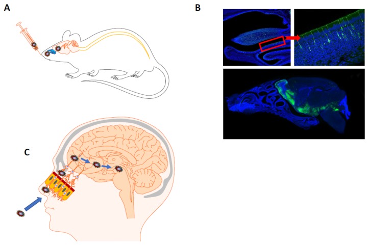
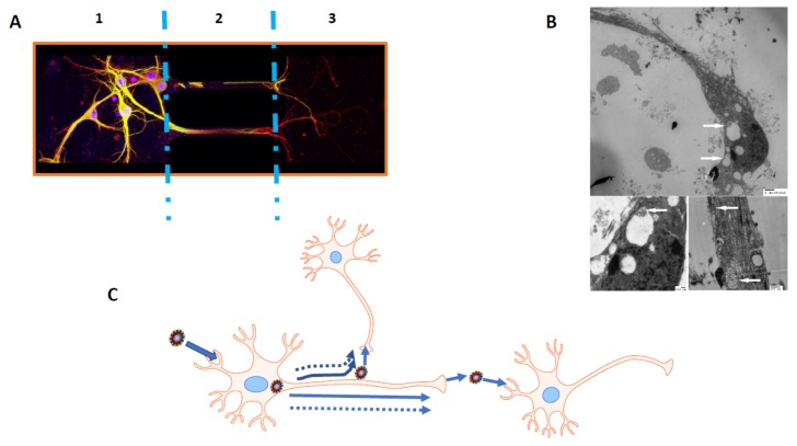
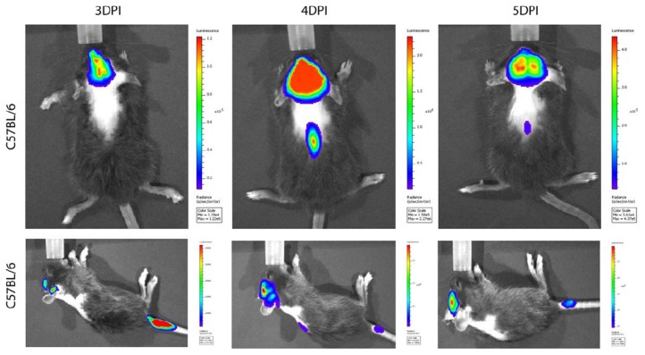
Verification
Claims the paper makes
The claims and statistics of this paper haven’t been checked yet.
Run the check — free, no account needed. It reads the open-access text and checks the sources it cites.
Claims other papers make about this one
Each is checked against this paper’s own text: does it say what the citing paper says it does?
In the citing paper
“Clinical, post-mortem, animal, in vitro, and cell culture studies demonstrated that coronaviruses are potentially neurotropic and can induce neuronal injuries (Desforges et al., 2019).”
From Anxiety and depression in COVID-19 survivors: role of inflammatory and clinical predictors (2020) · cites this paper as Desforges et al., 2019
In the citing paper
“As a consequence, CoV can enter the brain through the olfactory tract in the early stages of infection or nasal vaccination (Desforges et al., 2019, Mori, 2015).”
From Nervous system involvement after infection with COVID-19 and other coronaviruses (2020) · cites this paper as Desforges et al., 2019
In the citing paper
“Although there is rare evidence that CoV, especially SARS-CoV-2, invade the nervous system via the blood circulation pathway (Koyuncu et al., 2013, Desforges et al., 2019), subsequent studies are expected.”
From Nervous system involvement after infection with COVID-19 and other coronaviruses (2020) · cites this paper as Desforges et al., 2019
Self-check
Percentages, sums and reported differences re-computed from the paper’s own numbers.
The self-check needs the paper’s full text.
Citations
Cites
The environment and disease: association or causation?
Hill, A. B. (2015) · Journal of the Royal Society of Medicine · doi:10.1177/0141076814562718 · Cited by 7,804
The Environment and Disease: Association or Causation?
Hill, A. B. (1965) · Proceedings of the Royal Society of Medicine · doi:10.1177/003591576505800503 · Cited by 6,974
Isolation of a Novel Coronavirus from a Man with Pneumonia in Saudi Arabia
Zaki, A. M., van Boheemen, S., Bestebroer, T. M., Osterhaus, A. D. M. E., & Fouchier, R. A. M. (2012) · New England Journal of Medicine · doi:10.1056/nejmoa1211721 · Cited by 5,677
Identification of a novel coronavirus in patients with severe acute respiratory syndrome
Drosten, C., Günther, S., Preiser, W., van der Werf, S., Brodt, H.-R., Becker, S., Rabenau, H., Panning, M., Kolesnikova, L., Fouchier, R. A. M., et al. (2003) · New England Journal of Medicine · doi:10.1056/nejmoa030747 · Cited by 4,574
A Novel Coronavirus Associated with Severe Acute Respiratory Syndrome
Tg, K., Erdman, D. D., Cs, G., Zaki, Peret, T. C. T., Emery, S. L., Tong, S., Urbani, C., Comer, J. A., Lim, W., et al. (2003) · New England Journal of Medicine · doi:10.1056/nejmoa030781 · Cited by 4,353
Coronaviruses: An Overview of Their Replication and Pathogenesis
Fehr, A. R., & Perlman, S. M. (2015) · Methods in molecular biology · doi:10.1007/978-1-4939-2438-7_1 · Cited by 3,910
Nair, H., Nokes, D. J., Gessner, B. D., Dherani, M., Madhi, S. A., Singleton, R. J., O'Brien, K. L., Roca, A., Wright, P. F., Bruce, N., et al. (2010) · The Lancet · doi:10.1016/s0140-6736(10)60206-1 · Cited by 2,773
Bats Are Natural Reservoirs of SARS-Like Coronaviruses
Li, W., Shi, Z., Yu, M., Ren, W., Smith, C. S., Epstein, J. H., Wang, H., Crameri, G. S., Hú, Z., Zhang, H., et al. (2005) · Science · doi:10.1126/science.1118391 · Cited by 2,571
Galluzzi, L., Vitale, I., Abrams, J. M., Alnemri, E. S., Baehrecke, E. H., Blagosklonny, M. V., Dawson, T. M., Dawson, V. L., El-Deiry, W. S., Fulda, S., et al. (2012) · Cell Death & Differentiation · doi:10.1038/cdd.2011.96 · Cited by 2,421
Guan, Y., Zheng, B., He, Y., Liu, X. L., Zhuang, Z. X., Cheung, C. L., Luo, S., Li, P. H., Zhang, L., Guan, Y., et al. (2003) · Science · doi:10.1126/science.1087139 · Cited by 2,404
Cited by
Sorted by citations
Holmes, E. A., O'Connor, R. C., Perry, V. H., Tracey, I., Wessely, S., Arseneault, L., Ballard, C., Christensen, H., Cohen Silver, R., Everall, I., et al. (2020) · The Lancet Psychiatry · doi:10.1016/s2215-0366(20)30168-1 · Cited by 5,715
Rogers, J. P., Chesney, E., Oliver, D., Pollak, T. A., McGuire, P., Fusar-Poli, P., Zandi, M. S., Lewis, G., & David, A. S. (2020) · The Lancet Psychiatry · doi:10.1016/s2215-0366(20)30203-0 · Cited by 2,407
Neurological associations of COVID-19
Ellul, M. A., Benjamin, L., Singh, B., Lant, S., Michael, B. D., Easton, A., Kneen, R., Defres, S., Sejvar, J., & Solomon, T. (2020) · The Lancet Neurology · doi:10.1016/s1474-4422(20)30221-0 · Cited by 2,156
Nervous system involvement after infection with COVID-19 and other coronaviruses
Wu, Y., Xu, X., Chen, Z., Duan, J., Hashimoto, K., Yang, L., Liu, C., & Yang, C. (2020) · Brain, Behavior, and Immunity · doi:10.1016/j.bbi.2020.03.031 · Cited by 2,046
Anxiety and depression in COVID-19 survivors: role of inflammatory and clinical predictors
Mazza, M. G., De Lorenzo, R., Conte, C., Poletti, S., Vai, B., Bollettini, I., Melloni, E. M. T., Furlan, R., Ciceri, F., Rovere-Querini, P., et al. (2020) · Brain, Behavior, and Immunity · doi:10.1016/j.bbi.2020.07.037 · Cited by 1,804
Metadata
- Authors
- Desforges, M., Le Coupanec, A., Dubeau, P., Bourgouin, A., Lajoie, L., Dubé, M., & Talbot, P. J.
- Affiliations
- Institut National de la Recherche Scientifique (CA)
- Laboratory of Neuroimmunovirology, Institut national de la recherche scientifique (INRS)-Institut Armand-Frappier, Université du Québec, Laval, QC H7V 1B7, Canada
- Université de Sherbrooke (CA)
- Corresponding author
- Desforges, Marc
- Talbot, Pierre J.
- Venue
- Viruses
- Publisher
- MDPI AG
- Published
- 2019-12-20
- Volume
- 12
- Issue
- 1
- Pages
- 14
- ISSN
- 1999-4915 (ISSN-L)
- PMC
- PMC7020001
- OpenAlex
- W2995936855
- Cited by
- 1,048
- References
- 341
- Citations by year
- 2026: 16 · 2025: 32 · 2024: 59 · 2023: 101 · 2022: 200 · 2021: 323 · 2020: 314 · 2019: 3
- FWCI
- 29.51
- Type
- journal article
- Language
- en
- Open access
- gold
- dx.doi.org
- Licence
- Locations
- Viruses (publishedVersion, cc-by)
- Europe PMC (PubMed Central) (publishedVersion, cc-by)
- PubMed Central (publishedVersion, cc-by)
- PubMed (publishedVersion)
- EspaceINRS (National Institute for Scientific Research (Canada)) (submittedVersion)
- HAL (submittedVersion)
- Informationszentrum Lebenswissenschaften (ZB MED) (submittedVersion, cc-by)
- Funders
- Canadian Institutes of Health Research (MT-9203)
- Topics
- Health Sciences › Medicine › Infectious Diseases › Infectious Encephalopathies and Encephalitis
- Health Sciences › Medicine › Clinical Neurology › Long-Term Effects of COVID-19
- Health Sciences › Medicine › Epidemiology › Respiratory viral infections research
- Keywords
- human coronaviruses, respiratory viruses, central nervous system infection, encephalitis, neuropathology, opportunistic pathogens, emerging respiratory viruses, influenza A virus, SARS-CoV, autoimmunity, neurological disorders
- MeSH terms
- Animals, Central Nervous System, Humans, Nervous System Diseases, Respiratory Tract Infections, Virus Replication, Coronavirus, Coronavirus Infections, Encephalitis, Viral, Central Nervous System Viral Diseases
About these checks
Registry, retraction, Cites and statistics results are computed, not judged, with one exception: a printed reference that no index lists is read once by a model before it counts as a citation error. Claims are extracted by a model and kept only when their quote appears verbatim in the text; each cited source is then checked, with a model’s help, for whether it says what the claim relies on. Those verdicts can be wrong — read the claim’s quote and the cited source before relying on one.
Statistics and arithmetic are recomputed, not judged.
Confidence bands are shown only for fields calibrated on our annotated test set. None is calibrated yet, so no band is shown.
What counts as a claim ›How we measure confidence ›How we check this ›
Are you an author, or do you see a problem with this page? Request a review
