

The DOI matches its Crossref record; 47 of the 48 references it cites were found in a registry or an index.
Proinflammatory effects of S100A8/A9 via TLR4 and RAGE signaling pathways in BV-2 microglial cells
Open accessAbstract
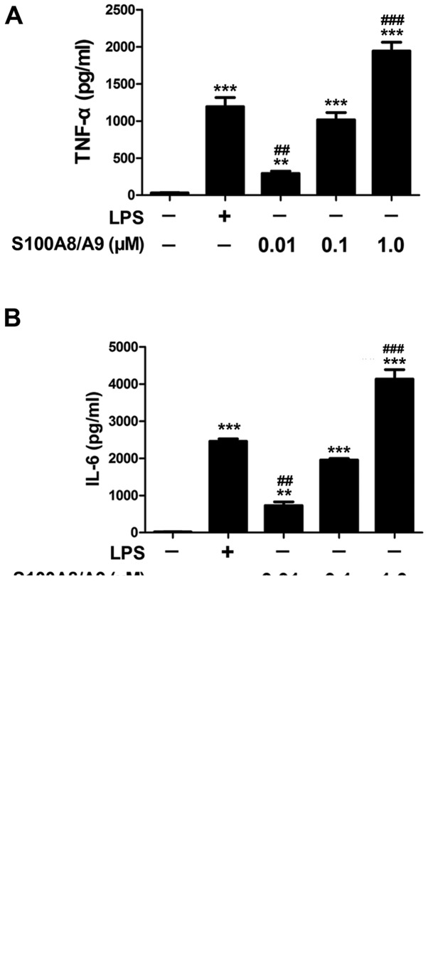
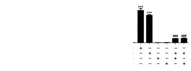
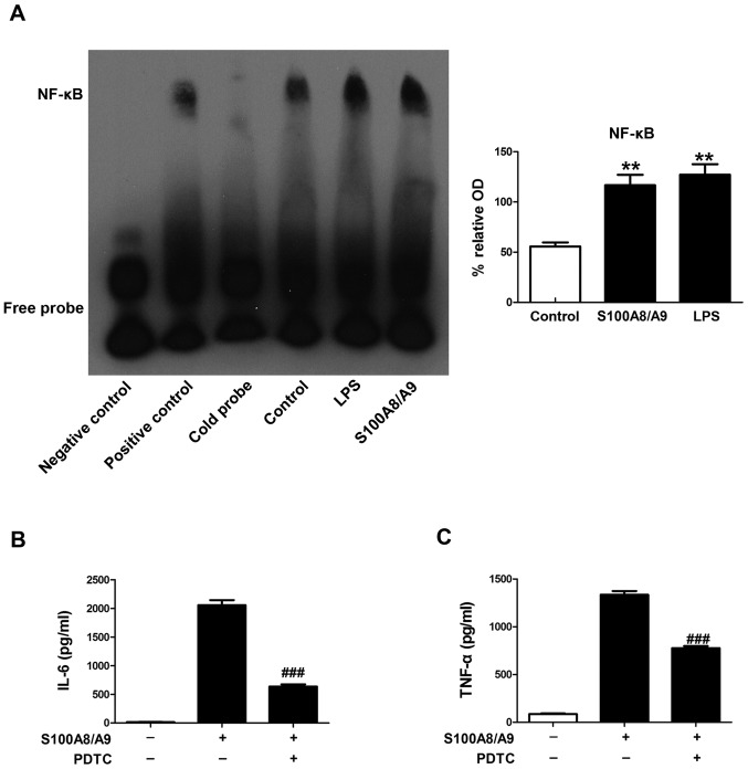
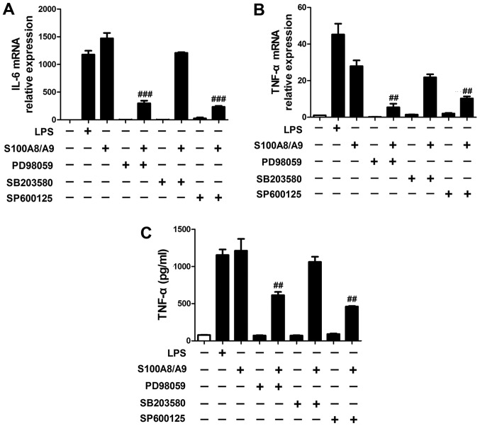
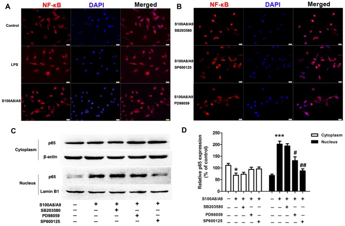
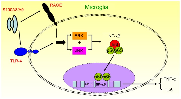
S100A8/A9, a heterodimer of the two calcium-binding proteins S100A8 and S100A9, has emerged as an important proinflammatory mediator in acute and chronic inflammation. However, whether S100A8/A9 is implicated in microglial‑induced neuroinflammatory response remains unclear. Here, we found that S100A8/A9 significantly increased the secretion of proinflammatory cytokines inclu-ding tumor necrosis factor-α (TNF-α) and interleukin-6 (IL-6) in cultured BV-2 microglial cells. Inhibition of the Toll-like receptor 4 (TLR4) and the receptor for advanced glycation end-products (RAGE) with C225 and a RAGE-blocking antibody, respectively significantly reduced the secretion of TNF-α and IL-6 from S100A8/A9-stimulated BV-2 microglial cells. Furthermore, S100A8/A9 markedly enhanced the nuclear translocation of NF-κB p65 and the DNA-binding activities of NF-κB in BV-2 microglial cells, and suppression of ERK and JNK/MAPK signaling pathways by PD98059 or SP600125 significantly inhibited NF-κB activity and the release of TNF-α and IL-6 in the S100A8/A9-treated BV-2 microglial cells. Our data also showed that inhibition of NF-κB with pyrrolidine dithiocarbamate (PDTC) significantly reduced the secretion of TNF-α and IL-6 from BV-2 microglial cells treated with S100A8/A9. Taken together, our data suggest that S100A8/A9 acts directly on BV-2 microglial cells via binding to TLR4 and RAGE on the membrane and then stimulates the secretion of proinflammatory cytokines through ERK and JNK-mediated NF-κB activity in BV-2 microglial cells. Targeting S100A8/A9 may provide a novel therapeutic strategy in microglial-induced neuroinflammatory diseases.
Overview
The claims and statistics of this paper haven’t been checked yet.
Run the check — free, no account needed. It reads the open-access text and checks the sources it cites.
Integrity signals
Registry
Cites
Statistics
Figures
Verification
Claims the paper makes
The claims and statistics of this paper haven’t been checked yet.
Run the check — free, no account needed. It reads the open-access text and checks the sources it cites.
Claims other papers make about this one
Each is checked against this paper’s own text: does it say what the citing paper says it does?
In the citing paper
“Tau-dependent microglial activation can enhance secretion of pro-inflammatory cytokines such as IL-1β, IL-6, and TNF-α [421–423].”
From Molecular and cellular mechanisms underlying the pathogenesis of Alzheimer’s disease (2020) · cites this paper as [422]
Self-check
Percentages, sums and reported differences re-computed from the paper’s own numbers.
The self-check needs the paper’s full text.
Citations
Cites
RAGE Mediates a Novel Proinflammatory Axis
Hofmann, M. A., Drury, S., Fu, C., Qu, W., Taguchi, A., Lu, Y., Avila, C., Kambham, N., Bierhaus, A., Nawroth, P., et al. (1999) · Cell · doi:10.1016/s0092-8674(00)80801-6 · Cited by 1,864 · cited 1×
“Previous studies have shown that activated microglia express high levels of TLR4 and RAGE in response to neuroinflammation (9-12).”
BackgroundNeumar, R. W., Nolan, J. P., Adrie, C., Aibiki, M., Berg, R. A., Böttiger, B. W., Callaway, C. W., Clark, R. S. B., Geocadin, R. G., Jauch, E. C., et al. (2008) · Circulation · doi:10.1161/circulationaha.108.190652 · Cited by 1,460 · cited 1×
“Microglial activation plays a pivotal role during the development and progression of neurodegenerative diseases based on its great capacity for secreting proinflammatory cytokines, such as tumor necrosis factor‑ α (TNF-α ) and interleukin‑6 (IL‑6), resulting in acute inflammation (13).”
BackgroundVogl, T., Tenbrock, K., Ludwig, S., Leukert, N., Ehrhardt, C., van Zoelen, M. A. D., Nacken, W., Foell, D., van der Poll, T., Sorg, C., et al. (2007) · Nature Medicine · doi:10.1038/nm1638 · Cited by 1,418 · cited 1×
“Recently, S100A8/A9, as the endogenous activator of TLR4 signaling, promotes lethal endotoxin‑induced shock (6).”
BackgroundUnderstanding RAGE, the receptor for advanced glycation end products
Bierhaus, A., Humpert, P. M., Morcos, M., Wendt, T., Chavakis, T., Arnold, B., Stern, D. M., & Nawroth, P. P. (2005) · Journal of Molecular Medicine · doi:10.1007/s00109-005-0688-7 · Cited by 1,297
Treatment of Septic Shock with the Tumor Necrosis Factor Receptor:Fc Fusion Protein
Fisher, C. J., Agosti, J. M., Opal, S. M., Lowry, S. F., BALK, R. A., SADOFF, J. C., Abraham, E. H., Schein, R. M. H., & Benjamin, E. (1996) · New England Journal of Medicine · doi:10.1056/nejm199606273342603 · Cited by 1,239 · cited 1×
“More studies also demonstrated that major risk factors for sepsis, systemic inflammatory response syndrome and septic shock are related to TNF‑ α ‑independent mechanisms (45,46).”
BackgroundLehnardt, S., Massillon, L., Follett, P., Jensen, F. E., Ratan, R., Rosenberg, P. A., Volpe, J. J., & Vartanian, T. (2003) · Proceedings of the National Academy of Sciences · doi:10.1073/pnas.1432609100 · Cited by 1,052 · cited 1×
“Thus, our data, which demonstrated that S100A8/A9 contributed to inflammation via TLR4, further revealed that neuroinflammation and ischemic brain injury is modulated by TLR4 (39-41).”
BackgroundNF-kB: a crucial transcription factor for glial and neuronal cell function
O'Neill, L. A. J., & Kaltschmidt, C. (1997) · Trends in Neurosciences · doi:10.1016/s0166-2236(96)01035-1 · Cited by 1,013
Intracellular and extracellular roles of S100 proteins
Donato, R. (2003) · Microscopy Research and Technique · doi:10.1002/jemt.10296 · Cited by 974 · cited 1×
“Previous studies have shown that activated microglia express high levels of TLR4 and RAGE in response to neuroinflammation (9-12).”
BackgroundEhrchen, J. M., Sunderkötter, C. H., Foell, D., Vogl, T., & Roth, J. (2009) · Journal of Leukocyte Biology · doi:10.1189/jlb.1008647 · Cited by 869 · cited 2×
“…the S100A8/A9 heterodimers are associ-/A9 heterodimers are associ-A9 heterodimers are associated with the pathogenesis of various diseases, and coupled with an inflammatory component, which is primarily released from activated or necrotic neutrophils and monocytes/macro-/macro-macro-phages (1,2).”
BackgroundRyckman, C., Vandal, K., Rouleau, P., Talbot, M., & Tessier, P. A. (2003) · The Journal of Immunology · doi:10.4049/jimmunol.170.6.3233 · Cited by 861 · cited 1×
“As an endogenous ligand of TLR4 and RAGE (14), S100A8/A9 also amplifies the production of proinflammatory cytokines and contributes to CNS injury (2,15).”
Background
Cited by
Sorted by citations
Wang, S., Song, R., Wang, Z., Jing, Z., Wang, S., & Ma, J. (2018) · Frontiers in Immunology · doi:10.3389/fimmu.2018.01298 · Cited by 1,450
“In BV-2 microglia, S100A8/A9 stimulates the production of TNF-α and IL-6 through ERK/NF-κB and JNK/ NF-κB signaling (72).”
BackgroundMolecular and cellular mechanisms underlying the pathogenesis of Alzheimer’s disease
Guo, T., Zhang, D., Zeng, Y., Huang, T. Y., Xu, H., & Zhao, Y. (2020) · Molecular Neurodegeneration · doi:10.1186/s13024-020-00391-7 · Cited by 994
Calprotectin: from biomarker to biological function
Jukic, A., Bakiri, L., Wagner, E. F., Tilg, H., & Adolph, T. E. (2021) · Gut · doi:10.1136/gutjnl-2021-324855 · Cited by 500
Transcriptional and Translational Differences of Microglia from Male and Female Brains
Guneykaya, D., Ivanov, A., Hernandez, D. P., Haage, V., Wojtas, B., Meyer, N., Maricos, M., Jordan, P., Buonfiglioli, A., Gielniewski, B., et al. (2018) · Cell Reports · doi:10.1016/j.celrep.2018.08.001 · Cited by 477
Calprotectin (S100A8/S100A9): a key protein between inflammation and cancer
Shabani, F., Farasat, A., Mahdavi, M., & Gheibi, N. (2018) · Inflammation Research · doi:10.1007/s00011-018-1173-4 · Cited by 251
Metadata
- Authors
- Ma, L., Sun, P., Zhang, J.-C., Zhang, Q., & Yao, S.-L.
- Affiliations
- Wuhan Union Hospital (CN)
- Corresponding author
- Zhang, Qing
- Venue
- International Journal of Molecular Medicine
- Publisher
- Spandidos Publications
- Published
- 2017-05-12
- Volume
- 40
- Issue
- 1
- Pages
- 31-38
- ISSN
- 1107-3756 (ISSN-L)
- 1791-244X
- PMC
- PMC5466387
- OpenAlex
- W2613996502
- Cited by
- 196
- References
- 48
- Citations by year
- 2026: 23 · 2025: 32 · 2024: 30 · 2023: 35 · 2022: 23 · 2021: 11 · 2020: 19 · 2019: 10 · 2018: 10 · 2017: 2
- FWCI
- 4.58
- Type
- journal article
- Language
- en
- Open access
- hybrid
- doi.org
- Licence
- cc-by-nc-nd
- Locations
- International Journal of Molecular Medicine (publishedVersion, cc-by-nc-nd)
- Europe PMC (PubMed Central) (publishedVersion, other-oa)
- PubMed Central (publishedVersion, cc-by-nc-nd)
- PubMed (publishedVersion)
- Funders
- National Natural Science Foundation of China (81101401)
- Topics
- Life Sciences › Biochemistry, Genetics and Molecular Biology › Molecular Biology › S100 Proteins and Annexins
- Life Sciences › Neuroscience › Neurology › Neuroinflammation and Neurodegeneration Mechanisms
- Life Sciences › Biochemistry, Genetics and Molecular Biology › Clinical Biochemistry › Advanced Glycation End Products research
- Keywords
- S100A8/A9, microglia, neuroinflammation, TLR4, RAGE, NF-κB, ERK signaling, JNK signaling, TNF-α, interleukin-6, inflammatory cytokines, pyrrolidine dithiocarbamate, C225, BV-2 cells
- MeSH terms
- Receptor for Advanced Glycation End Products, Animals, Cell Line, Transformed, Inflammation, Signal Transduction, Cytokines, Microglia, Inflammation Mediators, Calgranulin A, Calgranulin B, Toll-Like Receptor 4, Mice
About these checks
Registry, retraction, Cites and statistics results are computed, not judged, with one exception: a printed reference that no index lists is read once by a model before it counts as a citation error. Claims are extracted by a model and kept only when their quote appears verbatim in the text; each cited source is then checked, with a model’s help, for whether it says what the claim relies on. Those verdicts can be wrong — read the claim’s quote and the cited source before relying on one.
Statistics and arithmetic are recomputed, not judged.
Confidence bands are shown only for fields calibrated on our annotated test set. None is calibrated yet, so no band is shown.
What counts as a claim ›How we measure confidence ›How we check this ›
Are you an author, or do you see a problem with this page? Request a review
