

The DOI matches its Crossref record.
Downregulation of microRNA‑25‑3p inhibits the proliferation and promotes the apoptosis of multiple myeloma cells via targeting the PTEN/PI3K/AKT signaling pathway
Open accessAbstract
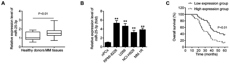
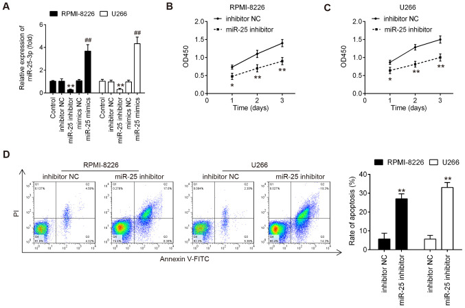
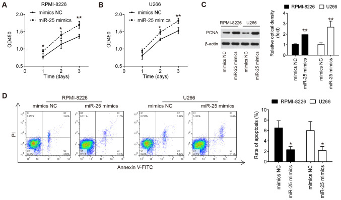
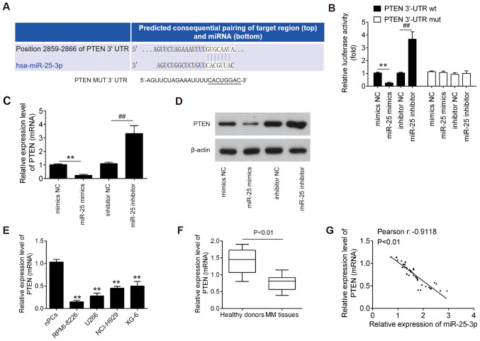
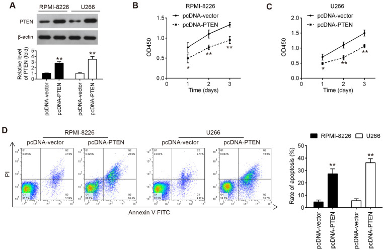
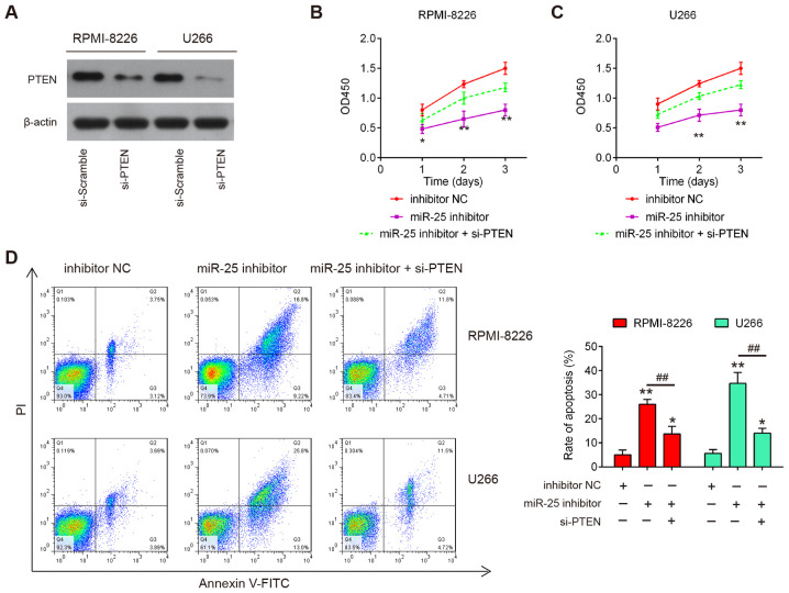
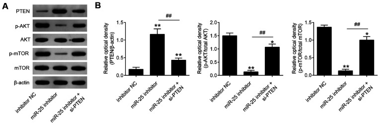
Numerous studies have confirmed that microRNAs (miRNAs or miRs) have important roles in cancer biogenesis and development including multiple myeloma (MM). MicroRNA‑25‑3p (miR‑25‑3p) has been proven to promote cancer progression, whereas its functions in MM has not yet been reported, at least to the best of our knowledge. Therefore, the present study aimed to investigate the function of miR‑25‑3p in MM and to identify the potential underlying mechanistic pathway. Herein, it was found that miR‑25‑3p expression was significantly increased in MM tissues and cell lines. The upregulation of miR‑25‑3p was closely associated with anemia, renal function impairment international staging system (ISS) staging and Durie‑Salmon (D‑S) staging. A high level of miR‑25‑3p was predictive of a poor prognosis of patients with MM. In vitro, the knockdown of miR‑25‑3p suppressed the proliferation and promoted the apoptosis of RPMI‑8226 and U266 cells, while the overexpression of miR‑25‑3p exerted opposite effects. In addition, phosphatase and tensin homolog deleted on chromosome 10 (PTEN), a well‑known tumor suppressor, was confirmed as a target of miR‑25‑3p in MM cells. Moreover, it was found that the PTEN expression levels were decreased, and inversely correlated with miR‑25‑3p expression levels in MM tissues. Further analyses revealed that the overexpression of PTEN exerted effects similar to those of miR‑25‑3p knockdown, whereas the knockdown of PTEN partially abolished the effects of miR‑25‑3p inhibitor on MM cells. Accompanied by PTEN induction, miR‑25‑3p promoted PI3K/AKT signaling pathway activation in MM cells. Collectively, these findings demonstrate critical roles for miR‑25‑3p in the pathogenesis of MM, and suggest that miR‑25‑3p may serve as a novel prognostic biomarker and therapeutic target of MM.
Overview
The claims and statistics of this paper haven’t been checked yet.
Run the check — free, no account needed. It reads the open-access text and checks the sources it cites.
Integrity signals
Registry
Cites
Statistics
Figures
Verification
Claims the paper makes
The claims and statistics of this paper haven’t been checked yet.
Run the check — free, no account needed. It reads the open-access text and checks the sources it cites.
Claims other papers make about this one
Each is checked against this paper’s own text: does it say what the citing paper says it does?
The source backs part of the claim, not all of it — or backs it for a different population, condition or comparison.
In the citing paper
“Zi Y et al. demonstrated that blocking the PI3K/AKT pathway could be achieved by knocking down miR-25-3p. This approach resulted in the inhibition of migration and progression of MM cells [71].”
From Multiple myeloma: signaling pathways and targeted therapy (2024) · cites this paper as [71]
Experimental — accuracy not yet measured
Self-check
Percentages, sums and reported differences re-computed from the paper’s own numbers.
The self-check needs the paper’s full text.
Citations
Cites
No references could be resolved yet.
Research works printed but not linked: 42
- [1]Terpos E Ntanasis-Stathopoulos I Gavriatopoulou M Dimopoulos MA Pathogenesis of bone disease in multiple myeloma: From bench to bedside Blood Cancer J 8 7 2018 10.1038/s41408-017-0037-4 29330358 PMC5802524 · DOI · Paper page
- [2]Nahi H Våtsveen TK Lund J Heeg BM Preiss B Alici E Møller MB Wader KF Møller HE Grøseth LA Proteasome inhibitors and IMiDs can overcome some high-risk cytogenetics in multiple myeloma but not gain 1q21 Eur J Haematol 96 46 54 2016 10.1111/ejh.12546 25779478 · DOI
- [3]Harding T Baughn L Kumar S Van Ness B The future of myeloma precision medicine: Integrating the compendium of known drug resistance mechanisms with emerging tumor profiling technologies Leukemia 33 863 883 2019 10.1038/s41375-018-0362-z 30683909 · DOI
- [4]Velez R Turesson I Landgren O Kristinsson SY Cuzick J Incidence of multiple myeloma in Great Britain, Sweden, and Malmö, Sweden: The impact of differences in case ascertainment on observed incidence trends BMJ Open 6 e009584 2016 10.1136/bmjopen-2015-009584 PMC4735168 26801465 · DOI
- [5]Chen D Zhou D Xu J Zhou R Ouyang J Chen B Prognostic value of 1q21 gain in multiple myeloma Clin Lymphoma Myeloma Leuk 19 e159 e164 2019 10.1016/j.clml.2018.12.002 30685183 · DOI
- [6]Bartel DP MicroRNAs: Target recognition and regulatory functions Cell 136 215 233 2009 10.1016/j.cell.2009.01.002 19167326 PMC3794896 · DOI · Paper page
- [7]Handa H Murakami Y Ishihara R Kimura-Masuda K Masuda Y The role and function of microRNA in the pathogenesis of multiple myeloma Cancers (Basel) 11 1378 2019 10.3390/cancers11111738 PMC6896016 31698726 · DOI
- [8]Adamia S Abiatari I Amin SB Fulciniti M Minvielle S Li C Moreau P Avet-Loiseau H Munshi NC Anderson KC The effects of MicroRNA deregulation on pre-RNA processing network in multiple myeloma Leukemia 34 167 179 2020 10.1038/s41375-019-0498-5 31182781 PMC6901818 · DOI
- [9]Xu YY Song YQ Huang ZM Zhang HB Chen M MicroRNA-26a inhibits multiple myeloma cell growth by suppressing cyclin-dependent kinase 6 expression Kaohsiung J Med Sci 35 277 283 2019 10.1002/kjm2.12057 30897301 PMC11900684 · DOI
- [10]Tian F Zhan Y Zhu W Li J Tang M Chen X Jiang J MicroRNA-497 inhibits multiple myeloma growth and increases susceptibility to bortezomib by targeting Bcl-2 Int J Mol Med 43 1058 1066 2019 30535471
- [11]Gupta N Kumar R Seth T Garg B Sati HC Sharma A Clinical significance of circulatory microRNA-203 in serum as novel potential diagnostic marker for multiple myeloma J Cancer Res Clin Oncol 145 1601 1611 2019 10.1007/s00432-019-02896-1 30891618 PMC11810419 · DOI
- [12]Zhang YK Wang H Leng Y Li ZL Yang YF Xiao FJ Li QF Chen XQ Wang LS Overexpression of microRNA-29b induces apoptosis of multiple myeloma cells through down regulating Mcl-1 Biochem Biophys Res Commun 414 233 239 2011 10.1016/j.bbrc.2011.09.063 21951844 · DOI
- [13]Jia CM Tian YY Quan LN Jiang L Liu AC miR-26b-5p suppresses proliferation and promotes apoptosis in multiple myeloma cells by targeting JAG1 Pathol Res Pract 214 1388 1394 2018 10.1016/j.prp.2018.07.025 30098829 · DOI
- [14]Zhang J Gong X Tian K Chen D Sun J Wang G Guo M miR-25 promotes glioma cell proliferation by targeting CDKN1C Biomed Pharmacother 71 7 14 2015 10.1016/j.biopha.2015.02.005 25960208 · DOI
- [15]Sanchez-Mejias A Kwon J Chew XH Siemens A Sohn HS Jing G Zhang B Yang H Tay Y A novel SOCS5/miR-18/miR-25 axis promotes tumorigenesis in liver cancer Int J Cancer 144 311 321 2019 10.1002/ijc.31857 30191950 · DOI
- [16]Navarro A Diaz T Tovar N Pedrosa F Tejero R Cibeira MT Magnano L Rosiñol L Monzó M Bladé J Fernández de Larrea C A serum microRNA signature associated with complete remission and progression after autologous stem-cell transplantation in patients with multiple myeloma Oncotarget 6 1874 1883 2015 10.18632/oncotarget.2761 25593199 PMC4359338 · DOI
- [17]Xiang T Hu AX Sun P Liu G Liu G Xiao Y Identification of four potential predicting miRNA biomarkers for multiple myeloma from published datasets PeerJ 5 e2831 2017 10.7717/peerj.2831 28168095 PMC5289111 · DOI
- [18]Palumbo A Avet-Loiseau H Oliva S Lokhorst HM Goldschmidt H Rosinol L Richardson P Caltagirone S Lahuerta JJ Facon T Revised international staging system for multiple myeloma: A report from international myeloma working group J Clin Oncol 33 2863 2869 2015 10.1200/JCO.2015.61.2267 26240224 PMC4846284 · DOI · Paper page
- [19]International Myeloma Working Group Criteria for the classification of monoclonal gammopathies, multiple myeloma and related disorders: A report of the international myeloma working group Br J Haematol 121 749 757 2003 10.1046/j.1365-2141.2003.04355.x 12780789 · DOI
- [20]Livak KJ Schmittgen TD Analysis of relative gene expression data using real-time quantitative PCR and the 2(-Delta Delta C(T)) method Methods 25 402 408 2001 10.1006/meth.2001.1262 11846609 · DOI · Paper page
- [21]Franke TF PI3K/Akt: Getting it right matters Oncogene 27 6473 6488 2008 10.1038/onc.2008.313 18955974 · DOI
- [22]Wang X Jiang X PTEN: A default gate-keeping tumor suppressor with a versatile tail Cell Res 18 807 816 2008 10.1038/cr.2008.83 18626510 · DOI
- [23]Sarbassov DD Guertin DA Ali SM Sabatini DM Phosphorylation and regulation of Akt/PKB by the rictor-mTOR complex Science 307 1098 1101 2005 10.1126/science.1106148 15718470 · DOI · Paper page
- [24]Razumilava N Bronk SF Smoot RL Fingas CD Werneburg NW Roberts LR Mott JL miR-25 targets TNF-related apoptosis inducing ligand (TRAIL) death receptor-4 and promotes apoptosis resistance in cholangiocarcinoma Hepatology 55 465 475 2012 10.1002/hep.24698 21953056 PMC3268937 · DOI
- [25]Feng S Pan W Jin Y Zheng J MiR-25 promotes ovarian cancer proliferation and motility by targeting LATS2 Tumour Biol 35 12339 12344 2014 10.1007/s13277-014-2546-0 25179841 · DOI
- [26]Boguslawska J Rodzik K Poplawski P Kędzierska H Rybicka B Sokół E Tański Z Piekiełko-Witkowska A TGF-beta1 targets a microRNA network that regulates cellular adhesion and migration in renal cancer Cancer Lett 412 155 169 2018 10.1016/j.canlet.2017.10.019 29079415 · DOI
- [27]Huo J Zhang Y Li R Wang Y Wu J Zhang D Upregulated MicroRNA-25 mediates the migration of melanoma cells by targeting DKK3 through the WNT/β-catenin pathway Int J Mol Sci 17 1124 2016 10.3390/ijms17111124 PMC5133768 27801786 · DOI
- [28]Ning L Zhang M Zhu Q Hao F Shen W Chen D miR-25-3p inhibition impairs tumorigenesis and invasion in gastric cancer cells in vitro and in vivo Bioengineered 11 81 90 2020 10.1080/21655979.2019.1710924 31909687 PMC6961587 · DOI
- [29]Liu H Ma L Wang J Overexpression of miR-25 is associated with progression and poor prognosis of cholangiocarcinoma Exp Ther Med 18 2687 2694 2019 31555370 PMC6755412
- [30]Chen H Pan H Qian Y Zhou W Liu X MiR-25-3p promotes the proliferation of triple negative breast cancer by targeting BTG2 Mol Cancer 17 4 2018 10.1186/s12943-017-0754-0 29310680 PMC5759260 · DOI
- [31]Wan W Wan W Long Y Li Q Jin X Wan G Zhang F Lv Y Zheng G Li Z Zhu Y MiR-25-3p promotes malignant phenotypes of retinoblastoma by regulating PTEN/Akt pathway Biomed Pharmacother 118 109111 2019 10.1016/j.biopha.2019.109111 31336343 · DOI
- [32]Peng G Yang C Liu Y Shen C miR-25-3p promotes glioma cell proliferation and migration by targeting FBXW7 and DKK3 Exp Ther Med 18 769 778 2019 31258712 PMC6566094
- [33]Kim YK Yu J Han TS Park SY Namkoong B Kim DH Hur K Yoo MW Lee HJ Yang HK Kim VN Functional links between clustered microRNAs: Suppression of cell-cycle inhibitors by microRNA clusters in gastric cancer Nucleic Acids Res 37 1672 1681 2009 10.1093/nar/gkp002 19153141 PMC2655672 · DOI
- [34]Li Y Tan W Neo TW Aung MO Wasser S Lim SG Tan TM Role of the miR-106b-25 microRNA cluster in hepatocellular carcinoma Cancer Sci 100 1234 1242 2009 10.1111/j.1349-7006.2009.01164.x 19486339 · DOI
- [35]Poliseno L Salmena L Riccardi L Fornari A Song MS Hobbs RM Sportoletti P Varmeh S Egia A Fedele G Identification of the miR-106b~25 microRNA cluster as a proto-oncogenic PTEN-targeting intron that cooperates with its host gene MCM7 in transformation Sci Signal 3 ra29 2010 10.1126/scisignal.2000594 20388916 PMC2982149 · DOI · Paper page
- [36]Carnero A Blanco-Aparicio C Renner O Link W Leal JF The PTEN/PI3K/AKT signalling pathway in cancer, therapeutic implications Curr Cancer Drug Targets 8 187 198 2008 10.2174/156800908784293659 18473732 · DOI
- [37]Jiang Y Chang H Chen G Effects of microRNA-20a on the proliferation, migration and apoptosis of multiple myeloma via the PTEN/PI3K/AKT signaling pathway Oncol Lett 15 10001 10007 2018 29963125 PMC6020174
- [38]Qu L Gao Y Sun H Wang H Liu X Sun D Role of PTEN-Akt-CREB signaling pathway in nervous system impairment of rats with chronic arsenite exposure Biol Trace Elem Res 170 366 372 2016 10.1007/s12011-015-0478-1 26296331 · DOI
- [39]Aziz AUR Farid S Qin K Wang H Liu B PIM kinases and their relevance to the PI3K/AKT/mTOR pathway in the regulation of ovarian cancer Biomolecules 8 7 2018 10.3390/biom8010007 PMC5871976 29401696 · DOI
- [40]Waniczek D Śnietura M Lorenc Z Nowakowska-Zajdel E Muc-Wierzgon M Assessment of PI3K/AKT/PTEN signaling pathway activity in colorectal cancer using quantum dot-conjugated antibodies Oncol Lett 15 1236 1240 2018 29422975 PMC5772923
- [41]Yu T Li L Liu W Ya B Cheng H Xin Q Silencing of NADPH oxidase 4 attenuates hypoxia resistance in neuroblastoma cells SH-SY5Y by inhibiting PI3K/Akt-dependent glycolysis Oncol Res 27 525 532 2019 10.3727/096504018X15179668157803 29426376 PMC7848326 · DOI
- [42]Xu H Li J Zhou ZG NEAT1 promotes cell proliferation in multiple myeloma by activating PI3K/AKT pathway Eur Rev Med Pharmacol Sci 22 6403 6411 2018 30338809
Cited by
Sorted by citations
Multiple myeloma: signaling pathways and targeted therapy
Lu, Q., Yang, D., Li, H., Niu, T., & Tong, A. (2024) · Molecular biomedicine · doi:10.1186/s43556-024-00188-w · Cited by 90
Metadata
- Authors
- Zi, Y., Zhang, Y., Wu, Y., Zhang, L., Yang, R., & Huang, Y.
- Affiliations
- First Affiliated Hospital of Xinxiang Medical University (CN)
- Central Laboratory, The First Affiliated Hospital of Xinxiang Medical University, Weihui, Henan 453100, P.R. China
- Corresponding author
- Zi, Youmei
- Venue
- International Journal of Molecular Medicine
- Publisher
- Spandidos Publications
- Published
- 2020-12-31
- Volume
- 47
- Issue
- 3
- ISSN
- 1107-3756 (ISSN-L)
- 1791-244X
- PMC
- PMC7834966
- OpenAlex
- W3116250464
- Cited by
- 40
- References
- 0
- Citations by year
- 2025: 10 · 2024: 14 · 2023: 6 · 2022: 8 · 2021: 2
- FWCI
- 2.53
- Type
- journal article
- Language
- en
- Open access
- hybrid
- doi.org
- Licence
- cc-by-nc-nd
- Locations
- International Journal of Molecular Medicine (publishedVersion, cc-by-nc-nd)
- Europe PMC (PubMed Central) (publishedVersion, other-oa)
- PubMed Central (publishedVersion, cc-by-nc-nd)
- PubMed (publishedVersion)
- Topics
- Health Sciences › Medicine › Hematology › Multiple Myeloma Research and Treatments
- Life Sciences › Biochemistry, Genetics and Molecular Biology › Cancer Research › MicroRNA in disease regulation
- Health Sciences › Medicine › Hematology › Chronic Lymphocytic Leukemia Research
- Keywords
- miR-25-3p, multiple myeloma, PTEN, PI3K/Akt signaling, cell proliferation, apoptosis, prognostic factors, therapeutic target, microRNA downregulation, tumor suppressor
- MeSH terms
- Aged, Female, Humans, Male, Middle Aged, Multiple Myeloma, RNA, Neoplasm, Signal Transduction, Down-Regulation, Gene Expression Regulation, Neoplastic, Apoptosis, Phosphatidylinositol 3-Kinases, MicroRNAs, Cell Line, Tumor, Proto-Oncogene Proteins c-akt, PTEN Phosphohydrolase
About these checks
Registry, retraction, Cites and statistics results are computed, not judged, with one exception: a printed reference that no index lists is read once by a model before it counts as a citation error. Claims are extracted by a model and kept only when their quote appears verbatim in the text; each cited source is then checked, with a model’s help, for whether it says what the claim relies on. Those verdicts can be wrong — read the claim’s quote and the cited source before relying on one.
Statistics and arithmetic are recomputed, not judged.
Confidence bands are shown only for fields calibrated on our annotated test set. None is calibrated yet, so no band is shown.
What counts as a claim ›How we measure confidence ›How we check this ›
Are you an author, or do you see a problem with this page? Request a review
How to cite
Cite CoChat for the checks on this page, and the paper for its own findings.
- This page
- CoChat. (2026). Citation check: Downregulation of microRNA‑25‑3p inhibits the proliferation and promotes the apoptosis of multiple myeloma cells via targeting the PTEN/PI3K/AKT signaling pathway. https://app.cochat.ai/papers/10.3892/ijmm.2020.4841
- The paper
- Zi, Y., Zhang, Y., Wu, Y., Zhang, L., Yang, R., & Huang, Y. (2020). Downregulation of microRNA‑25‑3p inhibits the proliferation and promotes the apoptosis of multiple myeloma cells via targeting the PTEN/PI3K/AKT signaling pathway. International Journal of Molecular Medicine. https://doi.org/10.3892/ijmm.2020.4841
