

The DOI matches its Crossref record; 122 of the 122 references it cites were found in a registry or an index.
Basic mechanisms of peripheral nerve injury and treatment via electrical stimulation
Open accessAbstract
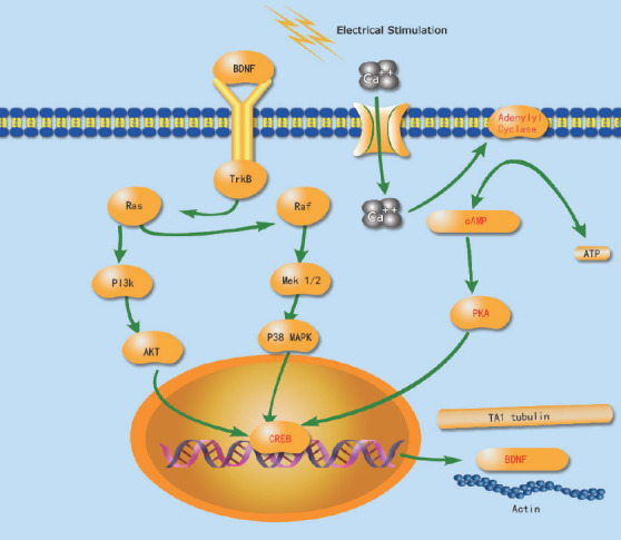
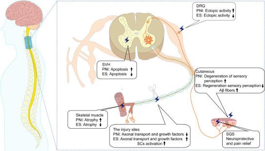
Previous studies on the mechanisms of peripheral nerve injury (PNI) have mainly focused on the pathophysiological changes within a single injury site. However, recent studies have indicated that within the central nervous system, PNI can lead to changes in both injury sites and target organs at the cellular and molecular levels. Therefore, the basic mechanisms of PNI have not been comprehensively understood. Although electrical stimulation was found to promote axonal regeneration and functional rehabilitation after PNI, as well as to alleviate neuropathic pain, the specific mechanisms of successful PNI treatment are unclear. We summarize and discuss the basic mechanisms of PNI and of treatment via electrical stimulation. After PNI, activity in the central nervous system (spinal cord) is altered, which can limit regeneration of the damaged nerve. For example, cell apoptosis and synaptic stripping in the anterior horn of the spinal cord can reduce the speed of nerve regeneration. The pathological changes in the posterior horn of the spinal cord can modulate sensory abnormalities after PNI. This can be observed in cases of ectopic discharge of the dorsal root ganglion leading to increased pain signal transmission. The injured site of the peripheral nerve is also an important factor affecting post-PNI repair. After PNI, the proximal end of the injured site sends out axial buds to innervate both the skin and muscle at the injury site. A slow speed of axon regeneration leads to low nerve regeneration. Therefore, it can take a long time for the proximal nerve to reinnervate the skin and muscle at the injured site. From the perspective of target organs, long-term denervation can cause atrophy of the corresponding skeletal muscle, which leads to abnormal sensory perception and hyperalgesia, and finally, the loss of target organ function. The mechanisms underlying the use of electrical stimulation to treat PNI include the inhibition of synaptic stripping, addressing the excessive excitability of the dorsal root ganglion, alleviating neuropathic pain, improving neurological function, and accelerating nerve regeneration. Electrical stimulation of target organs can reduce the atrophy of denervated skeletal muscle and promote the recovery of sensory function. Findings from the included studies confirm that after PNI, a series of physiological and pathological changes occur in the spinal cord, injury site, and target organs, leading to dysfunction. Electrical stimulation may address the pathophysiological changes mentioned above, thus promoting nerve regeneration and ameliorating dysfunction.
Overview
The claims and statistics of this paper haven’t been checked yet.
Run the check — free, no account needed. It reads the open-access text and checks the sources it cites.
Integrity signals
Registry
Cites
Statistics
Figures
Verification
Claims the paper makes
The claims and statistics of this paper haven’t been checked yet.
Run the check — free, no account needed. It reads the open-access text and checks the sources it cites.
Claims other papers make about this one
Each is checked against this paper’s own text: does it say what the citing paper says it does?
The cited source says what the claim attributes to it.
In the citing paper
“However, peripheral nerves have the potential to self-repair and regenerate, aided by conditions such as anatomical continuity of the injured site, myelination, and the extent of the injury [112].”
From Active Neural Interface Circuits and Systems for Selective Control of Peripheral Nerves: A Review (2024) · cites this paper as [112]
Self-check
Percentages, sums and reported differences re-computed from the paper’s own numbers.
The self-check needs the paper’s full text.
Citations
Cites
Antioxidants Maintain Cellular Redox Homeostasis by Elimination of Reactive Oxygen Species
He, L., He, T., Farrar, S., Ji, L., Liu, T., & Ma, X. (2017) · Cellular Physiology and Biochemistry · doi:10.1159/000485089 · Cited by 2,207
A CLASSIFICATION OF PERIPHERAL NERVE INJURIES PRODUCING LOSS OF FUNCTION
Sunderland, S. (1951) · Brain · doi:10.1093/brain/74.4.491 · Cited by 1,574
Neural plasticity after peripheral nerve injury and regeneration
Navarro, X., Vivó, M., & Valero‐Cabré, A. (2007) · Progress in Neurobiology · doi:10.1016/j.pneurobio.2007.06.005 · Cited by 908
Microglia in neuropathic pain: cellular and molecular mechanisms and therapeutic potential
Inoue, K., & Tsuda, M. (2018) · Nature Reviews Neuroscience · doi:10.1038/nrn.2018.2 · Cited by 908
Brief Electrical Stimulation Promotes the Speed and Accuracy of Motor Axonal Regeneration
Al‐Majed, A. A., Neumann, C. M., Brushart, T. M. E., & Gordon, T. (2000) · Journal of Neuroscience · doi:10.1523/jneurosci.20-07-02602.2000 · Cited by 851
Wallerian degeneration: an emerging axon death pathway linking injury and disease
Conforti, L., Gilley, J., & Coleman, M. P. (2014) · Nature reviews. Neuroscience · doi:10.1038/nrn3680 · Cited by 630
Adrenergic Excitation of Cutaneous Pain Receptors Induced by Peripheral Nerve Injury
Sato, J., & Perl, E. R. (1991) · Science · doi:10.1126/science.2011742 · Cited by 593
Al‐Majed, A. A., Brushart, T. M. E., & Gordon, T. (2000) · European Journal of Neuroscience · doi:10.1111/j.1460-9568.2000.01341.x · Cited by 484
Ploner, M., Sorg, C., & Groß, J. (2016) · Trends in Cognitive Sciences · doi:10.1016/j.tics.2016.12.001 · Cited by 472
Brushart, T. M. E., Hoffman, P. N., Royall, R. M., Murinson, B. B., Witzel, C., & Gordon, T. (2002) · Journal of Neuroscience · doi:10.1523/jneurosci.22-15-06631.2002 · Cited by 437
Research works printed but not linked: 1
- Gybels, 1990, The treatment of pain due to peripheral-nerve injury by electrical-stimulitation of the injured nerve, Adv Pain Res Ther, 13, 217
Cited by
Sorted by citations
Liu, M., Zhang, W., Han, S., Zhang, D., Zhou, X., Guo, X., Chen, H., Wang, H., Jin, L., Feng, S., et al. (2024) · Advanced Materials · doi:10.1002/adma.202313672 · Cited by 129
Hydrogel-based immunoregulation of macrophages for tissue repair and regeneration
Nie, R., Zhang, Q., Feng, Z.-Y., Huang, K., Zou, C., Fan, M., Zhang, Y., Zhang, J.-Y., Li‐Ling, J., Tan, B., et al. (2024) · International Journal of Biological Macromolecules · doi:10.1016/j.ijbiomac.2024.131643 · Cited by 75
Zhou, W., Rahman, M. S. U., Sun, C., Li, S., Zhang, N., Chen, H., Han, C. C., Xu, S., & Liu, Y. (2024) · Advanced Materials · doi:10.1002/adma.202307805 · Cited by 65
Wang, Q., Wang, H., Ma, Y., Cao, X., & Gao, H. (2022) · Biomaterials Science · doi:10.1039/d2bm01216b · Cited by 60
Wang, Q., Wei, Y., Yin, X., Zhan, G., Cao, X., & Gao, H. (2024) · Advanced Functional Materials · doi:10.1002/adfm.202400217 · Cited by 56
Metadata
- Authors
- Chu, X.-L., Song, X.-Z., Li, Q., Li, Y.-R., He, F., Gu, X.-S., & Ming, D.
- Affiliations
- Academy of Medical Engineering and Translational Medicine, Tianjin University, Tianjin, China
- Tianjin University (CN)
- Tianjin University of Sport (CN)
- Corresponding author
- He, Feng
- Gu, Xiao-Song
- Venue
- Neural Regeneration Research
- Published
- 2022-02-28
- Volume
- 17
- Issue
- 10
- Pages
- 2185-2193
- ISSN
- 1673-5374 (ISSN-L)
- 1876-7958
- PMC
- PMC9083151
- OpenAlex
- W4220934018
- Cited by
- 164
- References
- 122
- Citations by year
- 2026: 46 · 2025: 47 · 2024: 40 · 2023: 20 · 2022: 11
- FWCI
- 4.21
- Type
- journal article
- Language
- en
- Open access
- gold
- doi.org
- Licence
- cc-by-nc-sa
- Locations
- Neural Regeneration Research (publishedVersion, cc-by-nc-sa)
- PubMed Central (publishedVersion, cc-by-nc-sa)
- PubMed (publishedVersion)
- DOAJ (DOAJ: Directory of Open Access Journals) (submittedVersion, cc-by-sa)
- Topics
- Life Sciences › Neuroscience › Cellular and Molecular Neuroscience › Nerve injury and regeneration
- Health Sciences › Medicine › Surgery › Nerve Injury and Rehabilitation
- Life Sciences › Neuroscience › Cellular and Molecular Neuroscience › Neuroscience and Neural Engineering
- Keywords
- peripheral nerve injury, electrical stimulation, axon regeneration, neuropathic pain, spinal cord, synaptic stripping, dorsal root ganglia, ectopic discharges, denervation, skeletal muscle atrophy, hyperalgesia, functional rehabilitation, target organs
About these checks
Registry, retraction, Cites and statistics results are computed, not judged, with one exception: a printed reference that no index lists is read once by a model before it counts as a citation error. Claims are extracted by a model and kept only when their quote appears verbatim in the text; each cited source is then checked, with a model’s help, for whether it says what the claim relies on. Those verdicts can be wrong — read the claim’s quote and the cited source before relying on one.
Statistics and arithmetic are recomputed, not judged.
Confidence bands are shown only for fields calibrated on our annotated test set. None is calibrated yet, so no band is shown.
What counts as a claim ›How we measure confidence ›How we check this ›
Are you an author, or do you see a problem with this page? Request a review
