

The DOI matches its Crossref record; 50 of the 50 references it cites were found in a registry or an index.
Homozygous YME1L1 mutation causes mitochondriopathy with optic atrophy and mitochondrial network fragmentation
Open accessAbstract
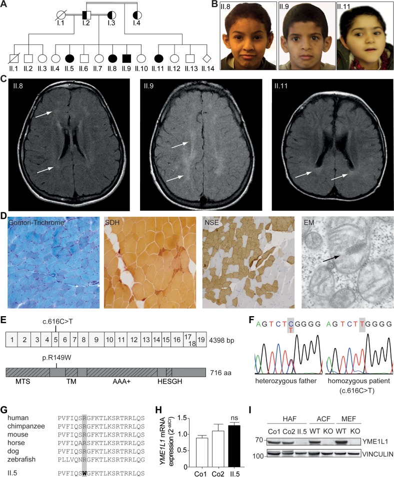
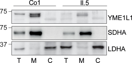
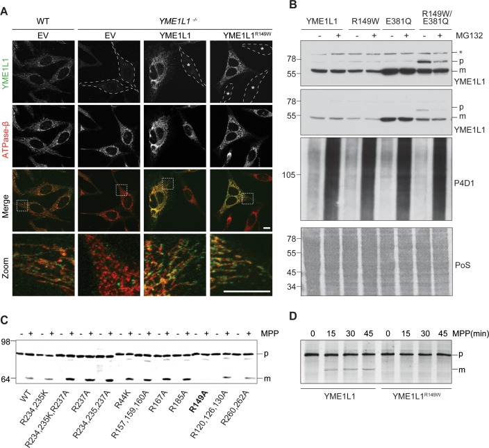
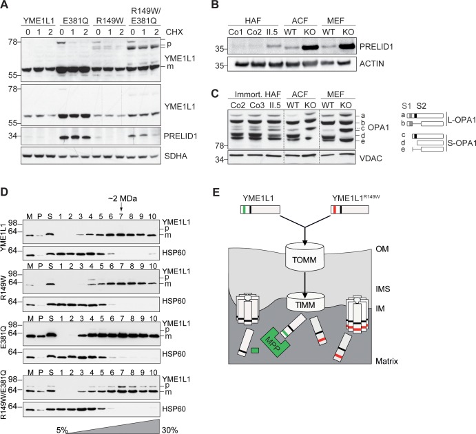
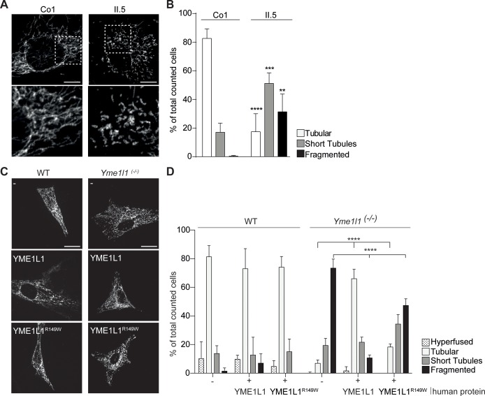
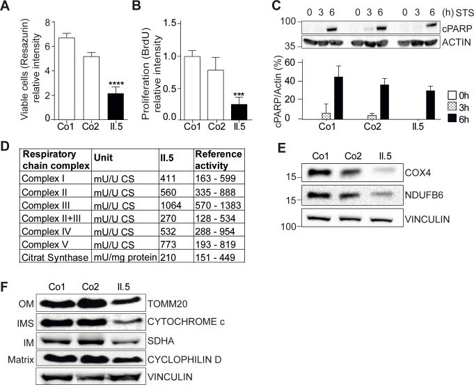
Mitochondriopathies often present clinically as multisystemic disorders of primarily high-energy consuming organs. Assembly, turnover, and surveillance of mitochondrial proteins are essential for mitochondrial function and a key task of AAA family members of metalloproteases. We identified a homozygous mutation in the nuclear encoded mitochondrial escape 1-like 1 gene YME1L1, member of the AAA protease family, as a cause of a novel mitochondriopathy in a consanguineous pedigree of Saudi Arabian descent. The homozygous missense mutation, located in a highly conserved region in the mitochondrial pre-sequence, inhibits cleavage of YME1L1 by the mitochondrial processing peptidase, which culminates in the rapid degradation of YME1L1 precursor protein. Impaired YME1L1 function causes a proliferation defect and mitochondrial network fragmentation due to abnormal processing of OPA1. Our results identify mutations in YME1L1 as a cause of a mitochondriopathy with optic nerve atrophy highlighting the importance of YME1L1 for mitochondrial functionality in humans.
Overview
The claims and statistics of this paper haven’t been checked yet.
Run the check — free, no account needed. It reads the open-access text and checks the sources it cites.
Integrity signals
Registry
Cites
Statistics
Figures
Verification
Claims the paper makes
The claims and statistics of this paper haven’t been checked yet.
Run the check — free, no account needed. It reads the open-access text and checks the sources it cites.
Claims other papers make about this one
Each is checked against this paper’s own text: does it say what the citing paper says it does?
In the citing paper
“Muscle biopsies from these patients display alterations in cristae morphology and paracrystalline inclusions and fiber-type grouping, indicating denervation [165].”
From The connection between the dynamic remodeling of the mitochondrial network and the regulation of muscle mass (2021) · cites this paper as [165]
In the citing paper
“The accumulation of damaged proteins due to a homozygous missense mutation that impairs the IMS protease maturation, YME1L1, causes infantile onset mitochondriopathy with severe intellectual disability, muscular impairments, and optic nerve atrophy.”
From The connection between the dynamic remodeling of the mitochondrial network and the regulation of muscle mass (2021) · cites this paper as [165]
Self-check
Percentages, sums and reported differences re-computed from the paper’s own numbers.
The self-check needs the paper’s full text.
Citations
Cites
Multiplex Genome Engineering Using CRISPR/Cas Systems
Cong, L., Ran, F. A., Cox, D., Lin, S., Barretto, R., Habib, N., Hsu, P. D., Wu, X., Jiang, W., Marraffini, L. A., et al. (2013) · Science · doi:10.1126/science.1231143 · Cited by 15,339
Mitochondria: In Sickness and in Health
Nunnari, J., & Suomalainen, A. (2012) · Cell · doi:10.1016/j.cell.2012.02.035 · Cited by 3,479
OPA1 Controls Apoptotic Cristae Remodeling Independently from Mitochondrial Fusion
Frezza, C., Cipolat, S., Martins de Brito, O., Micaroni, M., Beznoussenko, G. V., Rudka, T., Bartoli, D., Polishuck, R. S., Danial, N. N., De Strooper, B., et al. (2006) · Cell · doi:10.1016/j.cell.2006.06.025 · Cited by 1,673
Computational Method to Predict Mitochondrially Imported Proteins and their Targeting Sequences
Claros, M. G., & Vincens, P. (1996) · European Journal of Biochemistry · doi:10.1111/j.1432-1033.1996.00779.x · Cited by 1,650
Delettre, C., Lenaers, G., Griffoin, J.-M., Gigarel, N., Lorenzo, C., Belenguer, P., Pelloquin, L., Grosgeorge, J., Turc-Carel, C., Perret, E., et al. (2000) · Nature Genetics · doi:10.1038/79936 · Cited by 1,446
OPA1 requires mitofusin 1 to promote mitochondrial fusion
Cipolat, S., de Brito, O. M., Dal Zilio, B., & Scorrano, L. (2004) · Proceedings of the National Academy of Sciences · doi:10.1073/pnas.0407043101 · Cited by 1,300
Olichon, A., Baricault, L., Gas, N., Guillou, E., Valette, A., Belenguer, P., & Lenaers, G. (2003) · Journal of Biological Chemistry · doi:10.1074/jbc.c200677200 · Cited by 1,168
Deep sequencing reveals 50 novel genes for recessive cognitive disorders
Najmabadi, H., Hu, H., Garshasbi, M., Zemojtel, T., Abedini, S. S., Chen, W., Hosseini, M., Behjati, F., Haas, S., Jamali, P., et al. (2011) · Nature · doi:10.1038/nature10423 · Cited by 965
Self-citationSong, Z., Chen, H., Fiket, M., Alexander, C., & Chan, D. C. (2007) · The Journal of Cell Biology · doi:10.1083/jcb.200704110 · Cited by 818
The i -AAA protease YME1L and OMA1 cleave OPA1 to balance mitochondrial fusion and fission
Anand, R., Wai, T., Baker, M. J., Kladt, N., Schauss, A. C., Rugarli, E., & Langer, T. (2014) · Journal of Cell Biology · doi:10.1083/jcb.201308006 · Cited by 812
Self-citation
Cited by
Sorted by citations
Mitochondria dysfunction in the pathogenesis of Alzheimer’s disease: Recent advances.
Wang, W., Zhao, F., Ma, X., Perry, G., & Zhu, X. (2020) · Molecular Neurodegeneration · doi:10.1186/s13024-020-00376-6 · Cited by 1,274
Quality control of the mitochondrial proteome
Song, J., Herrmann, J. M., & Becker, T. (2020) · Nature Reviews Molecular Cell Biology · doi:10.1038/s41580-020-00300-2 · Cited by 501
Quality control of the mitochondrion
Ng, M. Y. W., Wai, T., & Simonsen, A. (2021) · Developmental Cell · doi:10.1016/j.devcel.2021.02.009 · Cited by 468
Self-citationMitochondrial dynamics in health and disease
Yapa, N. M. B., Lisnyak, V., Reljić, B., & Ryan, M. T. (2021) · FEBS Letters · doi:10.1002/1873-3468.14077 · Cited by 323
Mitochondrial Proteases: Multifaceted Regulators of Mitochondrial Plasticity
Deshwal, S., Fiedler, K. U., & Langer, T. (2020) · Annual Review of Biochemistry · doi:10.1146/annurev-biochem-062917-012739 · Cited by 250
Self-citationPuchades, C., Rampello, A. J., Shin, M., Giuliano, C. J., Wiseman, R. L., Glynn, S. E., & Lander, G. C. (2017) · Science · doi:10.1126/science.aao0464 · Cited by 223
Mitochondrial dynamics and their potential as a therapeutic target
Whitley, B. N., Engelhart, E. A., & Hoppins, S. C. (2019) · Mitochondrion · doi:10.1016/j.mito.2019.06.002 · Cited by 228
Romanello, V., & Sandri, M. (2021) · Cellular and Molecular Life Sciences · doi:10.1007/s00018-020-03662-0 · Cited by 197
Lipid signalling drives proteolytic rewiring of mitochondria by YME1L
MacVicar, T., Ohba, Y., Nolte, H., Mayer, F. C., Tatsuta, T., Sprenger, H., Lindner, B., Zhao, Y., Li, J., Bruns, C. J., et al. (2019) · Nature · doi:10.1038/s41586-019-1738-6 · Cited by 187
Self-citationCellular pyrimidine imbalance triggers mitochondrial DNA–dependent innate immunity
Sprenger, H., MacVicar, T., Bahat, A., Fiedler, K. U., Hermans, S., Ehrentraut, D., Ried, K., Milenkovic, D., Bonekamp, N. A., Larsson, N., et al. (2021) · Nature Metabolism · doi:10.1038/s42255-021-00385-9 · Cited by 170
Self-citationMitochondrial dynamics: Biological roles, molecular machinery, and related diseases
El‐Hattab, A. W., Suleiman, J., Almannai, M., & Scaglia, F. (2018) · Molecular Genetics and Metabolism · doi:10.1016/j.ymgme.2018.10.003 · Cited by 138
m-AAA proteases, mitochondrial calcium homeostasis and neurodegeneration
Patrón, M., Sprenger, H., & Langer, T. (2018) · Cell Research · doi:10.1038/cr.2018.17 · Cited by 134
Self-citationMitochondrial signalling and homeostasis: from cell biology to neurological disease
Collier, J. J., Oláhová, M., McWilliams, T. G., & Taylor, R. W. (2023) · Trends in Neurosciences · doi:10.1016/j.tins.2022.12.001 · Cited by 133
Mitochondrial Fission and Fusion: Molecular Mechanisms, Biological Functions, and Related Disorders
Ojaimi, M. A., Salah, A., & El‐Hattab, A. W. (2022) · Membranes · doi:10.3390/membranes12090893 · Cited by 130
Mitochondria as intracellular signaling platforms in health and disease
Tan, J. X., & Finkel, T. (2020) · The Journal of Cell Biology · doi:10.1083/jcb.202002179 · Cited by 123
Mitochondria-Associated Proteostasis
Ruan, L., Wang, Y., Zhang, X., Tomaszewski, A., McNamara, J. T., & Li, R. (2020) · Annual Review of Biophysics · doi:10.1146/annurev-biophys-121219-081604 · Cited by 95
The Maintenance of Mitochondrial DNA Integrity and Dynamics by Mitochondrial Membranes
Chapman, J., Ng, Y. S., & Nicholls, T. J. J. (2020) · Life · doi:10.3390/life10090164 · Cited by 100
Therapeutic Options in Hereditary Optic Neuropathies
Amore, G., Romagnoli, M., Carbonelli, M., Barboni, P., Carelli, V., & Morgia, C. L. (2020) · Drugs · doi:10.1007/s40265-020-01428-3 · Cited by 96
Rocha, S., Freitas, A., Guimarães, S., Vitorino, R. M. P., Aroso, M., & Gómez-Lázaro, M. (2017) · Antioxidants · doi:10.3390/antiox7010001 · Cited by 93
Liu, Y., Huang, X., Nguyen, D., Shammas, M. K., Wu, B. P., Dombi, E., Springer, D., Poulton, J., Sekine, S., & Narendra, D. P. (2020) · Human Molecular Genetics · doi:10.1093/hmg/ddaa077 · Cited by 73
AAA Proteases: Guardians of Mitochondrial Function and Homeostasis
Opalińska, M., & Jańska, H. (2018) · Cells · doi:10.3390/cells7100163 · Cited by 74
Havalová, H., Ondrovičová, G., Keresztesová, B., Bauer, J., Pevala, V., Kutějová, E., & Kunová, N. (2021) · International Journal of Molecular Sciences · doi:10.3390/ijms22158077 · Cited by 74
Ehmke, N., Graul-Neumann, L. M., Smorag, L., Koenig, R., Segebrecht, L., Magoulas, P. L., Scaglia, F., Kilic, E., Hennig, A. F., Adolphs, N., et al. (2017) · The American Journal of Human Genetics · doi:10.1016/j.ajhg.2017.09.016 · Cited by 65
Self-citationROMO1 is a constituent of the human presequence translocase required for YME1L protease import
Richter, F., Dennerlein, S., Nikolov, M., Jans, D. C., Naumenko, N., Aich, A., MacVicar, T., Linden, A., Jakobs, S., Urlaub, H., et al. (2018) · The Journal of Cell Biology · doi:10.1083/jcb.201806093 · Cited by 70
Self-citationMitochondrial DNA release and sensing in innate immune responses
VanPortfliet, J. J., Chute, C., Lei, Y., Shutt, T. E., & West, A. P. (2024) · Human Molecular Genetics · doi:10.1093/hmg/ddae031 · Cited by 70
Recent Advances in Understanding the Structural and Functional Evolution of FtsH Proteases
Yi, L., Liu, B., Nixon, P. J., Yu, J., & Chen, F. (2022) · Frontiers in Plant Science · doi:10.3389/fpls.2022.837528 · Cited by 58
Loss of the mitochondrial i‐AAA protease YME1L leads to ocular dysfunction and spinal axonopathy
Sprenger, H., Wani, G. A., Hesseling, A., König, T., Patrón, M., MacVicar, T., Ahola, S., Wai, T., Barth, E., Rugarli, E. I., et al. (2018) · EMBO Molecular Medicine · doi:10.15252/emmm.201809288 · Cited by 59
Self-citationMitochondrial quality control proteases and their modulation for cancer therapy
Zhang, J., Qiao, W., & Luo, Y. (2022) · Medicinal Research Reviews · doi:10.1002/med.21929 · Cited by 57
Metalloproteases of the Inner Mitochondrial Membrane
Levytskyy, R. M., Bohovych, I., & Khalimonchuk, O. (2017) · Biochemistry · doi:10.1021/acs.biochem.7b00663 · Cited by 58
Metabolic control of adult neural stem cell self-renewal by the mitochondrial protease YME1L
Wani, G. A., Sprenger, H., Ndoci, K., Chandragiri, S., Acton, R., Schatton, D., Kochan, S. M. V., Sakthivelu, V., Jevtic, M., Seeger, J. M., et al. (2022) · Cell Reports · doi:10.1016/j.celrep.2022.110370 · Cited by 54
Self-citationRegulation of mitochondrial plasticity by the i-AAA protease YME1L
Ohba, Y., MacVicar, T., & Langer, T. (2020) · Biological Chemistry · doi:10.1515/hsz-2020-0120 · Cited by 54
Self-citationATPase Domain AFG3L2 Mutations Alter OPA1 Processing and Cause Optic Neuropathy
Caporali, L., Magri, S., Legati, A., Dotto, V. D., Tagliavini, F., Balistreri, F., Nasca, A., Morgia, C. L., Carbonelli, M., Valentino, M. L., et al. (2020) · Annals of Neurology · doi:10.1002/ana.25723 · Cited by 53
Mitochondrial proteases in human diseases
Gala, M. G., & Vögtle, F. ‐. (2021) · FEBS Letters · doi:10.1002/1873-3468.14039 · Cited by 49
Nottia, M. D., Verrigni, D., Torraco, A., Rizza, T., Bertini, E., & Carrozzo, R. (2021) · Genes · doi:10.3390/genes12020247 · Cited by 51
Mitochondrial optic neuropathies
Carelli, V., Morgia, C. L., & Yu‐Wai‐Man, P. (2023) · Handbook of clinical neurology · doi:10.1016/b978-0-12-821751-1.00010-5 · Cited by 47
The relevance of mitochondrial morphology for human disease
Navaratnarajah, T., Anand, R., Reichert, A. S., & Distelmaier, F. (2021) · The International Journal of Biochemistry & Cell Biology · doi:10.1016/j.biocel.2021.105951 · Cited by 49
Mitochondrial calcium signalling and neurodegenerative diseases
Britti, E., Delaspre, F., Tamarit, J., & Ros, J. (2018) · Neuronal Signaling · doi:10.1042/ns20180061 · Cited by 47
Pareek, G., Thomas, R. E., & Pallanck, L. J. (2018) · Cell Death & Disease · doi:10.1038/s41419-018-0365-8 · Cited by 42
Panga, V., Kallor, A. A., Nair, A., Harshan, S., & Raghunathan, S. (2019) · PLoS ONE · doi:10.1371/journal.pone.0224632 · Cited by 41
Mitochondrial fragmentation and network architecture in degenerative diseases
Shah, S. I., Paine, J. G., Perez, C., & Ullah, G. (2019) · PLoS ONE · doi:10.1371/journal.pone.0223014 · Cited by 40
Mitochondrial Proteolysis and Metabolic Control
Ahola, S., Langer, T., & MacVicar, T. (2019) · Cold Spring Harbor Perspectives in Biology · doi:10.1101/cshperspect.a033936 · Cited by 37
Self-citationMitochondrial ATP-Dependent Proteases—Biological Function and Potential Anti-Cancer Targets
Feng, Y., Nouri, K., & Schimmer, A. D. (2021) · Cancers · doi:10.3390/cancers13092020 · Cited by 35
Cesnekova, J., Rodinová, M., Hansíková, H., Zeman, J., & Stibůrek, L. (2018) · International Journal of Molecular Sciences · doi:10.3390/ijms19123930 · Cited by 35
Disease-Associated Genetic Variation in Human Mitochondrial Protein Import
Nicolas, É., Tricarico, R., Savage, M. J., Golemis, E. A., & Hall, M. J. (2019) · The American Journal of Human Genetics · doi:10.1016/j.ajhg.2019.03.019 · Cited by 38
Mitochondrial biology and dysfunction in secondary mitochondrial disease
Baker, M. J., Crameri, J. J., Thorburn, D. R., Frazier, A. E., & Stojanovski, D. (2022) · Open Biology · doi:10.1098/rsob.220274 · Cited by 39
TIMM44 is a potential therapeutic target of human glioma
Guo, Y.-Z., Chen, G., Huang, M., Wang, Y., Liu, Y.-Y., Jiang, Q. L., Cao, C., & Liu, F. (2022) · Theranostics · doi:10.7150/thno.78616 · Cited by 32
The mitochondrial protein YME1 Like 1 is important for non-small cell lung cancer cell growth
Xia, Y., He, C., Hu, Z., Wu, Z., Hui, Y., Liu, Y.-Y., Mu, C., & Zha, J. (2023) · International Journal of Biological Sciences · doi:10.7150/ijbs.82217 · Cited by 29
Drosophila ADCK1 is critical for maintaining mitochondrial structures and functions in the muscle
Yoon, W., Hwang, S.-H., Lee, S.-H., & Chung, J. (2019) · PLoS Genetics · doi:10.1371/journal.pgen.1008184 · Cited by 29
Mitochondrial quality control in the brain: The physiological and pathological roles
Shen, X., Sun, P., Zhang, H., & Yang, H. (2022) · Frontiers in Neuroscience · doi:10.3389/fnins.2022.1075141 · Cited by 30
Cai, Y., Shen, H., Weng, H., Wang, Y.-Q., Cai, G., Chen, X., & Ye, Q. (2020) · Scientific Reports · doi:10.1038/s41598-020-67229-6 · Cited by 28
Cytosolic Self-DNA—A Potential Source of Chronic Inflammation in Aging
Akbari, M., Shanley, D. P., Bohr, V. A., & Rasmussen, L. J. (2021) · Cells · doi:10.3390/cells10123544 · Cited by 25
Shen, Y., Cheng, Z., Chen, S., Zhang, Y., Chen, Q., & Yi, S. (2021) · Molecular Neurobiology · doi:10.1007/s12035-021-02589-2 · Cited by 24
Borrelli, E., Bandello, F. M., Boon, C. J. F., Carelli, V., Lenaers, G., Reibaldi, M., Sadda, S. R., Sadun, A. A., Sarraf, D., Yu‐Wai‐Man, P., et al. (2024) · Progress in Retinal and Eye Research · doi:10.1016/j.preteyeres.2024.101264 · Cited by 25
Μitochondrial Membrane Dynamics and Inherited Optic Neuropathies
Bagli, E., Zikou, A. K., Agnantis, N. J., & Kitsos, G. (2017) · In Vivo · doi:10.21873/invivo.11090 · Cited by 24
Lebeau, J., Rainbolt, T. K., & Wiseman, R. L. (2018) · International review of cytology · doi:10.1016/bs.ircmb.2018.05.003 · Cited by 25
Mayr, S. J., Roeper, J., & Schwarz, G. (2020) · Journal of Biological Chemistry · doi:10.1074/jbc.ra119.010720 · Cited by 23
Chiaratti, M. R. (2021) · Biophysical Reviews · doi:10.1007/s12551-021-00891-w · Cited by 25
The Role of Mitochondria in Optic Atrophy With Autosomal Inheritance
Strachan, E. L., MacWhite-Begg, D., Crean, J. K. G., Reynolds, A., Kennedy, B. N., & O’Sullivan, N. C. (2021) · Frontiers in Neuroscience · doi:10.3389/fnins.2021.784987 · Cited by 22
Intraneuronal β-amyloid impaired mitochondrial proteostasis through the impact on LONP1
Wang, W., Ma, X., Bhatta, S., Shao, C., Zhao, F., Fujioka, H., Torres, S. L., Wu, F., & Zhu, X. (2023) · Proceedings of the National Academy of Sciences · doi:10.1073/pnas.2316823120 · Cited by 22
Targeting NAMPT‐OPA1 for treatment of senile osteoporosis
Bai, C., Tian, B., Zhang, M., Qin, Q., Shi, X., Yang, X., Gao, X., Zhou, X., Shan, H., & Bai, J. (2024) · Aging Cell · doi:10.1111/acel.14400 · Cited by 24
Bouzidi, A., Charoute, H., Charif, M., Amalou, G., Kandil, M., Barakat, A., & Lenaers, G. (2022) · Orphanet Journal of Rare Diseases · doi:10.1186/s13023-022-02340-7 · Cited by 20
Opalińska, M., Parys, K., Murcha, M. W., & Jańska, H. (2017) · Journal of Cell Science · doi:10.1242/jcs.200733 · Cited by 20
Opalińska, M., Parys, K., & Jańska, H. (2017) · International Journal of Molecular Sciences · doi:10.3390/ijms18112455 · Cited by 20
Li, J., Li, W., Gao, F., Qu, S., Hu, F., Zhang, S.-H., Li, L., Wang, Z., Qiu, Y., Wang, L.-S., et al. (2020) · Translational Vision Science & Technology · doi:10.1167/tvst.9.8.11 · Cited by 19
Recent Advances in Targeting Human Mitochondrial AAA+ Proteases to Develop Novel Cancer Therapeutics
Wong, K. S., & Houry, W. A. (2019) · Advances in experimental medicine and biology · doi:10.1007/978-981-13-8367-0_8 · Cited by 17
Molecular Mechanisms behind Inherited Neurodegeneration of the Optic Nerve
Maresca, A., & Carelli, V. (2021) · Biomolecules · doi:10.3390/biom11040496 · Cited by 20
Targeting the mitochondrial protein YME1L to inhibit osteosarcoma cell growth in vitro and in vivo
Sun, X., Shi, C., Dai, J., Zhang, M., Pei, D., & Yang, L. (2024) · Cell Death and Disease · doi:10.1038/s41419-024-06722-6 · Cited by 15
First TMEM126A missense mutation in an Italian proband with optic atrophy and deafness
Morgia, C. L., Caporali, L., Tagliavini, F., Palombo, F., Carbonelli, M., Liguori, R., Barboni, P., & Carelli, V. (2019) · Neurology Genetics · doi:10.1212/nxg.0000000000000329 · Cited by 15
Scott, G. K., Mahoney, S. A., Scott, M. K. D., Loureiro, A., Lopez‐Ramirez, A., Tanner, J. J., Ellerby, L., & Benz, C. C. (2021) · Amino Acids · doi:10.1007/s00726-021-03012-9 · Cited by 15
Proteolytic regulation of mitochondrial dynamics
Dietz, J. V., Bohovych, I., Viana, M. P., & Khalimonchuk, O. (2019) · Mitochondrion · doi:10.1016/j.mito.2019.04.008 · Cited by 15
AAA+ proteases: the first line of defense against mitochondrial damage
Pareek, G. (2022) · PeerJ · doi:10.7717/peerj.14350 · Cited by 14
Molecular pathways of mitochondrial outer membrane protein degradation
Zheng, J., Li, L., & Jiang, H. (2019) · Biochemical Society Transactions · doi:10.1042/bst20190275 · Cited by 13
Charif, M., Wong, Y. C., Kim, S., Guichet, A., Vignal‐Clermont, C., Zanlonghi, X., Bensaïd, P., Procaccio, V., Bonneau, D., Amati‐Bonneau, P., et al. (2021) · Molecular Neurodegeneration · doi:10.1186/s13024-021-00431-w · Cited by 13
Siblings with optic neuropathy and RTN4IP1 mutation
Okamoto, N., Miya, F., Hatsukawa, Y., Suzuki, Y., Kawato, K., Yamamoto, Y., Tsunoda, T., Kato, M., Saitoh, S., Yamasaki, M., et al. (2017) · Journal of Human Genetics · doi:10.1038/jhg.2017.68 · Cited by 13
Proteolytic Control of Lipid Metabolism
Sam, P. N., Avery, E., & Claypool, S. M. (2019) · ACS Chemical Biology · doi:10.1021/acschembio.9b00695 · Cited by 12
Kim, S., Nam, Y., Kim, M., Kwon, S., Jeon, J., Shin, S. J., Park, S., Chang, S., Kim, H. U., Lee, Y. Y., et al. (2022) · Journal of Ginseng Research · doi:10.1016/j.jgr.2022.09.008 · Cited by 12
Liao, W., Chu, P., Su, C.-C., Wu, C., & Li, C. (2022) · Pathology - Research and Practice · doi:10.1016/j.prp.2022.154215 · Cited by 12
Mitochondrial DNA release and inflammation in mitochondrial disease pathogenesis
Szabo, M., Lagos, D., Cross, E., Collier, J. J., & Horváth, R. (2026) · Brain · doi:10.1093/brain/awag037 · Cited by 11
A sibling study of isolated optic neuropathy associated with novel variants in the ACO2 gene
Kelman, J. C., Kamien, B., Murray, N., Goel, H., Fraser, C. L., & Grigg, J. (2018) · Ophthalmic Genetics · doi:10.1080/13816810.2018.1509353 · Cited by 11
Zou, X.-H., Guo, X.-X., Su, H., Wang, C., Dong, E., Wang, N., Chen, W., & Zhang, Q. (2019) · Journal of Molecular Neuroscience · doi:10.1007/s12031-019-01319-7 · Cited by 11
Cai, M., Wan, J., Cai, K., Li, S., Du, X., Song, H., Sun, W., & Hu, J. (2024) · International Journal of Obesity · doi:10.1038/s41366-024-01490-x · Cited by 11
Kan, K. T., Wilcock, J., & Lu, H. (2024) · Biochemical Society Transactions · doi:10.1042/bst20240450 · Cited by 12
Kumar, A., Waingankar, T. P., & D’Silva, P. (2023) · Journal of Cell Science · doi:10.1242/jcs.260060 · Cited by 11
Moisoi, N. (2024) · Biochimie · doi:10.1016/j.biochi.2024.06.005 · Cited by 8
Murray-Cors, S., Owens, M., Liew, Y.-T., Day, M., Cairns, W., & Prokop, A. (2025) · Frontiers in Neuroscience · doi:10.3389/fnins.2025.1631752 · Cited by 9
Leeming, P. J., Mercuri-Svik, J., & Stojanovski, D. (2025) · Molecular and Cellular Biology · doi:10.1080/10985549.2025.2566671 · Cited by 10
Expression and functional implications of YME1L in nasopharyngeal carcinoma
Cheng, F., Huang, H., Yin, S., Liu, J.-S., & Sun, P. (2024) · Cell Death and Disease · doi:10.1038/s41419-024-06811-6 · Cited by 8
A Conserved Mitochondrial Chaperone-Protease Complex Involved in Protein Homeostasis
Serricchio, M., & Bütikofer, P. (2021) · Frontiers in Molecular Biosciences · doi:10.3389/fmolb.2021.767088 · Cited by 7
Therapeutic potential of melatonin-induced mitophagy in the pathogenesis of Alzheimer’s disease
Goleij, P., Tabari, M. A. K., Poudineh, M., Sanaye, P. M., Khan, H. Z., Kumar, A. P., Larsen, D. S., & Daglia, M. (2025) · Inflammopharmacology · doi:10.1007/s10787-025-01859-y · Cited by 7
Quality control at the powerhouse: mitochondrial proteostasis dysfunction and disease
Baker, M. J., Yek, K. Q., & Stojanovski, D. (2025) · Biochemical Society Transactions · doi:10.1042/bst20253044 · Cited by 6
How much does a disrupted mitochondrial network influence neuronal dysfunction?
Chrzanowska‐Lightowlers, Z. M., & Lightowlers, R. N. (2018) · EMBO Molecular Medicine · doi:10.15252/emmm.201809899 · Cited by 6
Hereditary Optic Neuropathies: An Updated Review
Lee, S., Mura, C., Abreu, N. J., Rucker, J. C., Galetta, S. L., Balcer, L. J., & Grossman, S. N. (2024) · Journal of Clinical & Translational Ophthalmology · doi:10.3390/jcto2030006 · Cited by 5
Molecular mechanisms of mitochondrial AAA+ proteases
Currie, S., Goncalves, M. M., Schimmer, A. D., & Vahidi, S. (2026) · Journal of Biological Chemistry · doi:10.1016/j.jbc.2026.111264 · Cited by 5
OMA1 and YME1L as a Diagnostic Panel in Hepatocellular Carcinoma
Abass, S. A., Abdel‐Hamid, N. M., Elshazly, A. M., Abdo, W., & Zakaria, S. (2023) · The Yale Journal of Biology and Medicine · doi:10.59249/bwby8971 · Cited by 5
Song, J., Chen, Y., Chen, Y., Qiu, M., Xiang, W., Ke, B., & Fang, X. (2024) · Biochemical Genetics · doi:10.1007/s10528-024-10756-y · Cited by 4
A Perspective on Accelerated Aging Caused by the Genetic Deficiency of the Metabolic Protein, OPA1
Erchova, I. A., Sun, S., & Votruba, M. (2021) · Frontiers in Neurology · doi:10.3389/fneur.2021.641259 · Cited by 4
Inherited metabolic disorders in Cyprus
Georgiou, T., Petrou, P., Malekkou, A., Ioannou, I., Gavatha, M., Skordis, N., Nicolaidou, P., Savvidou, I., Athanasiou, E., Ourani, S., et al. (2024) · Molecular Genetics and Metabolism Reports · doi:10.1016/j.ymgmr.2024.101083 · Cited by 4
Alahmad, A., Muhammad, H., Pyle, A., Albash, B., McFarland, R., & Taylor, R. W. (2019) · Journal of Biochemical and Clinical Genetics · doi:10.24911/jbcgenetics/183-1548325196 · Cited by 3
Inactivation of PDH can Reduce Anaplastic Thyroid Cancer Cells’ Sensitivity to Artemisinin
Li, Y. (2021) · Anti-Cancer Agents in Medicinal Chemistry · doi:10.2174/1871520621666210910100803 · Cited by 3
Disorders of Mitochondrial Homeostasis, Dynamics, Protein Import, and Quality Control
de Boer, L., de Vries, M. C., Smeitink, J. A. M., & Koopman, W. J. H. (2022) · Springer eBooks · doi:10.1007/978-3-030-67727-5_46 · Cited by 3
Metadata
- Authors
- Hartmann, B., Wai, T., Hu, H., MacVicar, T., Musante, L., Fischer-Zirnsak, B., Stenzel, W., Gräf, R., van den Heuvel, L., Ropers, H.-H., Wienker, T. F., Hübner, C., Langer, T., & Kaindl, A. M.
- Affiliations
- Charité – Universitätsmedizin Berlin (DE)
- Cologne Excellence Cluster on Cellular Stress Responses in Aging Associated Diseases (DE)
- Guangzhou Women and Children Medical Center (CN)
- Max Planck Institute for Molecular Genetics (DE)
- Max Planck Institute for Molecular Genetics, Berlin, Germany
- University of Potsdam (DE)
- Nijmegen Center for Mitochondrial Disorders, Radboud University Medical Center, Nijmegen, Netherlands
- Corresponding author
- Kaindl, Angela M
- Venue
- eLife
- Publisher
- eLife Sciences Publications, Ltd
- Published
- 2016-08-06
- Volume
- 5
- ISSN
- 2050-084X (ISSN-L)
- PMC
- PMC4991934
- OpenAlex
- W2480597918
- Cited by
- 124
- References
- 50
- Citations by year
- 2026: 6 · 2025: 7 · 2024: 12 · 2023: 8 · 2022: 18 · 2021: 21 · 2020: 17 · 2019: 15 · 2018: 12 · 2017: 8
- FWCI
- 6.07
- Type
- journal article
- Language
- en
- Open access
- gold
- doi.org
- Licence
- Locations
- eLife (publishedVersion, cc-by)
- Europe PMC (PubMed Central) (publishedVersion, cc-by)
- PubMed Central (publishedVersion, cc-by)
- PubMed (publishedVersion)
- Kölner Universitäts PublikationsServer (Universität zu Köln) (publishedVersion)
- HAL (submittedVersion, other-oa)
- DOAJ (DOAJ: Directory of Open Access Journals) (submittedVersion, cc-by-sa)
- Max Planck Digital Library (submittedVersion, other-oa)
- publish.UP (University of Potsdam) (submittedVersion)
- MPG.PuRe (Max Planck Society) (submittedVersion, cc-by)
- Refubium (Universitätsbibliothek der Freien Universität Berlin) (submittedVersion, cc-by)
- Radboud Repository (Radboud University) (submittedVersion)
- Radboud Repository (Radboud University) (submittedVersion, other-oa)
- Universitätsbibliothek der FU Berlin Hochschulschriftenstelle u. Dokumentenserver (cc-by)
- Funders
- Sonnenfeld Stiftung
- Deutsche Gesellschaft für Muskelkranke e.V.
- Human Frontier Science Program
- Max-Planck-Gesellschaft
- Deutsche Forschungsgemeinschaft (KO2891/1-1, Reinhart Koselleck Grant, SFB665)
- National Center for Research Resources (P20 RR016453)
- EU FP 7 project GENCODYS (241995)
- Berlin Institute of Health (BIH, CRG1)
- Deutscher Akademischer Austauschdienst
- Topics
- Life Sciences › Biochemistry, Genetics and Molecular Biology › Molecular Biology › Mitochondrial Function and Pathology
- Life Sciences › Biochemistry, Genetics and Molecular Biology › Cell Biology › Autophagy in Disease and Therapy
- Life Sciences › Biochemistry, Genetics and Molecular Biology › Clinical Biochemistry › Metabolism and Genetic Disorders
- Keywords
- optic atrophy, mitochondrial processing peptidase, mitochondrial presequence, consanguineous pedigree, missense mutations, protein degradation
- MeSH terms
- ATPases Associated with Diverse Cellular Activities, Female, Homozygote, Humans, Male, Metalloendopeptidases, Mitochondria, Optic Atrophy, Mutation, Missense, Mitochondrial Proteins, Mitochondrial Diseases
About these checks
Registry, retraction, Cites and statistics results are computed, not judged, with one exception: a printed reference that no index lists is read once by a model before it counts as a citation error. Claims are extracted by a model and kept only when their quote appears verbatim in the text; each cited source is then checked, with a model’s help, for whether it says what the claim relies on. Those verdicts can be wrong — read the claim’s quote and the cited source before relying on one.
Statistics and arithmetic are recomputed, not judged.
Confidence bands are shown only for fields calibrated on our annotated test set. None is calibrated yet, so no band is shown.
What counts as a claim ›How we measure confidence ›How we check this ›
Are you an author, or do you see a problem with this page? Request a review
