

The DOI matches its Crossref record; 112 of the 113 references it cites were found in a registry or an index.
Degradation of engulfed mitochondria is rate-limiting in Optineurin-mediated mitophagy in neurons
Open accessAbstract
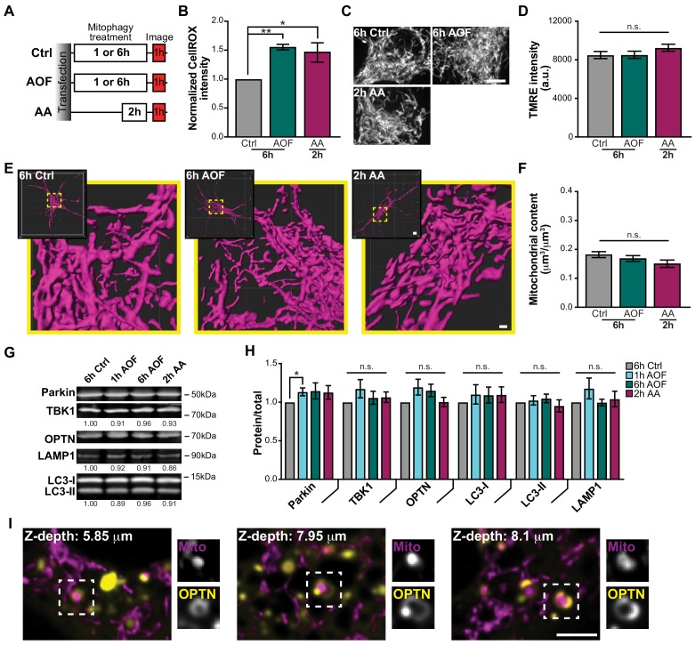
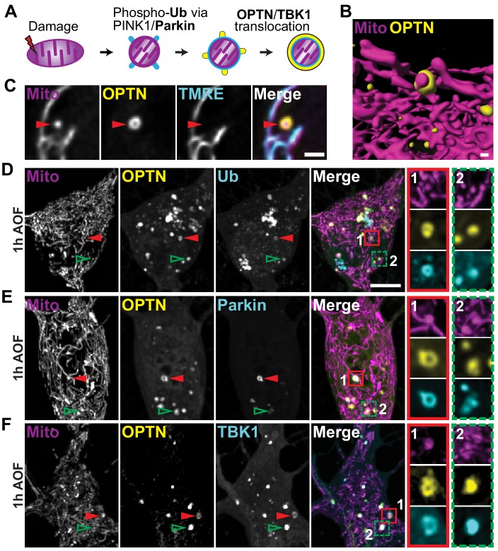
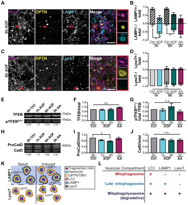
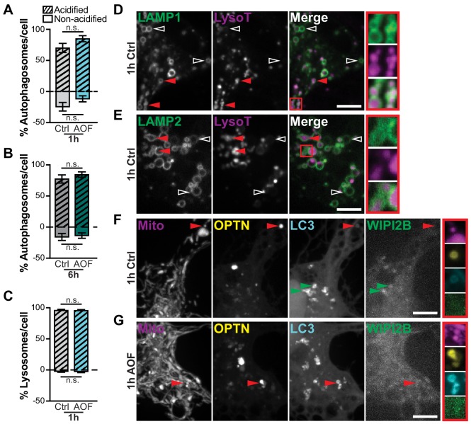
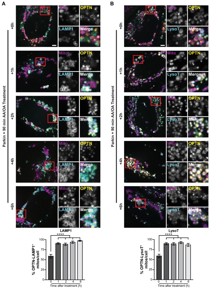
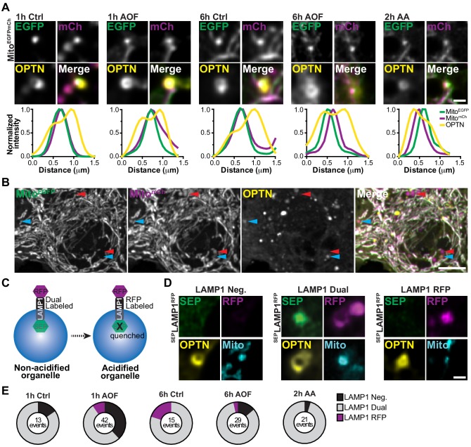
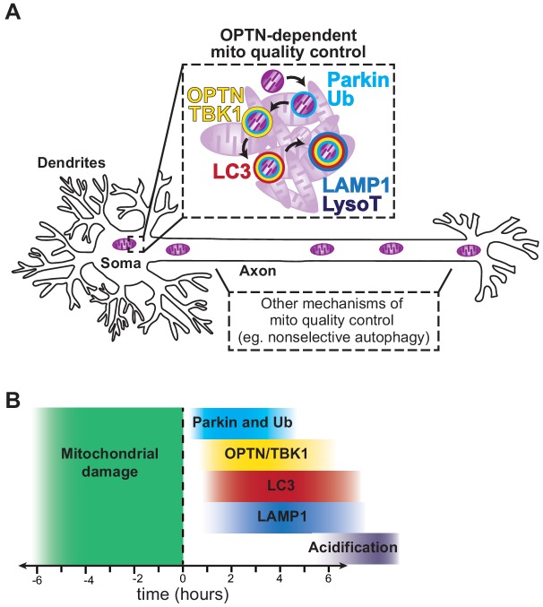
Mitophagy, the selective removal of damaged mitochondria, is thought to be critical to maintain neuronal homeostasis. Mutations of proteins in the pathway cause neurodegenerative diseases, suggesting defective mitochondrial turnover contributes to neurodegeneration. In primary rat hippocampal neurons, we developed a mitophagy induction paradigm where mild oxidative stress induced low levels of mitochondrial damage. Mitophagy-associated proteins were sequentially recruited to depolarized mitochondria followed by sequestration into autophagosomes. The localization of these mitophagy events had a robust somal bias. In basal and induced conditions, engulfed mitochondria remained in non-acidified organelles for hours to days, illustrating efficient autophagosome sequestration but delayed lysosomal fusion or acidification. Furthermore, expression of an ALS-linked mutation in the pathway disrupted mitochondrial network integrity and this effect was exacerbated by oxidative stress. Thus, age-related decline in neuronal health or expression of disease-associated mutations in the pathway may exacerbate the slow kinetics of neuronal mitophagy, leading to neurodegeneration.
Overview
The claims and statistics of this paper haven’t been checked yet.
Run the check — free, no account needed. It reads the open-access text and checks the sources it cites.
Integrity signals
Registry
Cites
Statistics
Figures
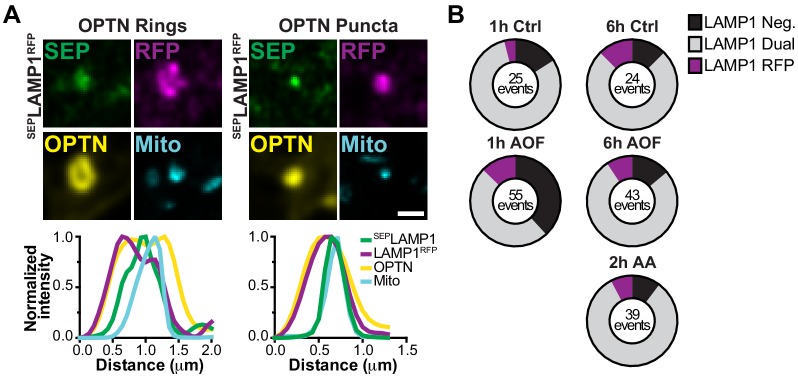
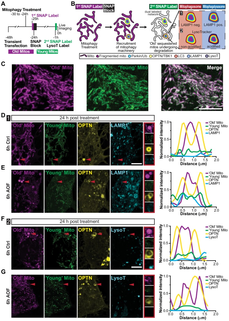
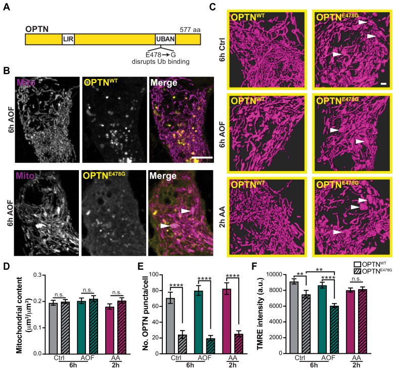
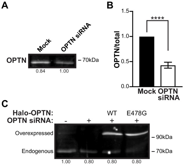
Verification
Claims the paper makes
The claims and statistics of this paper haven’t been checked yet.
Run the check — free, no account needed. It reads the open-access text and checks the sources it cites.
Claims other papers make about this one
Each is checked against this paper’s own text: does it say what the citing paper says it does?
In the citing paper
“the majority of pathogenic OPTN mutations are located in its UBAN domain and may impair ubiquitin binding [259]”
From Autophagy and ALS: mechanistic insights and therapeutic implications (2022) · cites this paper as [259]
Self-check
Percentages, sums and reported differences re-computed from the paper’s own numbers.
The self-check needs the paper’s full text.
Citations
Cites
1 retracted
RETRACTED ARTICLE: Parkin and PINK1 mitigate STING-induced inflammation
Sliter, D. A., Martinez, J., Hao, L., Chen, X., Sun, N., Fischer, T. D., Burman, J. L., Li, Y., Zhang, Z., Narendra, D. P., et al. (2018) · Nature · doi:10.1038/s41586-018-0448-9 · Cited by 1,287 · cited 2×
“Interestingly, recent work described an inflammatory phenotype in PINK1 and Parkin KO mice after acute or chronic mitochondrial stress that could be rescued by depletion of a regulator of the type 1 IFN signaling response (Sliter et al., 2018).”
BackgroundInfluentialRetractedFiji: an open-source platform for biological-image analysis
Schindelin, J., Arganda-Carreras, I., Frise, E., Kaynig, V., Longair, M., Pietzsch, T., Preibisch, S., Rueden, C., Saalfeld, S., Schmid, B., et al. (2012) · Nature Methods · doi:10.1038/nmeth.2019 · Cited by 66,890 · cited 6×
“Line scans In Fiji, line scans were generated using a pixel line width to trace OPTN-positive mitochondria and a three-pixel line width to trace axons and dendrites.”
MethodInfluentialRepertoires of Autophagy in the Pathogenesis of Ocular Diseases
Li, Y., Jiang, Q. L., Cao, G.-F., Yao, J., & Yan, B. (2015) · Cellular Physiology and Biochemistry · doi:10.1159/000373980 · Cited by 16,351
Mutations in the parkin gene cause autosomal recessive juvenile parkinsonism
Kitada, T., Asakawa, S., Hattori, N., Matsumine, H., Yamamura, Y., Minoshima, S., Yokochi, M., Mizuno, Y., & Shimizu, N. (1998) · Nature · doi:10.1038/33416 · Cited by 5,227 · cited 1×
“Mitophagy is primarily regulated by two proteins linked to familial PD, PTEN-Induced Putative Kinase 1 (PINK1) and Parkin (Kitada et al., 1998; Narendra et al., 2008; Narendra et al., 2010b; Valente et al., 2004; Wang and Klionsky, 2011; Youle and Narendra, 2011).”
BackgroundParkin is recruited selectively to impaired mitochondria and promotes their autophagy
Narendra, D., Tanaka, A., Suen, D.-F., & Youle, R. J. (2008) · The Journal of Cell Biology · doi:10.1083/jcb.200809125 · Cited by 4,026 · cited 6×
“PINK1 recruits Parkin to phospho-ubiquitinate outer mitochondrial membrane proteins, which act as a specific cue for organelle degradation (Geisler et al., 2010; Kane et al., 2014; Kazlauskaite et al., 2014; Koyano et al., 2014; Matsuda et al., 2010; Narendra et al., 2008; Narendra et al., 2010b; Ordureau et al., 2015…”
BackgroundInfluentialHereditary Early-Onset Parkinson's Disease Caused by Mutations in PINK1
Valente, E. M., Abou-Sleiman, P. M., Caputo, V., Muqit, M. M. K., Harvey, K., Gispert, S., Ali, Z., Del Turco, D., Bentivoglio, A. R., Healy, D. G., et al. (2004) · Science · doi:10.1126/science.1096284 · Cited by 3,481 · cited 1×
“Mitophagy is primarily regulated by two proteins linked to familial PD, PTEN-Induced Putative Kinase 1 (PINK1) and Parkin (Kitada et al., 1998; Narendra et al., 2008; Narendra et al., 2010b; Valente et al., 2004; Wang and Klionsky, 2011; Youle and Narendra, 2011).”
BackgroundYoule, R. J., & Narendra, D. P. (2011) · Nature Reviews Molecular Cell Biology · doi:10.1038/nrm3028 · Cited by 3,273
Fission and selective fusion govern mitochondrial segregation and elimination by autophagy
Twig, G., Elorza, A., Molina, A. J. A., Mohamed, H., Wikstrom, J. D., Walzer, G., Stiles, L., Haigh, S. E., Katz, S., Las, G., et al. (2008) · The EMBO Journal · doi:10.1038/sj.emboj.7601963 · Cited by 3,082 · cited 1×
“Since rounding and fragmentation are hallmarks of mitochondrial damage (Twig et al., 2008; Westermann, 2010; Youle and Narendra, 2011), we examined morphology following treatments by either staining with tetramethyl rhodamine ethyl ester (TMRE), a vital dye used to measure mitochondrial potential (Figure 1C–D), or by…”
MethodPINK1 Is Selectively Stabilized on Impaired Mitochondria to Activate Parkin
Narendra, D. P., Jin, S. M., Tanaka, A., Suen, D.-F., Gautier, C. A., Shen, J., Cookson, M. R., & Youle, R. J. (2010) · PLoS Biology · doi:10.1371/journal.pbio.1000298 · Cited by 2,944
The ubiquitin kinase PINK1 recruits autophagy receptors to induce mitophagy
Lazarou, M., Sliter, D. A., Kane, L. A., Sarraf, S. A., Wang, C., Burman, J. L., Sideris, D. P., Fogel, A. I., & Youle, R. J. (2015) · Nature · doi:10.1038/nature14893 · Cited by 2,774 · cited 7×
“In non-neuronal cells, OPTN, NDP52, and TAX1BP1 are proposed to mediate the autophagic clearance of damaged mitochondria, while p62 clusters organelles without directly facilitating mitophagy (Heo et al., 2015; Lazarou et al., 2015; Moore and Holzbaur, 2016; Narendra et al., 2010a; Wong and Holzbaur, 2014a).”
BackgroundInfluential
Cited by
Sorted by citations
The mechanisms and roles of selective autophagy in mammals
Vargas, J. N. S., Hamasaki, M., Kawabata, T., Youle, R. J., & Yoshimori, T. (2022) · Nature Reviews Molecular Cell Biology · doi:10.1038/s41580-022-00542-2 · Cited by 1,088
Mitochondria ROS and mitophagy in acute kidney injury
Su, L., Zhang, J., Gomez, H., Kellum, J. A., & Peng, Z. (2022) · Autophagy · doi:10.1080/15548627.2022.2084862 · Cited by 822
The role of PINK1–Parkin in mitochondrial quality control
Narendra, D. P., & Youle, R. J. (2024) · Nature Cell Biology · doi:10.1038/s41556-024-01513-9 · Cited by 600
“In cultured cells, acidification and degradation of mitochondria in the soma may be rate-limiting steps, suggesting that neurons may have comparatively low rates of mitophagic flux 137 .”
Quality control of the mitochondrion
Ng, M. Y. W., Wai, T., & Simonsen, A. (2021) · Developmental Cell · doi:10.1016/j.devcel.2021.02.009 · Cited by 468
Defective mitophagy in Alzheimer’s disease
Pradeepkiran, J. A., & Reddy, P. H. (2020) · Ageing Research Reviews · doi:10.1016/j.arr.2020.101191 · Cited by 367
“A uthor M anuscript A uthor M anuscript A uthor M anuscript A uthor M anuscript mitochondrial content determines the markers of mitochondrial quality (Evans and Holzbaur, 2020; Calkins et al., 2011; Wang et al., 2009).”
BackgroundProgramming axonal mitochondrial maintenance and bioenergetics in neurodegeneration and regeneration
Cheng, X., Huang, N., & Sheng, Z. (2022) · Neuron · doi:10.1016/j.neuron.2022.03.015 · Cited by 257
Autophagy and ALS: mechanistic insights and therapeutic implications
Chua, J. P., De Calbiac, H., Kabashi, E., & Barmada, S. J. (2022) · Autophagy · doi:10.1080/15548627.2021.1926656 · Cited by 189
BNIP3L/NIX-mediated mitophagy protects against glucocorticoid-induced synapse defects
Choi, G. E., Lee, H. J., Chae, C. W., Cho, J. H., Jung, Y. H., Kim, J. S., Kim, S. Y., Lim, J. R., & Han, H. J. (2021) · Nature Communications · doi:10.1038/s41467-020-20679-y · Cited by 187
Mitochondrial quality control in human health and disease
Liu, B., Xu, C.-Z., Liu, Y., Lu, Z., Fu, T.-L., Li, G., Deng, Y., Luo, G., Ding, S. Z., Li, N., et al. (2024) · Military Medical Research · doi:10.1186/s40779-024-00536-5 · Cited by 211
Borcherding, N. C., Jia, W., Giwa, R., Field, R. L., Moley, J. R., Kopecky, B. J., Chan, M. M., Yang, B. Q., Sabio, J. M., Walker, E. C., et al. (2022) · Cell Metabolism · doi:10.1016/j.cmet.2022.08.010 · Cited by 175
Boecker, C. A., Goldsmith, J., Dou, D., Cajka, G. G., & Holzbaur, E. L. F. (2021) · Current Biology · doi:10.1016/j.cub.2021.02.061 · Cited by 172
Self-citationTargeting mitophagy in neurodegenerative diseases
Antico, O., Thompson, P. W., Hertz, N. T., Muqit, M. M. K., & Parton, L. E. (2025) · Nature Reviews Drug Discovery · doi:10.1038/s41573-024-01105-0 · Cited by 187
“Although most mitochondria are distributed in the distal neuronal compartments 5 , mitophagy in neurons has been detected mainly in the soma (cell bodies), correlating with where the majority of active lys osomes are situated 80–83 .”
Eapen, V. V., Swarup, S., Hoyer, M., Paulo, J. A., & Harper, J. W. (2021) · eLife · doi:10.7554/elife.72328 · Cited by 162
Neuronal autophagy and mitophagy in Parkinson's disease
Lizama, B. N., & Chu, C. T. (2021) · Molecular Aspects of Medicine · doi:10.1016/j.mam.2021.100972 · Cited by 152
Targeting autophagy in prostate cancer: preclinical and clinical evidence for therapeutic response
Ashrafizadeh, M., Paskeh, M. D. A., Mirzaei, S., Gholami, M. H., Zarrabi, A., Hashemi, F., Hushmandi, K., Hashemi, M., Nabavi, N., Crea, F., et al. (2022) · Journal of Experimental & Clinical Cancer Research · doi:10.1186/s13046-022-02293-6 · Cited by 150
Emerging views of OPTN (optineurin) function in the autophagic process associated with disease
Qiu, Y., Wang, J., Li, H., Yang, B., Wang, J., He, Q., & Weng, Q. (2021) · Autophagy · doi:10.1080/15548627.2021.1908722 · Cited by 143
Motor proteins at the mitochondria–cytoskeleton interface
Kruppa, A. J., & Buß, F. (2021) · Journal of Cell Science · doi:10.1242/jcs.226084 · Cited by 127
The multifaceted role of mitochondria in autism spectrum disorder
Khaliulin, I., Hamoudi, W., & Amal, H. (2024) · Molecular Psychiatry · doi:10.1038/s41380-024-02725-z · Cited by 128
“This results in the translocation of Parkin to the mitochondria and initiation of mitochondrial degradation [322, 323], which helps to maintain neuronal homeostasis [324].”
Agujetas, V. R., Barbero‐Camps, E., de Dios, C., Podlesniy, P., Abadin, X., Morales, A., Marı́, M., Trullás, R., & Colell, A. (2021) · Molecular Neurodegeneration · doi:10.1186/s13024-021-00435-6 · Cited by 113
“This apparent discrepancy may be likely related to the inducer used and the speci c time course of mitophagy in neurons, which is longer compared with non-neuronal cells [68, 69].”
Goldsmith, J., Ordureau, A., Harper, J. W., & Holzbaur, E. L. F. (2022) · Neuron · doi:10.1016/j.neuron.2021.12.029 · Cited by 112
Self-citationAgarwal, S., & Muqit, M. M. K. (2021) · Current Opinion in Neurobiology · doi:10.1016/j.conb.2021.09.005 · Cited by 104
ALS- and FTD-associated missense mutations in TBK1 differentially disrupt mitophagy
Harding, O. J., Evans, C. S., Ye, J., Cheung, J., Maniatis, T., & Holzbaur, E. L. F. (2021) · Proceedings of the National Academy of Sciences · doi:10.1073/pnas.2025053118 · Cited by 99
Self-citationGlucocorticoid impairs mitochondrial quality control in neurons
Choi, G. E., & Han, H. J. (2021) · Neurobiology of Disease · doi:10.1016/j.nbd.2021.105301 · Cited by 95
Lie, P. P. Y., Yang, D.-S., Stavrides, P., Goulbourne, C. N., Zheng, P., Mohan, P. S., Cataldo, A. M., & Nixon, R. A. (2021) · Cell Reports · doi:10.1016/j.celrep.2021.109034 · Cited by 97
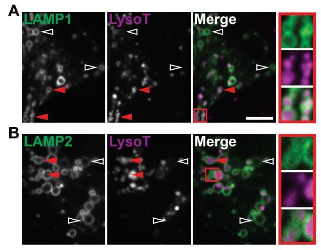
“, 2010), and LAMP1 acquisition by autophagosomes is temporally separate from acidification (Evans and Holzbaur, 2020).”
BackgroundMechanisms underlying ubiquitin-driven selective mitochondrial and bacterial autophagy
Goodall, E. A., Kraus, F., & Harper, J. W. (2022) · Molecular Cell · doi:10.1016/j.molcel.2022.03.012 · Cited by 86
Phospholipase D3 degrades mitochondrial DNA to regulate nucleotide signaling and APP metabolism
Acker, Z. P. V., Perdok, A., Hellemans, R., North, K., Vorsters, I., Cappel, C. C., Dehairs, J., Swinnen, J. V., Sannerud, R., Bretou, M., et al. (2023) · Nature Communications · doi:10.1038/s41467-023-38501-w · Cited by 76
“The latter may be particularly relevant to neurons with their high energetic activity and associated high mitochondrial turnover 43 .”
Antico, O., Ordureau, A., Stevens, M., Singh, F., Nirujogi, R. S., Gierliński, M., Barini, E., Rickwood, M. L., Prescott, A. R., Toth, R. L., et al. (2021) · Science Advances · doi:10.1126/sciadv.abj0722 · Cited by 68
“…the autophagy receptor proteins NDP52, p62, OPTN, Tax1bp, and Next to BRCA1 gene 1 protein (NBR1), and several studies suggests that NDP52 and OPTN are the main ubiquitin-dependent receptors for efficient mitophagic clearance of damaged mitochondria both in HeLa cells and neurons ( 23 , 24 , 49 ).”
Wrighton, P. J., Shwartz, A., Heo, J., Quenzer, E. D., LaBella, K. A., Harper, J. W., & Goessling, W. (2021) · Journal of Cell Science · doi:10.1242/jcs.256255 · Cited by 61
Rojas-Charry, L., Nardi, L., Methner, A., & Schmeißer, M. J. (2020) · Journal of Molecular Medicine · doi:10.1007/s00109-020-02018-2 · Cited by 62
Role of Autophagy Pathway in Parkinson’s Disease and Related Genetic Neurological Disorders
Themistokleous, C., Bagnoli, E., Parulekar, R., & Muqit, M. M. K. (2023) · Journal of Molecular Biology · doi:10.1016/j.jmb.2023.168144 · Cited by 51
Giovedı̀, S., Ravanelli, M. M., Parisi, B., Bettegazzi, B., & Guarnieri, F. C. (2020) · Frontiers in Cellular Neuroscience · doi:10.3389/fncel.2020.602116 · Cited by 53
“Similarly, ALS-linked mutations in optineurin interfere with the clearance of protein inclusions and of damaged mitochondria in cell lines and neurons (Wong and Holzbaur, 2014b; Shen et al., 2015; Evans and Holzbaur, 2020).”
BackgroundNeuronal Mitophagy: Friend or Foe?
Doxaki, C., & Palikaras, K. (2021) · Frontiers in Cell and Developmental Biology · doi:10.3389/fcell.2020.611938 · Cited by 48
Impaired Mitophagy in Neurons and Glial Cells during Aging and Age-Related Disorders
Sukhorukov, V. S., Воронков, Д. Н., Baranich, T. I., Mudzhiri, N. M., Magnaeva, A., & Illarioshkin, S. N. (2021) · International Journal of Molecular Sciences · doi:10.3390/ijms221910251 · Cited by 50
Decreasing pdzd8-mediated mito–ER contacts improves organismal fitness and mitigates Aβ 42 toxicity
Hewitt, V. L., Miller‐Fleming, L., Twyning, M. J., Andreazza, S., Mattedi, F., Prudent, J., Polleux, F., Vagnoni, A., & Whitworth, A. J. (2022) · Life Science Alliance · doi:10.26508/lsa.202201531 · Cited by 45
Organization of the autophagy pathway in neurons
Sidibe, D. K., Vogel, M. C., & Maday, S. (2022) · Current Opinion in Neurobiology · doi:10.1016/j.conb.2022.102554 · Cited by 39
Mitophagy and Neurodegeneration: Between the Knowns and the Unknowns
Jetto, C. T., Nambiar, A., & Manjithaya, R. R. (2022) · Frontiers in Cell and Developmental Biology · doi:10.3389/fcell.2022.837337 · Cited by 44
Li, H., Doric, Z., Berthet, A., Jorgens, D. M., Nguyen, M. K., Hsieh, I., Margulis, J., Fang, R., Debnath, J., Sesaki, H., et al. (2021) · Science Advances · doi:10.1126/sciadv.abf6580 · Cited by 44
“Notably, these findings differ from those of a recent study that used LysoTracker, superecliptic pHluorin (SEP)–LAMP1-RFP, and mitochondria-targeted EGFP-mCherry reporters to conclude that mitochondrial acidification is a very slow process that takes several hours following mitophagosome formation and LAMP1 accumulati…”
ResultZaninello, M., Palikaras, K., Sotiriou, A., Tavernarakis, N., & Scorrano, L. (2021) · Cell Death and Differentiation · doi:10.1038/s41418-021-00847-3 · Cited by 40
Mitophagy in Neurons: Mechanisms Regulating Mitochondrial Turnover and Neuronal Homeostasis
Basak, B., & Holzbaur, E. L. F. (2025) · Journal of Molecular Biology · doi:10.1016/j.jmb.2025.169161 · Cited by 44
Self-citationRole of mitophagy in the neurodegenerative diseases and its pharmacological advances: A review
Wang, Q., Xue, H., Yue, Y., Hao, S., Huang, S., & Zhang, Z. (2022) · Frontiers in Molecular Neuroscience · doi:10.3389/fnmol.2022.1014251 · Cited by 38
“Interestingly, OPTN-mediated mitophagy appears to be spatially confined to neuronal cell bodies, with just a few mitophagy events observed in axons or dendrites (Evans and Holzbaur, 2020).”
BackgroundPARK15/FBXO7 is dispensable for PINK1/Parkin mitophagy in iNeurons and HeLa cell systems
Kraus, F., Goodall, E. A., Smith, I. R. L., Jiang, Y., Paoli, J. C., Adolf, F., Zhang, J., Paulo, J. A., Schulman, B. A., & Harper, J. W. (2023) · EMBO Reports · doi:10.15252/embr.202256399 · Cited by 36
“…recruitment of selective autophagy cargo receptors including OPTN, CALCOCO2 (also called NDP52), and SQSTM1 (also called p62) to facilitate the assembly of an autophagosome around the ubiquitylated organelle (Wong & Holz-baur, 2014; Heo et al , 2015; Lazarou et al , 2015; Evans & Holzbaur, 2020).”
Hallmarks and Molecular Tools for the Study of Mitophagy in Parkinson’s Disease
Goiran, T., Eldeeb, M. A., Zorca, C. E., & Fon, E. A. (2022) · Cells · doi:10.3390/cells11132097 · Cited by 38
Mitophagy in relation to chronic inflammation/ROS in aging
Kong, L., Li, S., Fu, Y., Cai, Q., Du, X., Liang, J., & Ma, T. (2024) · Molecular and Cellular Biochemistry · doi:10.1007/s11010-024-05042-9 · Cited by 40
“Mitophagy restricts mtROS production, by removing aging and damaged mitochondria, hindering the progression of this process [89].”
Liu, W., Ren, Y., Wang, T., Wang, M., Xu, Y., Zhang, J., Bi, J., Wu, Z., Zhang, Y., & Wu, R. (2024) · Cell Death Discovery · doi:10.1038/s41420-024-01923-6 · Cited by 33
“These results reveal a novel CIRP-mediated acute pancreatitis injury pathway and provide a new target for future treatment.”
BackgroundMethodUpregulated pexophagy limits the capacity of selective autophagy
Germain, K., So, R. W. L., DiGiovanni, L. F., Watts, J. C., Bandsma, R., & Kim, P. K. (2024) · Nature Communications · doi:10.1038/s41467-023-44005-4 · Cited by 34
Optineurin-facilitated axonal mitochondria delivery promotes neuroprotection and axon regeneration
Liu, D., Webber, H. C., Bian, F., Xu, Y., Prakash, M., Feng, X., Yang, M., Yang, H., You, I.-J., Li, L., et al. (2025) · Nature Communications · doi:10.1038/s41467-025-57135-8 · Cited by 34
“The OPTN E478G mutant found in ALS patients loses the interaction between the OPTN UBAN domain and ubiquitinated cargos, which disrupts mitophagy and causes cell death in cultured neurons 59 .”
Shen, J., Xie, P., Wang, J., Yang, F., Li, S., Jiang, H., Wu, X., Zhou, F., & Li, J. (2024) · Redox Biology · doi:10.1016/j.redox.2024.103196 · Cited by 31
“Typically, mitophagy contributes to neuronal cell viability and offers protection through various mechanisms, including the removal of damaged mitochondria, which are primary sources of ROS production [51,52].”
BackgroundSelective Autophagy in Hyperglycemia-Induced Microvascular and Macrovascular Diseases
Bharath, L. P., Rockhold, J. D., & Conway, R. (2021) · Cells · doi:10.3390/cells10082114 · Cited by 31
“It is also known that mutations of proteins involved in mitochondrial turnover promote diabetic neurological diseases [86].”
BackgroundAutophagy in the eye: from physiology to pathophysiology
Liton, P. B., Boesze‐Battaglia, K., Boulton, M. E., Boya, P., Ferguson, T. A., Ganley, I. G., Kauppinen, A., Laurie, G. W., Mizushima, N., Morishita, H., et al. (2023) · Autophagy Reports · doi:10.1080/27694127.2023.2178996 · Cited by 31
Liu, M., Wang, Q., Wu, W., Chen, M., Zhang, P., Guo, M., Lin, H., Ma, Z., Zhou, H., & Fan, H. (2022) · PLoS Pathogens · doi:10.1371/journal.ppat.1010912 · Cited by 28
ROCK2 inhibition: A futuristic approach for the management of Alzheimer’s disease
Mani, S., Jindal, D., Chopra, H., Jha, S. K., Singh, S. K., Ashraf, G. M., Kamal, M., Iqbal, D., Chellappan, D. K., Dey, A., et al. (2022) · Neuroscience & Biobehavioral Reviews · doi:10.1016/j.neubiorev.2022.104871 · Cited by 29
Evans, C. S., & Holzbaur, E. L. F. (2020) · Autophagy · doi:10.1080/15548627.2020.1734330 · Cited by 28
Self-citationA Healthy Heart and a Healthy Brain: Looking at Mitophagy
Luo, H., Zhang, R., Krigman, J., McAdams, A., Ozgen, S., & Sun, N. (2020) · Frontiers in Cell and Developmental Biology · doi:10.3389/fcell.2020.00294 · Cited by 25
TSG101 negatively regulates mitochondrial biogenesis in axons
Lin, T.-H., Bis‐Brewer, D. M., Sheehan, A. E., Townsend, L., Maddison, D. C., Zuchner, S. L., Smith, G. A., & Freeman, M. R. (2021) · Proceedings of the National Academy of Sciences · doi:10.1073/pnas.2018770118 · Cited by 24
Advances in SIRT3 involvement in regulating autophagy-related mechanisms
Xi, S., Chen, W., & Ke, Y. (2024) · Cell Division · doi:10.1186/s13008-024-00124-y · Cited by 27
“Mitochondrial autophagy is considered a bona fide strategy to limit mitochondrial ROS production by specifically isolating and phagocytosing aged and damaged mitochondria in lysosomes [87].”
BackgroundEndo-IP and lyso-IP toolkit for endolysosomal profiling of human-induced neurons
Hundley, F. V., Gonzalez‐Lozano, M. A., Gottschalk, L. M., Cook, A. N. K., Zhang, J., Paulo, J. A., & Harper, J. W. (2024) · Proceedings of the National Academy of Sciences · doi:10.1073/pnas.2419079121 · Cited by 26
“…with unique properties ( 35 , 44 – 46 ). iNeurons maintain many features of animal - derived neurons, including intact axonal and dendritic trafficking pathways, and reliance on organelle quality control systems such as autophagy to remove damaged or superfluous organelles ( 16 , 47 , 48 ).”
Juncker, M. A., Kim, C., Reed, R. E., Haas, A. L., Schwartzenburg, J., & Desai, S. D. (2021) · Biochimica et Biophysica Acta (BBA) - Molecular Basis of Disease · doi:10.1016/j.bbadis.2021.166102 · Cited by 26
Candelise, N., Salvatori, I., Scaricamazza, S., Nesci, V., Zenuni, H., Ferri, A., & Valle, C. (2022) · Metabolites · doi:10.3390/metabo12030233 · Cited by 25
“The ALS-linked mutation OPTNE478G was demonstrated to be sufficient to disrupt mitophagy and to cause neurodegeneration in primary neurons [65].”
BackgroundProspects for gene replacement therapies in amyotrophic lateral sclerosis
Giovannelli, I., Higginbottom, A., Kirby, J., Azzouz, M., & Shaw, P. J. (2022) · Nature Reviews Neurology · doi:10.1038/s41582-022-00751-5 · Cited by 23
Gene therapy breakthroughs in ALS: a beacon of hope for 20% of ALS patients
Xie, Q., Li, K., Chen, Y., Li, Y., Jiang, W., Cao, W., Yu, H., Fan, D., & Deng, B. (2025) · Translational Neurodegeneration · doi:10.1186/s40035-025-00477-6 · Cited by 27
“OPTN, as a selective autophagy receptor, identifies and binds ubiquitinated proteins and damaged organelles such as mitochondria, transporting them to autophago-somes for degradation [156].”
Peripheral neuropathy in mitochondrial disease
Horváth, R., Medina, J. M., Reilly, M. M., Shy, M. E., & Zuchner, S. L. (2023) · Handbook of clinical neurology · doi:10.1016/b978-0-12-821751-1.00014-2 · Cited by 22
PINK1 regulated mitophagy is evident in skeletal muscles
Singh, F., Wilhelm, L. P., Prescott, A. R., Ostacolo, K., Zhao, J., Ogmundsdottir, M. H., & Ganley, I. G. (2024) · Autophagy Reports · doi:10.1080/27694127.2024.2326402 · Cited by 21
Mitochondrial damage triggers the concerted degradation of negative regulators of neuronal autophagy
Basak, B., & Holzbaur, E. L. F. (2025) · Nature Communications · doi:10.1038/s41467-025-62379-5 · Cited by 22
“Previous work from our lab has shown that fusion of mitopha-gosomes with lysosomes is slow, and in fact is the rate-limiting step in neuronal mitophagy 36 .”
Self-citationVevea, J., & Chapman, E. R. (2023) · Journal of Neuroscience · doi:10.1523/jneurosci.1356-22.2023 · Cited by 19
“Low doses of AA ( , 10 n M ) increased retrograde mitochondrial transport (M. Y. Lin et al., 2017) or had no effect on motility but induced mitophagy in the soma (Evans and Holzbaur, 2020).”
Stavropoulos, F., Sargiannidou, I., Potamiti, L., Kagiava, A., Panayiotidis, M. I., Bae, J. H., Yeom, S. C., Lee, J. Y., & Kleopa, K. A. (2021) · International Journal of Molecular Sciences · doi:10.3390/ijms222111569 · Cited by 21
“Moreover, mitophagy is a much slower process in neurons compared to non-neuronal cells [43,44], and this may be another factor rendering peripheral nerves more susceptible to MFN2 mutations, as their greater axonal length seems to be a rate-limiting factor for mitophagy.”
BackgroundMitophagy mechanisms in neuronal physiology and pathology during ageing
Markaki, M., Tsagkari, D., & Tavernarakis, N. (2021) · Biophysical Reviews · doi:10.1007/s12551-021-00894-7 · Cited by 18
“Specifically, autophagosome fusion with lysosome and acidification occur hours after the stimulus of mitophagy has emerged (Evans and Holzbaur 2020b).”
BackgroundHuang, K.-C., Gomes, C., Shiga, Y., Belforte, N., VanderWall, K. B., Lavekar, S. S., Fligor, C. M., Harkin, J., Hetzer, S. M., Patil, S., et al. (2024) · Acta Neuropathologica Communications · doi:10.1186/s40478-024-01872-2 · Cited by 18
“During the process of autophagy, OPTN is necessary to recruit the microtubule-associated protein light chain 3 (LC3) for the engulfment of protein aggregates and/ or damaged organelles by formation of the autophago-some [29, 43].”
BackgroundLi, D., Geng, D., & Wang, M. (2024) · The FASEB Journal · doi:10.1096/fj.202401409r · Cited by 18
“192 ,193”
The compartmentalised nature of neuronal mitophagy: molecular insights and implications
Borbolis, F., & Palikaras, K. (2022) · Expert Reviews in Molecular Medicine · doi:10.1017/erm.2022.31 · Cited by 16
“142).”
Defective PTEN-induced kinase 1/Parkin mediated mitophagy and neurodegenerative diseases
Braun, M. M., & Puglielli, L. (2022) · Frontiers in Cellular Neuroscience · doi:10.3389/fncel.2022.1031153 · Cited by 16
“However, it is unclear if this increase results in increased degradation of mitochondria since TDP-43 expression also increases the percentage of stationary mitochondria in axons and neurites (Cai et al., 2012; Wang et al., 2013; Evans and Holzbaur, 2020a).”
BackgroundMethodInfluentialHuynh, D. T., Tsolova, K. N., Watson, A. J., Khal, S. K., Green, J. R., Li, D., Hu, J., Soderblom, E. J., Chi, J., Evans, C. S., et al. (2023) · Nature Communications · doi:10.1038/s41467-023-42227-0 · Cited by 15
“Therefore, we used live-cell fl uorescence microscopy 75 to test whether NF-L O-GlcNAcylation impacts organelle motility.”
Self-citationAltered Mitochondrial Protein Homeostasis and Proteinopathies
Jishi, A., & Qi, X. (2022) · Frontiers in Molecular Neuroscience · doi:10.3389/fnmol.2022.867935 · Cited by 17
“Mitochondria are engulfed in mitophagosomes and degraded, though the process of their degradation is rather inefficient due to the slow acidification of these structures (Liu et al., 2018; Evans and Holzbaur, 2020).”
BackgroundOptineurin Deficiency and Insufficiency Lead to Higher Microglial TDP-43 Protein Levels
Prtenjača, N., Rob, M., Alam, M. S., Markovinović, A., Stuani, C., Buratti, E., & Munitić, I. (2022) · International Journal of Molecular Sciences · doi:10.3390/ijms23126829 · Cited by 13
“Notably, the majority of results on the mechanistic link between optineurin and autophagy were obtained on immortalized cell lines rather than the relevant CNS cells [28,29,35], and few recent studies have been performed in neurons or neuronal cell lines [33,36].”
BackgroundWen, D., Ji, Y., Li, Y., Duan, W., Wang, Y., Li, Z., Tao, M., & Liu, Y. (2023) · FEBS Journal · doi:10.1111/febs.17009 · Cited by 18
“These organelles carry out cellular functions, including Ca 2 + buffering and energy generation, via the MMP. Mitochondrial depolarization or damage impairs cell function and results in the transmission of signals to maintenance pathways to repair or eliminate dysfunctional organelles [35].”
BackgroundLi, Y., Li, Z., Grillo, E., Desler, C., Navarro, C., Bohr, V. A., Berliocchi, L., & Rasmussen, L. J. (2024) · Free Radical Biology and Medicine · doi:10.1016/j.freeradbiomed.2024.07.013 · Cited by 14
Jeong, Y., Davis, C.-H. O., Muscarella, A. M., Navarro, H. H., Deshpande, V. A., Moore, L. G., Keun-young Kim, A., Ellisman, M. H., & Marsh‐Armstrong, N. (2025) · eLife · doi:10.7554/elife.103844 · Cited by 12
“…( Xie and Klionsky, 2007 ; Mizushima and Komatsu, 2011 ; Fu et al., 2014 ; Wong and Holzbaur, 2014b ; Tammineni et al., 2017 ; Stavoe et al., 2019 ; Evans and Holzbaur, 2020 ; Boecker et al., 2021 ; Kuijpers et al., 2021 ), we used the EGFP-LC3b signal to create a mask to demark the RGC axons, so…”
InfluentialYi, S., Zheng, Y., Yi, Z., Wang, Y., Jiang, Y., Ouyang, J., Li, S., Xiao, X., Sun, W., Wang, P., et al. (2023) · Genes · doi:10.3390/genes14040952 · Cited by 12
“As a multifactorial mitochondrial disorder, it is assumed to be affected by poor maternal nutrition or hazardous exposure prior to and during pregnancy, as the bio-energetically impaired mitochondria are likely present in the ovum and transmits to zygote, which act as an additive hit to the mitochondrial dysfunction c…”
BackgroundSung, H., & Lloyd, T. E. (2023) · Autophagy · doi:10.1080/15548627.2023.2249750 · Cited by 11
“The soma is thought to be the primary site of major degradative pathways and is enriched in acidified lysosomes [12–15], whereas presynaptic terminals, responsible for signal transmission, are the primary site of autophagy initiation and autophagosome formation [9,16–18].”
BackgroundDrosophila Primary Neuronal Cultures as a Useful Cellular Model to Study and Image Axonal Transport
Voelzmann, A., & Sánchez‐Soriano, N. (2022) · Methods in molecular biology · doi:10.1007/978-1-0716-1990-2_23 · Cited by 11
Enhancing CNS mitophagy: drug development and disease-relevant models
Dhar, K. S., Townsend, B., Montgomery, A. P., Danon, J. J., Pagan, J. K., & Kassiou, M. (2024) · Trends in Pharmacological Sciences · doi:10.1016/j.tips.2024.09.002 · Cited by 11
Mitochondrial motility modulators coordinate quality control dynamics to promote neuronal health
Jenkins, J. E., Fazli, M., & Evans, C. S. (2024) · Current Opinion in Cell Biology · doi:10.1016/j.ceb.2024.102383 · Cited by 9
Self-citationChen, K., Padilla, C. G., Kiselyov, K. I., & Kozai, T. D. Y. (2023) · Biomaterials · doi:10.1016/j.biomaterials.2023.122316 · Cited by 9
Lu, Y., Yang, Q., Yang, S., Zhang, J., Zhang, Y., Lu, J., Li, Z., Shen, Y., Yan, W., Jiang, Q., et al. (2025) · Nano Research · doi:10.26599/nr.2025.94907463 · Cited by 8
Li, W., Shao, G., & Qi, R. (2025) · Cell Regeneration · doi:10.1186/s13619-025-00268-4 · Cited by 9
“Mitochondria are central to cellular energy production via oxidative phosphorylation and the regulation of intra-cellular signaling cascades (Evans and Holzbaur 2020).”
ALS-Linked VapB P56S Mutation Alters Neuronal Mitochondrial Turnover at the Synapse
Wong, H.-T., Lang, A. E., Stein, C., & Drerup, C. M. (2024) · Journal of Neuroscience · doi:10.1523/jneurosci.0879-24.2024 · Cited by 9
Presynapses are mitophagy pit stops that prevent axon degenerationPreprint
Lam, W. K., Lindblom, R. S. J., Milky, B., Mazzachi, P., Hadian-Jazi, M., Küng, C. J., Khuu, G., Uoselis, L., Nguyen, T. N., Skulsuppaisarn, M., et al. (2024) · bioRxiv (Cold Spring Harbor Laboratory) · doi:10.1101/2024.09.09.611943 · Cited by 10
Morphological multiparameter filtration and persistent homology in mitochondrial image analysis
Chung, Y.-M., Hu, C.-S., Sun, E., & Tseng, H. C. (2024) · PLoS ONE · doi:10.1371/journal.pone.0310157 · Cited by 7
Guo, Y., Xia, P., Tian, Y., Fu, D., Hu, X., Xie, K., Dong, W., Zhang, W., Liu, D., Shen, R., et al. (2025) · Cell Death Discovery · doi:10.1038/s41420-025-02641-3 · Cited by 8
Chun, C., Lee, J. H., Bothwell, M. A., Nghiem, P., Smith, A. S. T., & Mack, D. L. (2024) · Journal of Neuroscience · doi:10.1523/jneurosci.1787-23.2024 · Cited by 7
Probing β-Cell Biology in Space and Time
Drigo, R. A. E. (2021) · Diabetes · doi:10.2337/dbi21-0008 · Cited by 6
“Age mosaicism has recently been observed in the vastly different lifetimes of biological elements and systems, from the varying half-lives of NAD(1) across tissues (79) to the long-term association of transcription factors with the chromatin to determine cell phenotype (80), to the longevity of NPCs, histones, and mit…”
BackgroundRadiation and Diabetic Retinopathy: A Dark Synergy
Gardiner, T. A., ARCHER, D. B., Silvestri, G., & Amoaku, W. M. K. (2023) · International Journal of Translational Medicine · doi:10.3390/ijtm3010011 · Cited by 6
Liu, Y., Li, J., Xiong, Y., Tan, C., Li, C., Cao, Y., Xie, W., & Deng, Z. (2024) · Scientific Reports · doi:10.1038/s41598-024-62748-y · Cited by 6
The Unified Theory of Neurodegeneration Pathogenesis Based on Axon Deamidation
Joseph, D. (2025) · International Journal of Molecular Sciences · doi:10.3390/ijms26094143 · Cited by 6
“Regarding oxidative stress, Evans et al. [134] reported that oxidative stress renders mitophagy unable to maintain the health of the neuron, leading to neurodegeneration.”
Correcting abnormal mitochondrial dynamics to facilitate tumor treatment
Yang, B., Lin, Y., & Shen, Y. (2023) · Mitochondrial Communications · doi:10.1016/j.mitoco.2023.07.001 · Cited by 7
Sun, L., 宁波大学农产品质量安全危害因子与风险防控国家重点实验室, 浙. 宁. 3., Dai, Y.-F., You, M.-X., Li, C., & 青岛海洋科学与技术国家实验室海洋渔业科学与食物产出过程功能实验室, 山. 青. 2. (2023) · 动物学研究 · doi:10.24272/j.issn.2095-8137.2023.106 · Cited by 6
Molecular aspects of cytoprotection by Optineurin during stress and disease
Swarup, G., Medchalmi, S., Ramachandran, G., & Sayyad, Z. (2025) · Biochimica et Biophysica Acta (BBA) - Molecular Cell Research · doi:10.1016/j.bbamcr.2024.119895 · Cited by 6
Li, Z., Liu, J., Ju, J., Peng, X., Zhao, W., Ren, J., Jia, X., Wang, J., Tu, Y., & Gao, F. (2025) · British Journal of Pharmacology · doi:10.1111/bph.70048 · Cited by 5
“Damaged mitochondria are trafficked back to the soma (retrograde) for degradation, avoiding the local accumulation of these damaged mitochondria (Evans & Holzbaur, 2020).”
Li, N., Hou, H., Su, D., Yang, B., Ni, R., Ma, G., Sun, S., Ma, Q., Peng, Q., Chen, S., et al. (2025) · Autophagy · doi:10.1080/15548627.2025.2572528 · Cited by 5
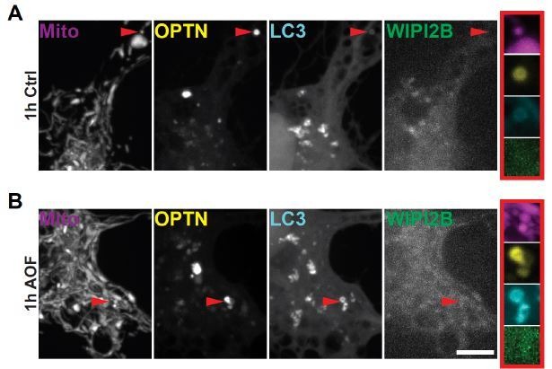
“During PINK1-PRKN-mediated mitophagy, LC3B is recruited to damaged mitochondria after the mitochondrial translocation of the E3 ubiquitin ligase PRKN and mitophagy receptor OPTN [31].”
Pabon, A., Bhupana, J. N., & Wong, C. (2024) · Neural Regeneration Research · doi:10.4103/nrr.nrr-d-23-02095 · Cited by 6
“Damaged distal mitochondria are transported to neuronal soma for mitophagy (Zheng et al., 2019; Evans and Holzbaur, 2020).”
BackgroundGuttipatti, P., Saadallah, N., Ji, R., Avula, U. M. R., Goulbourne, C. N., & Wan, E. Y. (2024) · Journal of Structural Biology · doi:10.1016/j.jsb.2024.108110 · Cited by 6
Metadata
- Authors
- Evans, C. S., & Holzbaur, E. L.
- Affiliations
- University of Pennsylvania (US)
- Corresponding author
- Holzbaur, Erika LF
- Venue
- eLife
- Publisher
- eLife Sciences Publications, Ltd
- Published
- 2020-01-14
- Volume
- 9
- ISSN
- 2050-084X (ISSN-L)
- PMC
- PMC6959996
- OpenAlex
- W2999206621
- Cited by
- 137
- References
- 113
- Citations by year
- 2026: 13 · 2025: 20 · 2024: 26 · 2023: 20 · 2022: 26 · 2021: 25 · 2020: 7
- FWCI
- 13.99
- Type
- journal article
- Language
- en
- Open access
- gold
- doi.org
- Licence
- Locations
- eLife (publishedVersion, cc-by)
- PubMed Central (publishedVersion, cc-by)
- PubMed (publishedVersion)
- DOAJ (DOAJ: Directory of Open Access Journals) (submittedVersion, cc-by-sa)
- Funders
- National Institute of Neurological Disorders and Stroke (R37 NS060698)
- Howard Hughes Medical Institute (Hanna H Gray Fellowship)
- Topics
- Life Sciences › Biochemistry, Genetics and Molecular Biology › Cell Biology › Autophagy in Disease and Therapy
- Life Sciences › Biochemistry, Genetics and Molecular Biology › Molecular Biology › Mitochondrial Function and Pathology
- Health Sciences › Medicine › Clinical Neurology › Parkinson's Disease Mechanisms and Treatments
- Keywords
- mitophagy, optineurin, mitochondrial degradation, lysosome fusion, lysosomal acidification, oxidative stress, mitochondrial depolarization, amyotrophic lateral sclerosis, neuronal health, neurodegeneration, hippocampal neurons, age-related decline
- MeSH terms
- Autophagosomes, Animals, Autophagy, Axons, HeLa Cells, Hippocampus, Humans, Kinetics, Lysosomes, Mitochondria, Mutation, Neurons, Phagosomes, Protein Kinases, Rats, Sprague-Dawley, Reactive Oxygen Species, Oxidative Stress, Neurodegenerative Diseases, RNA, Small Interfering, Transcription Factor TFIIIA, Rats, Membrane Potential, Mitochondrial, Mitophagy
About these checks
Registry, retraction, Cites and statistics results are computed, not judged, with one exception: a printed reference that no index lists is read once by a model before it counts as a citation error. Claims are extracted by a model and kept only when their quote appears verbatim in the text; each cited source is then checked, with a model’s help, for whether it says what the claim relies on. Those verdicts can be wrong — read the claim’s quote and the cited source before relying on one.
Statistics and arithmetic are recomputed, not judged.
Confidence bands are shown only for fields calibrated on our annotated test set. None is calibrated yet, so no band is shown.
What counts as a claim ›How we measure confidence ›How we check this ›
Are you an author, or do you see a problem with this page? Request a review
