Cited by 40

The DOI matches its Crossref record; 3 of the 3 references it cites were found in a registry or an index.

A role for CIM6P/IGF2 receptor in memory consolidation and enhancement

Yu, Xiao-Wen, Pandey, Kiran, Katzman, Aaron C, Alberini, Cristina M·2020·eLife

Abstract

Cation-independent mannose-6-phosphate receptor, also called insulin-like growth factor two receptor (CIM6P/IGF2R), plays important roles in growth and development, but is also extensively expressed in the mature nervous system, particularly in the hippocampus, where its functions are largely unknown. One of its major ligands, IGF2, is critical for long-term memory formation and strengthening. Using CIM6P/IGF2R inhibition in rats and neuron-specific knockdown in mice, here we show that hippocampal CIM6P/IGF2R is necessary for hippocampus-dependent memory consolidation, but dispensable for learning, memory retrieval, and reconsolidation. CIM6P/IGF2R controls the training-induced upregulation of de novo protein synthesis, including increase of Arc, Egr1, and c-Fos proteins, without affecting their mRNA induction. Hippocampal or systemic administration of mannose-6-phosphate, like IGF2, significantly enhances memory retention and persistence in a CIM6P/IGF2R-dependent manner. Thus, hippocampal CIM6P/IGF2R plays a critical role in memory consolidation by controlling the rate of training-regulated protein metabolism and is also a target mechanism for memory enhancement.

Overview

The claims and statistics of this paper haven’t been checked yet.

Run the check — free, no account needed. It reads the open-access text and checks the sources it cites.

A few free checks a day · papers already verified open instantly · results are public

Integrity signals

Registry

Crossref · 10.7554/elife.54781

Cites

3 of 3 references verified0 citation errors · 0 not checkable

Statistics

Figures

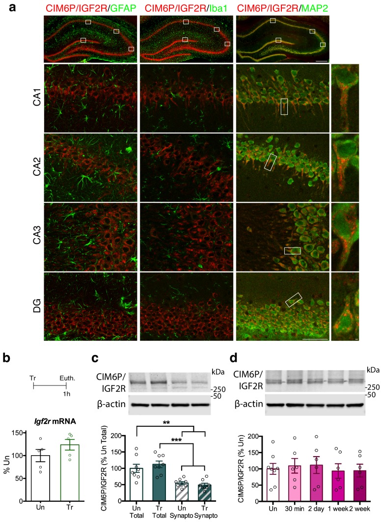
Figure 1. CIM6P/IGF2R is expressed in rat hippocampal neurons and mostly localizes to the somatic compartment. (a) Immunofluorescence co-staining of CIM6P/IGF2R and GFAP, Iba1, or MAP2. Upper panels: representative composite tile scans of whole hippocampus (scale bar, 500 μm). Lower panels: CA1, CA2, CA3, and DG (scale bar, 50 μm). Far right panels: zoomed images showing co-localization of MAP2 with CIM6P/IGF2R (scale bar, 1 μm). (b) Rats were trained on IA (Tr) or remained in their home cages (untrained, Un) and euthanized 1 hr after training. Igf2r mRNA levels (n = 5, two independent experiments). (c) Western blot analyses comparing total and synaptoneurosomal extracts (n = 8, two independent experiments). (d) Total extracts from rats euthanized at various time points after training (30 min, 2 days, 1 week, and 2 weeks) (n = 6–8, four independent experiments). Two-tailed Student t-test or one-way ANOVA followed by Tukey’s post-hoc tests. **p<0.01 and ***p<0.001; see Source data one for detailed statistical information.
Figure 1. CIM6P/IGF2R is expressed in rat hippocampal neurons and mostly localizes to the somatic compartment. (a) Immunofluorescence co-staining of CIM6P/IGF2R and GFAP, Iba1, or MAP2. Upper panels: representative composite tile scans of whole hippocampus (scale bar, 500 μm). Lower panels: CA1, CA2, CA3, and DG (scale bar, 50 μm). Far right panels: zoomed images showing co-localization of MAP2 with CIM6P/IGF2R (scale bar, 1 μm). (b) Rats were trained on IA (Tr) or remained in their home cages (untrained, Un) and euthanized 1 hr after training. Igf2r mRNA levels (n = 5, two independent experiments). (c) Western blot analyses comparing total and synaptoneurosomal extracts (n = 8, two independent experiments). (d) Total extracts from rats euthanized at various time points after training (30 min, 2 days, 1 week, and 2 weeks) (n = 6–8, four independent experiments). Two-tailed Student t-test or one-way ANOVA followed by Tukey’s post-hoc tests. **p<0.01 and ***p<0.001; see Source data one for detailed statistical information.CC BY · PubMed Central
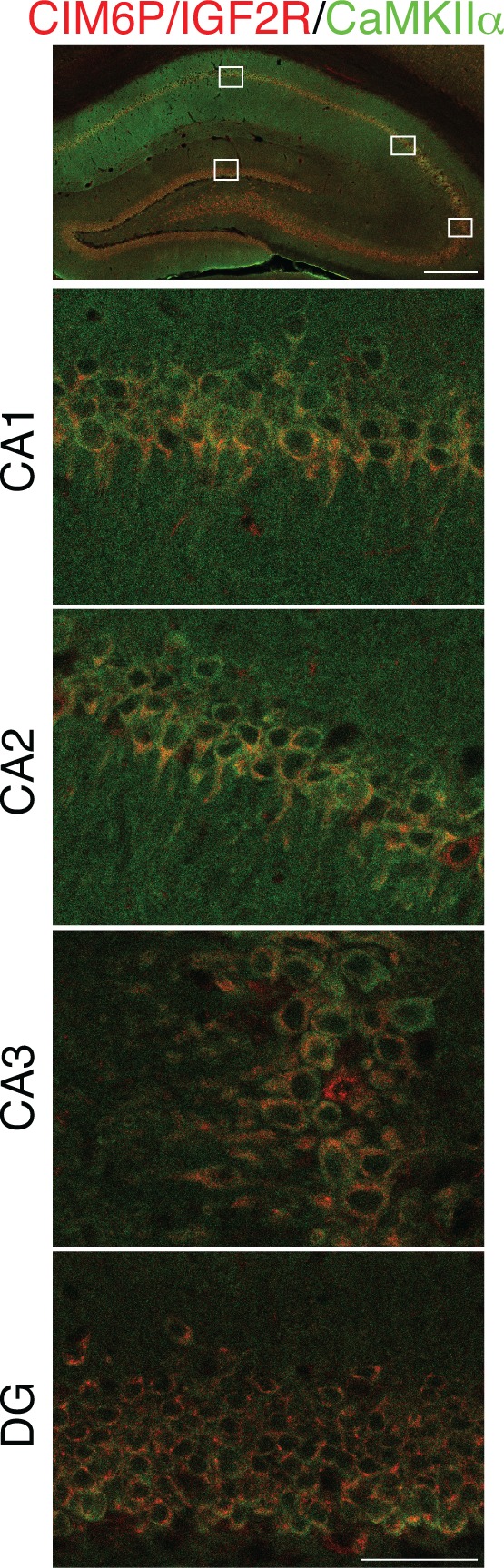
Figure 1—figure supplement 1. CIM6P/IGF2R is expressed in CaMKIIα neurons of rat hippocampus. Immunofluorescence co-staining of CIM6P/IGF2R and CaMKIIα. Upper panels: representative composite tile scans of whole hippocampus (scale bar, 500 μm). Lower panels: CA1, CA2, CA3, and DG (scale bar, 50 μm).
Figure 1—figure supplement 1. CIM6P/IGF2R is expressed in CaMKIIα neurons of rat hippocampus. Immunofluorescence co-staining of CIM6P/IGF2R and CaMKIIα. Upper panels: representative composite tile scans of whole hippocampus (scale bar, 500 μm). Lower panels: CA1, CA2, CA3, and DG (scale bar, 50 μm).CC BY · PubMed Central
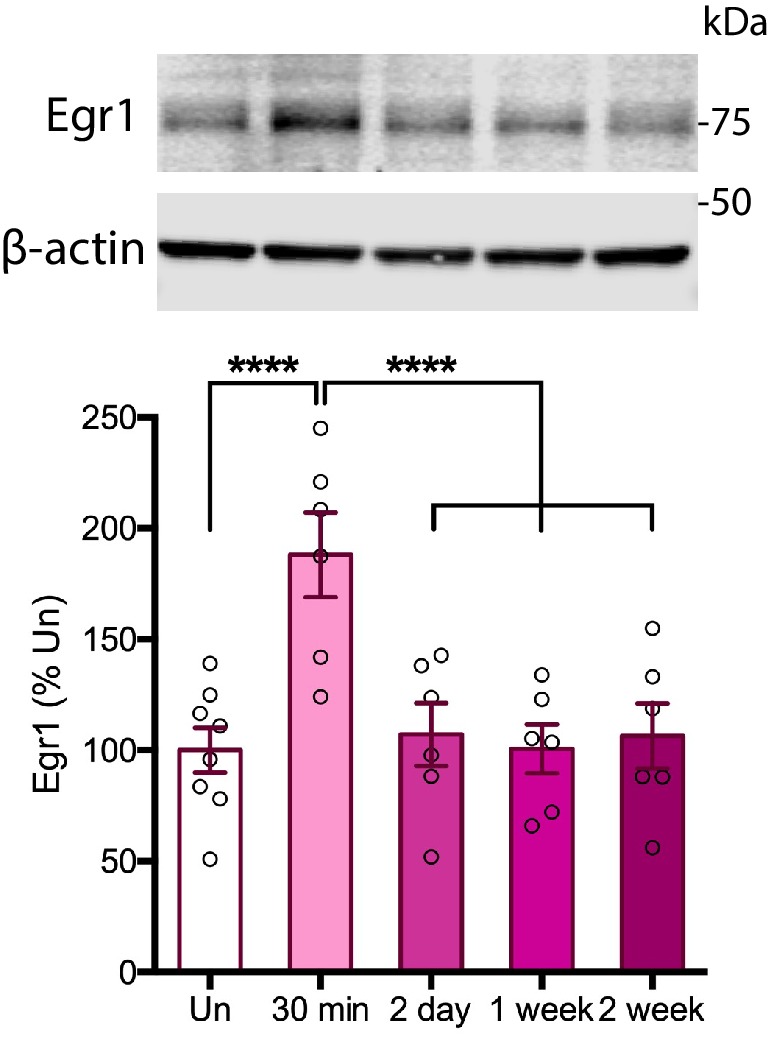
Figure 1—figure supplement 2. Time course of Egr1 protein induction following IA training in rats. Cohorts of rats were trained on IA, and euthanized 30 min, 2 days, 1 week, or 2 weeks later. Total homogenates were analyzed by western blot for Egr1 protein levels. Egr1 was induced at 30 min after training (Tr), as compared to untrained controls (Un) (n = 6–8, four independent experiments). One-way ANOVA followed by Tukey’s post hoc test. ****p<0.0001; see Source data one for detailed statistical information.
Figure 1—figure supplement 2. Time course of Egr1 protein induction following IA training in rats. Cohorts of rats were trained on IA, and euthanized 30 min, 2 days, 1 week, or 2 weeks later. Total homogenates were analyzed by western blot for Egr1 protein levels. Egr1 was induced at 30 min after training (Tr), as compared to untrained controls (Un) (n = 6–8, four independent experiments). One-way ANOVA followed by Tukey’s post hoc test. ****p<0.0001; see Source data one for detailed statistical information.CC BY · PubMed Central
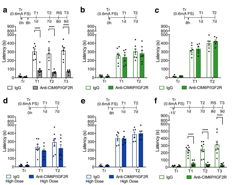
Figure 2. In rats, CIM6P/IGF2R is rapidly recruited by learning and required for memory consolidation within a limited temporal window. Experimental timelines are shown above graphs. Rats were injected with IgG or anti-CIM6P/IGF2R antibody before or after IA training (↑). IA acquisition (Tr) and memory retention are expressed as mean latency ± SEM (in seconds). (a) The effect of two injections of either IgG control or anti-CIM6P/IGF2R, given immediately and 8 hr after training (Tr), on IA memory tested 1 day (1 d) and 1 week after training, as well as after a reminder shock (RS; n = 6, two independent experiments). (b and c) The effect of a single injection of anti-CIM6P/IGF2R, given immediately or 8 hr after training, on memory tested 1 day and 1 week after training (n = 6–7, two independent experiments). (d and e) The effect of a single 10-fold higher dose of IgG or anti-CIM6P/IGF2R, given at the same time points, on memory tested 1 day and 1 week later (n = 6–7, two independent experiments). (f) The effect of a single injection, given 15 min before training, on memory tested 1 day and 1 week after training, as well as after a reminder shock (n = 7–8, two independent experiments). Two-way repeated measures ANOVA followed by Sidak’s post-hoc tests. ****p<0.0001; see Source data one for detailed statistical information.
Figure 2. In rats, CIM6P/IGF2R is rapidly recruited by learning and required for memory consolidation within a limited temporal window. Experimental timelines are shown above graphs. Rats were injected with IgG or anti-CIM6P/IGF2R antibody before or after IA training (↑). IA acquisition (Tr) and memory retention are expressed as mean latency ± SEM (in seconds). (a) The effect of two injections of either IgG control or anti-CIM6P/IGF2R, given immediately and 8 hr after training (Tr), on IA memory tested 1 day (1 d) and 1 week after training, as well as after a reminder shock (RS; n = 6, two independent experiments). (b and c) The effect of a single injection of anti-CIM6P/IGF2R, given immediately or 8 hr after training, on memory tested 1 day and 1 week after training (n = 6–7, two independent experiments). (d and e) The effect of a single 10-fold higher dose of IgG or anti-CIM6P/IGF2R, given at the same time points, on memory tested 1 day and 1 week later (n = 6–7, two independent experiments). (f) The effect of a single injection, given 15 min before training, on memory tested 1 day and 1 week after training, as well as after a reminder shock (n = 7–8, two independent experiments). Two-way repeated measures ANOVA followed by Sidak’s post-hoc tests. ****p<0.0001; see Source data one for detailed statistical information.CC BY · PubMed Central
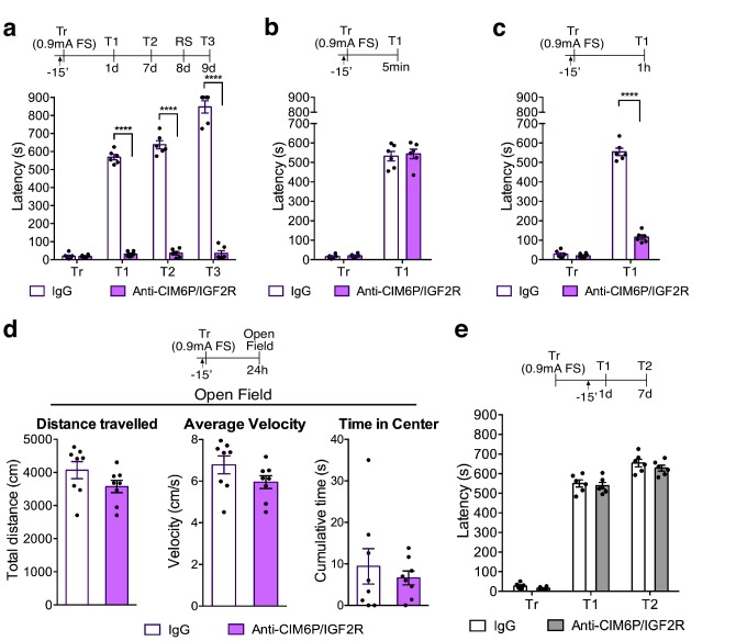
Figure 3. In rats, CIM6P/IGF2R is required for memory consolidation, but not learning or memory retrieval. Experimental timelines are shown above graphs. Rats were injected with IgG or anti-CIM6P/IGF2R antibody 15 min before IA training or testing (↑). Inhibitory avoidance training and memory retention are expressed as mean latency ± SEM (in seconds). (a) The effect of a single injection of anti-CIM6P/IGF2R on memory retention in animals trained (Tr) with a stronger, (0.9 mA) shock intensity (n = 6, two independent experiments). (b) The effect of anti-CIM6P/IGF2R on memory tested 5 min after training (n = 6, two independent experiments), or 1 hr after training (c) n = 6, two independent experiments). (d) Open field test was conducted 24 hr after IA training; total distance travelled, average travel velocity, and cumulative time spent in the center of the arena were recorded (n = 8, two independent experiments). (e) The effect of anti-CIM6P/IGF2R on memory retrieval: anti-CIM6P/IGF2R was injected 15 min before memory test given 1 day after IA training (n = 6, two independent experiments). Two-way repeated measures ANOVA followed by Sidak’s post hoc tests. ****p<0.0001; see Source data one for detailed statistical information.
Figure 3. In rats, CIM6P/IGF2R is required for memory consolidation, but not learning or memory retrieval. Experimental timelines are shown above graphs. Rats were injected with IgG or anti-CIM6P/IGF2R antibody 15 min before IA training or testing (↑). Inhibitory avoidance training and memory retention are expressed as mean latency ± SEM (in seconds). (a) The effect of a single injection of anti-CIM6P/IGF2R on memory retention in animals trained (Tr) with a stronger, (0.9 mA) shock intensity (n = 6, two independent experiments). (b) The effect of anti-CIM6P/IGF2R on memory tested 5 min after training (n = 6, two independent experiments), or 1 hr after training (c) n = 6, two independent experiments). (d) Open field test was conducted 24 hr after IA training; total distance travelled, average travel velocity, and cumulative time spent in the center of the arena were recorded (n = 8, two independent experiments). (e) The effect of anti-CIM6P/IGF2R on memory retrieval: anti-CIM6P/IGF2R was injected 15 min before memory test given 1 day after IA training (n = 6, two independent experiments). Two-way repeated measures ANOVA followed by Sidak’s post hoc tests. ****p<0.0001; see Source data one for detailed statistical information.CC BY · PubMed Central
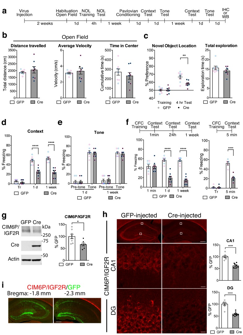
Figure 4. Hippocampal neuronal knockdown of CIM6P/IGF2R in mice selectively impairs hippocampus-dependent memories. (a) Experimental timeline for b–e and g–i. Data are expressed as mean ± SEM, with blue datapoints representing male mice, and pink datapoints representing female mice. Igf2r-floxed mice received bilateral hippocampal injections of AAV-hSyn-Cre-eGFP (Cre) or control AAV-hSyn-eGFP (GFP), 2 weeks prior to behavioral experiments. (b) Open field of GFP- and Cre-injected mice measured total distance travelled, average travel velocity, and cumulative time spent in the center of the area (n = 8, four independent experiments). (c) Novel objection location memory and total exploration time, tested 4 hr after training, (n = 8, four independent experiments). (d) Percent (%) of time spent freezing in mice tested in the training context 1 day (1 d) and 1 week after Pavlovian conditioning (n = 8, four independent experiments). (e) Percent (%) of time spent freezing to a new context prior to onset of tone, and during the tone (n = 8, four independent experiments). (f) Contextual fear conditioning expressed as percent (%) of time spent freezing. Left: % of time spent freezing in mice tested 1 min, 1 day, and 1 week after training (n = 8, four independent experiments). Right: % of time spent freezing in mice tested 5 min after training. (g) Representative western blot and quantification obtained from dorsal hippocampi homogenates from Cre- or GFP-injected mice stained for CIM6P/IGF2R, Cre recombinase (Cre), and actin. Actin-normalized values were expressed as mean percentage ± SEM (n = 7–8, four independent experiments). (h) Upper panels: representative dorsal hippocampus composite tile scans in GFP-injected or Cre-injected mice immunostained for CIM6P/IGF2R (scale bar, 500 μm). Middle and lower panels: CA1 and DG (scale bar, 10 μm) are shown. Bar graphs on the right report immunostaining intensity quantifications for each sub-region (n = 8, four independent experiments). (i) Representative images of GFP- or Cre-injected mice, composite tile scans of dorsal hippocampus double staining with anti-GFP and anti-CIM6P/IGF2R antibodies (scale bar 500 μm). Two-tailed Student’s t-test or two-way repeated measures ANOVA followed by Sidak’s post-hoc tests. *p<0.05, ****p<0.0001; see Source data one for detailed statistical information.
Figure 4. Hippocampal neuronal knockdown of CIM6P/IGF2R in mice selectively impairs hippocampus-dependent memories. (a) Experimental timeline for b–e and g–i. Data are expressed as mean ± SEM, with blue datapoints representing male mice, and pink datapoints representing female mice. Igf2r-floxed mice received bilateral hippocampal injections of AAV-hSyn-Cre-eGFP (Cre) or control AAV-hSyn-eGFP (GFP), 2 weeks prior to behavioral experiments. (b) Open field of GFP- and Cre-injected mice measured total distance travelled, average travel velocity, and cumulative time spent in the center of the area (n = 8, four independent experiments). (c) Novel objection location memory and total exploration time, tested 4 hr after training, (n = 8, four independent experiments). (d) Percent (%) of time spent freezing in mice tested in the training context 1 day (1 d) and 1 week after Pavlovian conditioning (n = 8, four independent experiments). (e) Percent (%) of time spent freezing to a new context prior to onset of tone, and during the tone (n = 8, four independent experiments). (f) Contextual fear conditioning expressed as percent (%) of time spent freezing. Left: % of time spent freezing in mice tested 1 min, 1 day, and 1 week after training (n = 8, four independent experiments). Right: % of time spent freezing in mice tested 5 min after training. (g) Representative western blot and quantification obtained from dorsal hippocampi homogenates from Cre- or GFP-injected mice stained for CIM6P/IGF2R, Cre recombinase (Cre), and actin. Actin-normalized values were expressed as mean percentage ± SEM (n = 7–8, four independent experiments). (h) Upper panels: representative dorsal hippocampus composite tile scans in GFP-injected or Cre-injected mice immunostained for CIM6P/IGF2R (scale bar, 500 μm). Middle and lower panels: CA1 and DG (scale bar, 10 μm) are shown. Bar graphs on the right report immunostaining intensity quantifications for each sub-region (n = 8, four independent experiments). (i) Representative images of GFP- or Cre-injected mice, composite tile scans of dorsal hippocampus double staining with anti-GFP and anti-CIM6P/IGF2R antibodies (scale bar 500 μm). Two-tailed Student’s t-test or two-way repeated measures ANOVA followed by Sidak’s post-hoc tests. *p<0.05, ****p<0.0001; see Source data one for detailed statistical information.CC BY · PubMed Central
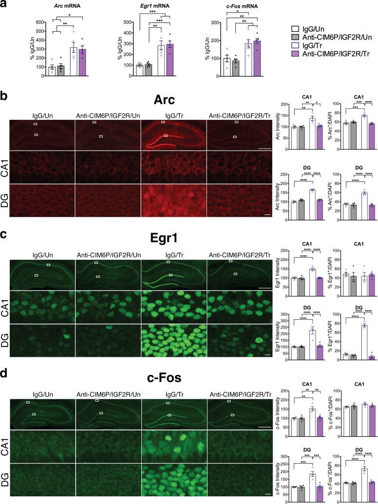
Figure 5. CIM6P/IGF2R is required for learning-induced IEG protein induction in rats. Rats were injected with IgG or anti-CIM6P/IGF2R, and then were either trained on IA 15 min later (Tr) or remained Untrained (Un). To examine mRNA levels, rats were euthanized 1 hr after training and hippocampi flash-frozen. (a) qPCR of Arc, Egr1, and c-Fos performed on dorsal hippocampal extracts obtained from rats injected with either IgG or anti-CIM6P/IGF2R and euthanized 1 hr after IA training (n = 5, two independent experiments). (b–d) Rats underwent IA training and were perfused 1 hr later; coronal brain sections were immunostained for Arc (b), Egr1 (c), and c-Fos (d). Upper panels: representative composite tile scan of dorsal hippocampus (scale bar, 500 μm). Middle and lower panels: CA1 and DG (scale bar, 10 μm). Bar graphs shown at right show quantifications for normalized intensity and percentage of cells positive for IEGs, for each sub-region (n = 4, two independent experiments). Two-way ANOVA followed by Tukey’s post hoc tests. *p<0.05, **p<0.01, ***p<0.001, ****p<0.0001; see Source data one for detailed statistical information.
Figure 5. CIM6P/IGF2R is required for learning-induced IEG protein induction in rats. Rats were injected with IgG or anti-CIM6P/IGF2R, and then were either trained on IA 15 min later (Tr) or remained Untrained (Un). To examine mRNA levels, rats were euthanized 1 hr after training and hippocampi flash-frozen. (a) qPCR of Arc, Egr1, and c-Fos performed on dorsal hippocampal extracts obtained from rats injected with either IgG or anti-CIM6P/IGF2R and euthanized 1 hr after IA training (n = 5, two independent experiments). (b–d) Rats underwent IA training and were perfused 1 hr later; coronal brain sections were immunostained for Arc (b), Egr1 (c), and c-Fos (d). Upper panels: representative composite tile scan of dorsal hippocampus (scale bar, 500 μm). Middle and lower panels: CA1 and DG (scale bar, 10 μm). Bar graphs shown at right show quantifications for normalized intensity and percentage of cells positive for IEGs, for each sub-region (n = 4, two independent experiments). Two-way ANOVA followed by Tukey’s post hoc tests. *p<0.05, **p<0.01, ***p<0.001, ****p<0.0001; see Source data one for detailed statistical information.CC BY · PubMed Central
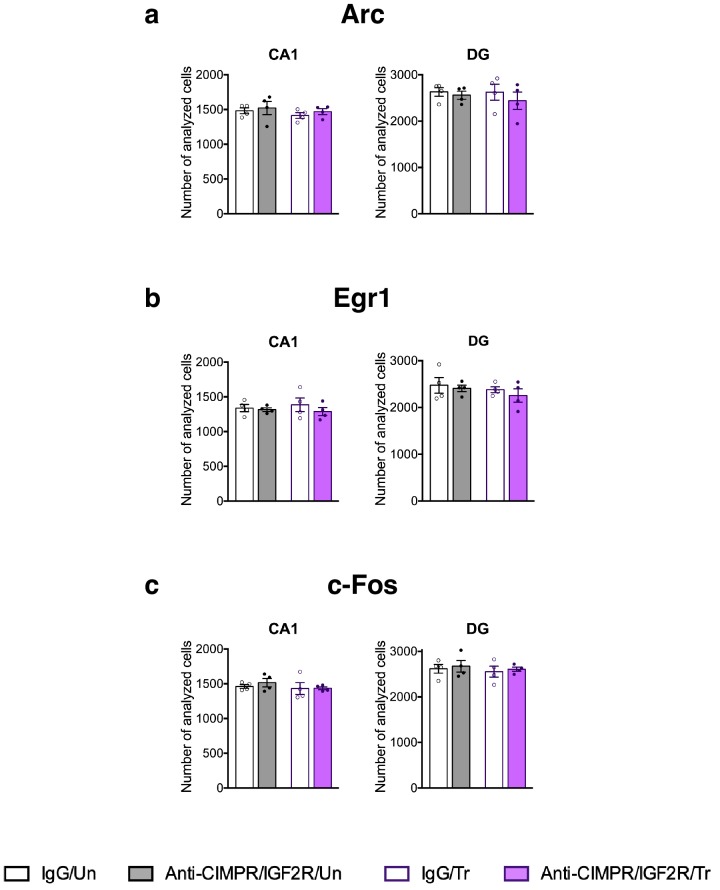
Figure 5—figure supplement 1. Total number of cells assessed by DAPI staining in the rat dorsal hippocampus fields used in the IEGs immunofluorescent analyses. The number of total cells detected in our fields of analyses (DAPI-positive cells) used for (a) Arc, (b) Egr1, and (c) c-Fos, for each sub-region (n = 4, two independent experiments). Two-way ANOVA followed by Tukey’s post hoc tests. See Source data one for detailed statistical information.
Figure 5—figure supplement 1. Total number of cells assessed by DAPI staining in the rat dorsal hippocampus fields used in the IEGs immunofluorescent analyses. The number of total cells detected in our fields of analyses (DAPI-positive cells) used for (a) Arc, (b) Egr1, and (c) c-Fos, for each sub-region (n = 4, two independent experiments). Two-way ANOVA followed by Tukey’s post hoc tests. See Source data one for detailed statistical information.CC BY · PubMed Central
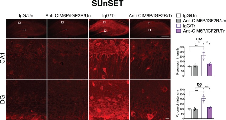
Figure 6. CIM6P/IGF2R is required for learning-induced de novo protein synthesis in rats. SUnSET was employed to quantify de novo protein synthesis in the rat hippocampus. Puromycin was co-injected with IgG or anti-CIM6P/IGF2R prior to training, and rats were perfused 2 hr later. Upper panels: representative images of anti-puromycin immunostaining, composite tile scans of whole hippocampus (scale bar, 500 μm). Middle and lower panels: CA1 and DG (scale bar 10 μm). Bar graphs at right show immunostaining intensity quantifications for each sub-region (n = 4, two independent experiments). Two-way ANOVA followed by Tukey’s post hoc tests. **p<0.01, ***p<0.001; see Source data one for detailed statistical information.
Figure 6. CIM6P/IGF2R is required for learning-induced de novo protein synthesis in rats. SUnSET was employed to quantify de novo protein synthesis in the rat hippocampus. Puromycin was co-injected with IgG or anti-CIM6P/IGF2R prior to training, and rats were perfused 2 hr later. Upper panels: representative images of anti-puromycin immunostaining, composite tile scans of whole hippocampus (scale bar, 500 μm). Middle and lower panels: CA1 and DG (scale bar 10 μm). Bar graphs at right show immunostaining intensity quantifications for each sub-region (n = 4, two independent experiments). Two-way ANOVA followed by Tukey’s post hoc tests. **p<0.01, ***p<0.001; see Source data one for detailed statistical information.CC BY · PubMed Central
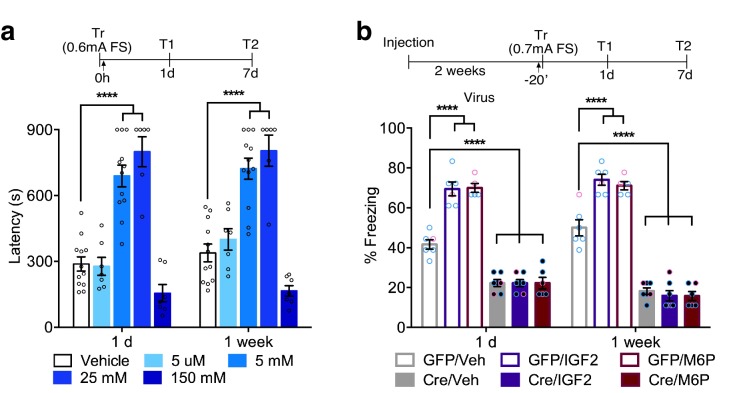
Figure 7. In rats and mice, M6P, like IGF2, enhances memories via CIM6P/IGF2R in hippocampal neurons. Experimental timelines are shown above graphs. (a) Rats were injected bilaterally into the dorsal hippocampus with vehicle or the indicated concentrations of mannose-6-phosphate (M6P) immediately after IA training (↑), and memory was tested 1 day and 1 week later (n = 6–12, five independent experiments). (b) Vehicle, IGF2 (30 μg/kg), or M6P (850 μg/kg) was administered systemically (s.c., ↑) 20 min before contextual fear conditioning training to GFP- or Cre-injected mice. Percent (%) of time spent freezing were measured 1 day or 1 week after training (n = 5–7, five independent experiments). Two-way repeated measures ANOVA followed by Sidak’s post hoc tests. ****p<0.0001; see Source data one for detailed statistical information.
Figure 7. In rats and mice, M6P, like IGF2, enhances memories via CIM6P/IGF2R in hippocampal neurons. Experimental timelines are shown above graphs. (a) Rats were injected bilaterally into the dorsal hippocampus with vehicle or the indicated concentrations of mannose-6-phosphate (M6P) immediately after IA training (↑), and memory was tested 1 day and 1 week later (n = 6–12, five independent experiments). (b) Vehicle, IGF2 (30 μg/kg), or M6P (850 μg/kg) was administered systemically (s.c., ↑) 20 min before contextual fear conditioning training to GFP- or Cre-injected mice. Percent (%) of time spent freezing were measured 1 day or 1 week after training (n = 5–7, five independent experiments). Two-way repeated measures ANOVA followed by Sidak’s post hoc tests. ****p<0.0001; see Source data one for detailed statistical information.CC BY · PubMed Central

Verification

Claims the paper makes

The claims and statistics of this paper haven’t been checked yet.

Run the check — free, no account needed. It reads the open-access text and checks the sources it cites.

A few free checks a day · papers already verified open instantly · results are public

Claims other papers make about this one

Each is checked against this paper’s own text: does it say what the citing paper says it does?

Self-check

Percentages, sums and reported differences re-computed from the paper’s own numbers.

The self-check needs the paper’s full text.

Citations

Cites

Show all 3 references

Research works printed but not linked: 78

  1. Agis-Balboa, 2011, A hippocampal insulin-growth factor 2 pathway regulates the extinction of fear memories, The EMBO Journal, 30, 4071, 10.1038/emboj.2011.293 · DOI
  2. Alberini, 2009, Transcription factors in long-term memory and synaptic plasticity, Physiological Reviews, 89, 121, 10.1152/physrev.00017.2008 · DOI · Paper page
  3. Alberini, 2011, The role of reconsolidation and the dynamic process of long-term memory formation and storage, Frontiers in Behavioral Neuroscience, 5, 10.3389/fnbeh.2011.00012 · DOI
  4. Alberini, 2015, The regulation of transcription in memory consolidation, Cold Spring Harbor Perspectives in Biology, 7, 10.1101/cshperspect.a021741 · DOI · Paper page
  5. Arguello, 2013, CCAAT enhancer binding protein δ plays an essential role in memory consolidation and reconsolidation, Journal of Neuroscience, 33, 3646, 10.1523/JNEUROSCI.1635-12.2013 · DOI
  6. Bambah-Mukku, 2014, A positive autoregulatory BDNF feedback loop via C/EBPβ mediates hippocampal memory consolidation, Journal of Neuroscience, 34, 12547, 10.1523/JNEUROSCI.0324-14.2014 · DOI · Paper page
  7. Barzilai, 1989, 5-HT modulates protein synthesis and the expression of specific proteins during long-term facilitation in Aplysia sensory neurons, Neuron, 2, 1577, 10.1016/0896-6273(89)90046-9 · DOI
  8. Chen, 2011, A critical role for IGF-II in memory consolidation and enhancement, Nature, 469, 491, 10.1038/nature09667 · DOI
  9. Chorney, 1998, A Quantitative Trait Locus Associated With Cognitive Ability in Children, Psychological Science, 9, 159, 10.1111/1467-9280.00032 · DOI
  10. Cioni, 2019, Late endosomes act as mRNA translation platforms and sustain mitochondria in axons, Cell, 176, 56, 10.1016/j.cell.2018.11.030 · DOI
  11. Costa-Mattioli, 2009, Translational control of long-lasting synaptic plasticity and memory, Neuron, 61, 10, 10.1016/j.neuron.2008.10.055 · DOI · Paper page
  12. De Souza, 1995, M6P/IGF2R gene is mutated in human hepatocellular carcinomas with loss of heterozygosity, Nature Genetics, 11, 447, 10.1038/ng1295-447 · DOI
  13. El-Shewy, 2006, Insulin-like growth factors mediate heterotrimeric G protein-dependent ERK1/2 activation by transactivating sphingosine 1-phosphate receptors, Journal of Biological Chemistry, 281, 31399, 10.1074/jbc.M605339200 · DOI
  14. El-Shewy, 2009, Chapter 24 insulin‐like growth factor‐2/Mannose‐6 phosphate receptors, Vitamins & Hormones, 80, 667, 10.1016/S0083-6729(08)00624-9 · DOI
  15. Fernández-Monreal, 2012, The balance between receptor recycling and trafficking toward lysosomes determines synaptic strength during long-term depression, Journal of Neuroscience, 32, 13200, 10.1523/JNEUROSCI.0061-12.2012 · DOI
  16. Gallo, 2018, Immediate early genes, memory and psychiatric disorders: focus on c-Fos, Egr1 and arc, Frontiers in Behavioral Neuroscience, 12, 10.3389/fnbeh.2018.00079 · DOI
  17. Garcia-Osta, 2006, MuSK expressed in the brain mediates cholinergic responses, synaptic plasticity, and memory formation, Journal of Neuroscience, 26, 7919, 10.1523/JNEUROSCI.1674-06.2006 · DOI
  18. Ghosh, 2003, Mannose 6-phosphate receptors: new twists in the tale, Nature Reviews Molecular Cell Biology, 4, 202, 10.1038/nrm1050 · DOI
  19. Goo, 2017, Activity-dependent trafficking of lysosomes in dendrites and dendritic spines, Journal of Cell Biology, 216, 2499, 10.1083/jcb.201704068 · DOI · Paper page
  20. Goodman, 2011, Novel insights into the regulation of skeletal muscle protein synthesis as revealed by a new nonradioactive in vivo technique, The FASEB Journal, 25, 1028, 10.1096/fj.10-168799 · DOI
  21. Hankins, 1996, M6P/IGF2 receptor: a candidate breast tumor suppressor gene, Oncogene, 12, 2003
  22. Hartig, 2013, Basic Image Analysis and Manipulation in ImageJ, Current Protocols in Molecular Biology, 102, 14.15.1, 10.1002/0471142727.mb1415s102 · DOI
  23. Hawkes, 2006, Single transmembrane domain insulin-like growth factor-II/mannose-6-phosphate receptor regulates central cholinergic function by activating a G-protein-sensitive, protein kinase C-dependent pathway, Journal of Neuroscience, 26, 585, 10.1523/JNEUROSCI.2730-05.2006 · DOI
  24. Hawkes, 2003, Insulin-like growth factor-II/mannose-6-phosphate receptor: widespread distribution in neurons of the central nervous system including those expressing cholinergic phenotype, The Journal of Comparative Neurology, 458, 113, 10.1002/cne.10578 · DOI
  25. Hawkes, 2004, The insulin-like growth factor-II/mannose-6-phosphate receptor: structure, distribution and function in the central nervous system, Brain Research Reviews, 44, 117, 10.1016/j.brainresrev.2003.11.002 · DOI
  26. Ikezu, 1995, In vivo Coupling of Insulin-like Growth Factor II/Mannose 6-Phosphate Receptor to Heteromeric G Proteins, Journal of Biological Chemistry, 270, 29224, 10.1074/jbc.270.49.29224 · DOI
  27. Jedlicka, 2015, Neuroligin-1 regulates excitatory synaptic transmission, LTP and EPSP-spike coupling in the dentate gyrus in vivo, Brain Structure and Function, 220, 47, 10.1007/s00429-013-0636-1 · DOI
  28. Kar, 1993, Quantitative autoradiographic localization of [125I]insulin-like growth factor I, [125I]insulin-like growth factor II, and [125I]insulin receptor binding sites in developing and adult rat brain, The Journal of Comparative Neurology, 333, 375, 10.1002/cne.903330306 · DOI
  29. Kar, 2006, Cellular distribution of insulin-like growth factor-II/mannose-6-phosphate receptor in normal human brain and its alteration in Alzheimer's disease pathology, Neurobiology of Aging, 27, 199, 10.1016/j.neurobiolaging.2005.03.005 · DOI
  30. Kim, 1992, Modality-specific retrograde amnesia of fear, Science, 256, 675, 10.1126/science.1585183 · DOI · Paper page
  31. Kong, 2000, M6P/IGF2R is mutated in squamous cell carcinoma of the lung, Oncogene, 19, 1572, 10.1038/sj.onc.1203437 · DOI
  32. Körner, 1995, Mannose 6-Phosphate/Insulin-like Growth Factor II Receptor Fails to Interact with G-proteins, Journal of Biological Chemistry, 270, 287, 10.1074/jbc.270.1.287 · DOI
  33. Lau, 1994, Loss of the imprinted IGF2/cation-independent mannose 6-phosphate receptor results in fetal overgrowth and perinatal lethality, Genes & Development, 8, 2953, 10.1101/gad.8.24.2953 · DOI
  34. Lesniak, 1988, Receptors for insulin-like growth factors I and II: autoradiographic localization in rat brain and comparison to receptors for insulin, Endocrinology, 123, 2089, 10.1210/endo-123-4-2089 · DOI
  35. Liu, 2012, Neuronal excitability and calcium/calmodulin-dependent protein kinase type II: Location, location, location, Epilepsia, 53, 45, 10.1111/j.1528-1167.2012.03474.x · DOI · Paper page
  36. Lonergan, 2010, Time-Dependent expression of arc and Zif268 after acquisition of fear conditioning, Neural Plasticity, 2010, 1, 10.1155/2010/139891 · DOI
  37. Lovett-Barron, 2014, Dendritic inhibition in the Hippocampus supports fear learning, Science, 343, 857, 10.1126/science.1247485 · DOI · Paper page
  38. Ludwig, 1996, Mouse mutants lacking the type 2 IGF receptor (IGF2R) are rescued from perinatal lethality in Igf2 and Igf1r null backgrounds, Developmental Biology, 177, 517, 10.1006/dbio.1996.0182 · DOI
  39. Martin-Montañez, 2014, Involvement of IGF-II receptors in the antioxidant and neuroprotective effects of IGF-II on adult cortical neuronal cultures, Biochimica Et Biophysica Acta (BBA) - Molecular Basis of Disease, 1842, 1041, 10.1016/j.bbadis.2014.03.010 · DOI
  40. McKinnon, 2001, Stimulation of human extravillous trophoblast migration by IGF-II is mediated by IGF type 2 receptor involving inhibitory G protein(s) and phosphorylation of MAPK, The Journal of Clinical Endocrinology & Metabolism, 86, 3665, 10.1210/jcem.86.8.7711 · DOI
  41. Mellott, 2014, IGF2 ameliorates amyloidosis, increases cholinergic marker expression and raises BMP9 and neurotrophin levels in the Hippocampus of the APPswePS1dE9 alzheimer's disease model mice, PLOS ONE, 9, 10.1371/journal.pone.0094287 · DOI
  42. Mellott, 2017, Perinatal choline supplementation reduces amyloidosis and increases choline acetyltransferase expression in the Hippocampus of the APPswePS1dE9 alzheimer's Disease Model Mice, PLOS ONE, 12, 10.1371/journal.pone.0170450 · DOI
  43. Mendelsohn, 1988, Autoradiographic localization of insulin-like growth factor II receptors in cerebellar cortex of Weaver and purkinje cell degeneration mutant mice, Brain Research, 458, 361, 10.1016/0006-8993(88)90479-9 · DOI
  44. Minatohara, 2016, Role of Immediate-Early genes in synaptic plasticity and neuronal ensembles underlying the memory trace, Frontiers in Molecular Neuroscience, 8, 10.3389/fnmol.2015.00078 · DOI
  45. Morgan, 1987, Insulin-like growth factor II receptor as a multifunctional binding protein, Nature, 329, 301, 10.1038/329301a0 · DOI
  46. Nishimoto, 1989, Possible direct linkage of insulin-like growth factor-II receptor with guanine nucleotide-binding proteins, The Journal of Biological Chemistry, 264, 14029, 10.1016/S0021-9258(18)71637-2 · DOI
  47. Nixon, 2005, Endosome function and dysfunction in Alzheimer's disease and other neurodegenerative diseases, Neurobiology of Aging, 26, 373, 10.1016/j.neurobiolaging.2004.09.018 · DOI
  48. Nolan, 1987, Antibody to the phosphomannosyl receptor inhibits recycling of receptor in fibroblasts, Journal of Cellular Biochemistry, 35, 137, 10.1002/jcb.240350207 · DOI
  49. Oka, 1985, Direct demonstration of rapid insulin-like growth factor II receptor internalization and recycling in rat adipocytes insulin stimulates 125I-insulin-like growth factor II degradation by modulating the IGF-II receptor recycling process, The Journal of Biological Chemistry, 260, 9435, 10.1016/S0021-9258(17)39385-7 · DOI
  50. Okamoto, 1990, A simple structure encodes G protein-activating function of the IGF-II/mannose 6-phosphate receptor, Cell, 62, 709, 10.1016/0092-8674(90)90116-V · DOI
  51. Ouyang, 1997, The insulin-like growth factor II receptor gene is mutated in genetically unstable cancers of the endometrium, stomach, and colorectum, Cancer Research, 57, 1851
  52. Pascual‐Lucas, 2014, Insulin‐like growth factor 2 reverses memory and synaptic deficits in APP transgenic mice, EMBO Molecular Medicine, 6, 1246, 10.15252/emmm.201404228 · DOI
  53. Phillips, 1992, Differential contribution of amygdala and Hippocampus to cued and contextual fear conditioning, Behavioral Neuroscience, 106, 274, 10.1037/0735-7044.106.2.274 · DOI · Paper page
  54. Richter, 2009, Making synaptic plasticity and memory last mechanisms of translational regulation, Genes & Development, 23, 1, 10.1101/gad.1735809 · DOI
  55. Roy, 2016, Memory retrieval by activating Engram cells in mouse models of early Alzheimer's disease, Nature, 531, 508, 10.1038/nature17172 · DOI
  56. Sakano, 1991, The design, expression, and characterization of human insulin-like growth factor II (IGF-II) mutants specific for either the IGF-II/cation-independent mannose 6-phosphate receptor or IGF-I receptor, The Journal of Biological Chemistry, 266, 20626, 10.1016/S0021-9258(18)54755-4 · DOI
  57. Santini, 2013, Exaggerated translation causes synaptic and behavioural aberrations associated with autism, Nature, 493, 411, 10.1038/nature11782 · DOI
  58. Santini, 2014, Mechanisms of translation control underlying long-lasting synaptic plasticity and the consolidation of long-term memory, Progress in Molecular Biology and Translational Science, 122, 131, 10.1016/B978-0-12-420170-5.00005-2 · DOI
  59. Schmidt, 2009, SUnSET, a nonradioactive method to monitor protein synthesis, Nature Methods, 6, 275, 10.1038/nmeth.1314 · DOI · Paper page
  60. Schrick, 2007, N-cadherin regulates cytoskeletally associated IQGAP1/ERK signaling and memory formation, Neuron, 55, 786, 10.1016/j.neuron.2007.07.034 · DOI
  61. Smith, 1988, The insulin-like growth factor-II (IGF-II) receptor of rat brain: regional distribution visualized by autoradiography, Brain Research, 445, 241, 10.1016/0006-8993(88)91185-7 · DOI
  62. Steinmetz, 2016, Insulin-like growth factor 2 rescues aging-related memory loss in rats, Neurobiology of Aging, 44, 9, 10.1016/j.neurobiolaging.2016.04.006 · DOI
  63. Steinmetz, 2018, Insulin-Like growth factor II targets the mTOR pathway to reverse Autism-Like phenotypes in mice, The Journal of Neuroscience, 38, 1015, 10.1523/JNEUROSCI.2010-17.2017 · DOI
  64. Stern, 2014, Enhancement of memories by systemic administration of insulin-like growth factor II, Neuropsychopharmacology, 39, 2179, 10.1038/npp.2014.69 · DOI
  65. Stork, 1999, Memory formation and the regulation of gene expression, Cellular and Molecular Life Sciences, 55, 575, 10.1007/s000180050316 · DOI
  66. Sutton, 2006, Dendritic protein synthesis, synaptic plasticity, and memory, Cell, 127, 49, 10.1016/j.cell.2006.09.014 · DOI
  67. Taubenfeld, 2001, The consolidation of new but not reactivated memory requires hippocampal C/EBPβ, Nature Neuroscience, 4, 813, 10.1038/90520 · DOI
  68. Tischmeyer, 1999, Activation of immediate early genes and memory formation, Cellular and Molecular Life Sciences, 55, 564, 10.1007/s000180050315 · DOI
  69. Vasileiou, 2016, Toward Contactless Biology: Acoustophoretic DNA Transfection, Scientific Reports, 6, 1, 10.1038/srep20023 · DOI
  70. Veyrac, 2014, The transcription factor Zif268/Egr1, brain plasticity, and memory, Progress in Molecular Biology and Translational Science, 122, 89, 10.1016/B978-0-12-420170-5.00004-0 · DOI
  71. Wang, 1994, Regulation of embryonic growth and lysosomal targeting by the imprinted Igf2/Mpr gene, Nature, 372, 464, 10.1038/372464a0 · DOI
  72. Wang, 2009, Synapse- and stimulus-specific local translation during long-term neuronal plasticity, Science, 324, 1536, 10.1126/science.1173205 · DOI
  73. Wang, 2015, Overexpression of the Insulin-Like growth factor II receptor increases β-Amyloid production and affects cell viability, Molecular and Cellular Biology, 35, 2368, 10.1128/MCB.01338-14 · DOI
  74. Wang, 2017, Insulin-Like growth Factor-II/Cation-Independent mannose 6-Phosphate receptor in neurodegenerative diseases, Molecular Neurobiology, 54, 2636, 10.1007/s12035-016-9849-7 · DOI
  75. Wylie, 2003, Tissue-specific inactivation of murine M6P/IGF2R, The American Journal of Pathology, 162, 321, 10.1016/S0002-9440(10)63823-0 · DOI
  76. Ye, 2017, Direct dorsal hippocampal-prelimbic cortex connections strengthen fear memories, Nature Neuroscience, 20, 52, 10.1038/nn.4443 · DOI
  77. Zhang, 2005, Mapping neuronal activation and the influence of adrenergic signaling during contextual memory retrieval, Learning & Memory, 12, 239, 10.1101/lm.90005 · DOI
  78. Zhou, 1995, Cloning and expression of the cDNA of chicken cation-independent mannose-6-phosphate receptor, PNAS, 92, 9762, 10.1073/pnas.92.21.9762 · DOI

Cited by

Sorted by citations

Metadata

Authors
Yu, X.-W., Pandey, K., Katzman, A. C., & Alberini, C. M.
Affiliations
Center for Neural Science, New York University, New York, United States
Venue
eLife
Published
2020-05-05
Volume
9
ISSN
2050-084X
Cited by
40
References
3
Language
en
Funders
National Institute of Mental Health (MH065635)

About these checks

Registry, retraction, Cites and statistics results are computed, not judged, with one exception: a printed reference that no index lists is read once by a model before it counts as a citation error. Claims are extracted by a model and kept only when their quote appears verbatim in the text; each cited source is then checked, with a model’s help, for whether it says what the claim relies on. Those verdicts can be wrong — read the claim’s quote and the cited source before relying on one.

Statistics and arithmetic are recomputed, not judged.

Confidence bands are shown only for fields calibrated on our annotated test set. None is calibrated yet, so no band is shown.

What counts as a claim ›How we measure confidence ›How we check this ›

Are you an author, or do you see a problem with this page? Request a review