Cited by 70

The DOI matches its Crossref record; 1 of the 1 references it cites were found in a registry or an index.

Biofilms deform soft surfaces and disrupt epithelia

Cont, Alice, Rossy, Tamara, Al-Mayyah, Zainebe, Persat, Alexandre·2020·eLife

Published version of a preprint

Abstract

During chronic infections and in microbiota, bacteria predominantly colonize their hosts as multicellular structures called biofilms. A common assumption is that biofilms exclusively interact with their hosts biochemically. However, the contributions of mechanics, while being central to the process of biofilm formation, have been overlooked as a factor influencing host physiology. Specifically, how biofilms form on soft, tissue-like materials remains unknown. Here, we show that biofilms of the pathogens Vibrio cholerae and Pseudomonas aeruginosa can induce large deformations of soft synthetic hydrogels. Biofilms buildup internal mechanical stress as single cells grow within the elastic matrix. By combining mechanical measurements and mutations in matrix components, we found that biofilms deform by buckling, and that adhesion transmits these forces to their substrates. Finally, we demonstrate that V. cholerae biofilms can generate sufficient mechanical stress to deform and even disrupt soft epithelial cell monolayers, suggesting a mechanical mode of infection.

Overview

The claims and statistics of this paper haven’t been checked yet.

Run the check — free, no account needed. It reads the open-access text and checks the sources it cites.

A few free checks a day · papers already verified open instantly · results are public

Integrity signals

Registry

Crossref · 10.7554/elife.56533

Cites

1 of 1 references verified0 citation errors · 0 not checkable

Statistics

Figures

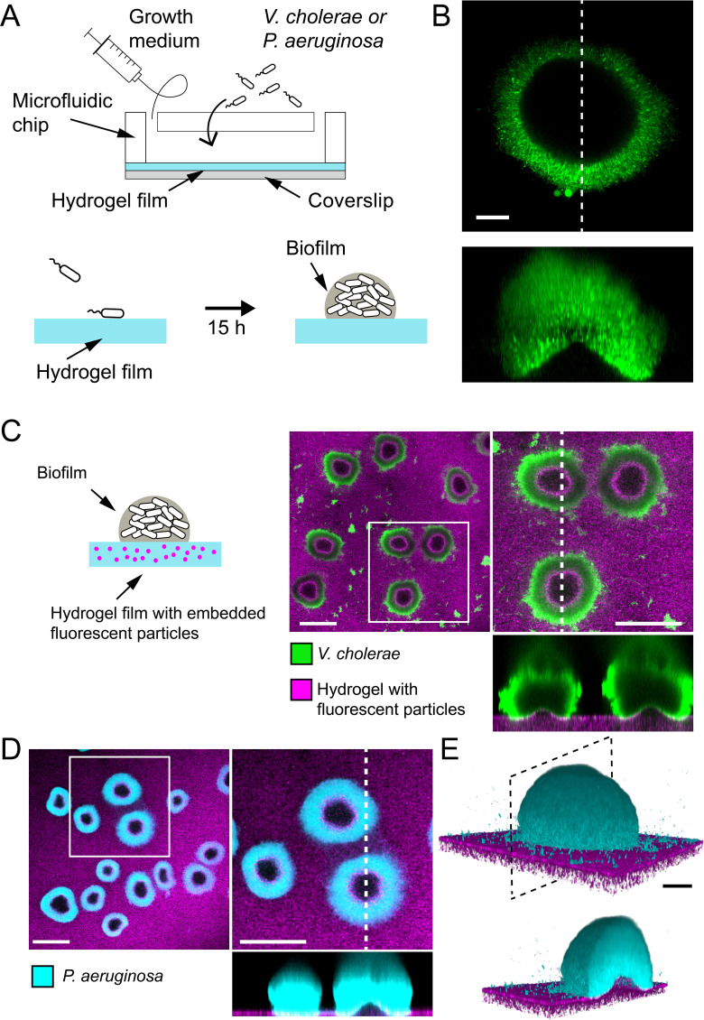
Figure 1. Biofilms deform soft substrates. (A) Illustration of experimental setup where we generate thin hydrogel films at the bottom surface of microchannels. These devices allow us to study biofilm formation on hydrogels reproducing mechanical properties of host tissues. (B) In-plane and cross-sectional confocal visualizations show that V. cholerae Rg biofilms growing on hydrogels display large gaps at their core. (C) Embedding fluorescence tracer particle in the hydrogel films allow for visualization of deformations. V. cholerae Rg biofilms formed at the surface of the films deform the substrate. (D) P. aeruginosa Rg biofilms similarly deform the soft substrates. Hydrogel elastic modulus: (B and C) E = 12 kPa, (D and E) E = 38 kPa. Scale bars: (C and D) 100 µm, (B and E) 20 µm.
Figure 1. Biofilms deform soft substrates. (A) Illustration of experimental setup where we generate thin hydrogel films at the bottom surface of microchannels. These devices allow us to study biofilm formation on hydrogels reproducing mechanical properties of host tissues. (B) In-plane and cross-sectional confocal visualizations show that V. cholerae Rg biofilms growing on hydrogels display large gaps at their core. (C) Embedding fluorescence tracer particle in the hydrogel films allow for visualization of deformations. V. cholerae Rg biofilms formed at the surface of the films deform the substrate. (D) P. aeruginosa Rg biofilms similarly deform the soft substrates. Hydrogel elastic modulus: (B and C) E = 12 kPa, (D and E) E = 38 kPa. Scale bars: (C and D) 100 µm, (B and E) 20 µm.CC BY · PubMed Central
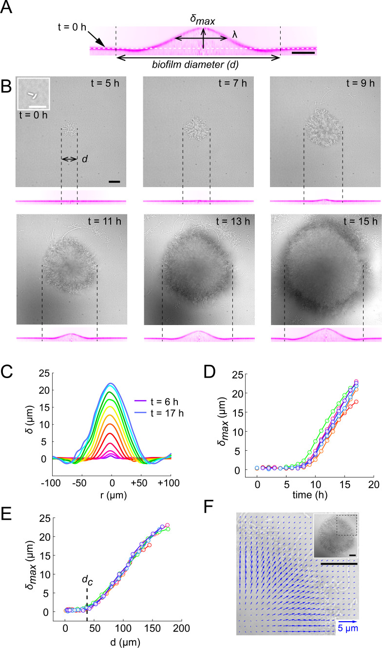
Figure 2. Biofilms deform their substrate by buckling. (A) Morphological parameters δmax (maximum deformation amplitude) and λ (half max full width) computed from resliced deformation profiles. Dashed line indicates the baseline position of the gel surface. (B) Timelapse visualization of V. cholerae Rg biofilm growth (brightfield, top) with deformation (reslice, bottom). Dashed lines indicate biofilm position and size on the corresponding hydrogel profile. (C) Superimposition of these profiles shows the rapid deformation and the emergence of a recess at biofilm edges. Each color corresponds to the same biofilm at different times. (D) Time evolution of δmax shows a rapid increase after 6 to 7 hr of growth. (E) The dependence of δmax on biofilm diameter highlights a critical biofilm diameter dc above which deformation occurs. For D and E, each line color corresponds to a different biofilm. (F) Hydrogel strain field computed by digital volume correlation between 11 hr and 12 hr of growth. We superimposed the vector strain field with a brightfield image of the biofilm. For visualization purposes we only display data for the top right quarter of the biofilm shown in inset (dashed lines). E = 38 kPa. Scale bar: 10 µm for inset t = 0 hr in (B), else 20 µm.
Figure 2. Biofilms deform their substrate by buckling. (A) Morphological parameters δmax (maximum deformation amplitude) and λ (half max full width) computed from resliced deformation profiles. Dashed line indicates the baseline position of the gel surface. (B) Timelapse visualization of V. cholerae Rg biofilm growth (brightfield, top) with deformation (reslice, bottom). Dashed lines indicate biofilm position and size on the corresponding hydrogel profile. (C) Superimposition of these profiles shows the rapid deformation and the emergence of a recess at biofilm edges. Each color corresponds to the same biofilm at different times. (D) Time evolution of δmax shows a rapid increase after 6 to 7 hr of growth. (E) The dependence of δmax on biofilm diameter highlights a critical biofilm diameter dc above which deformation occurs. For D and E, each line color corresponds to a different biofilm. (F) Hydrogel strain field computed by digital volume correlation between 11 hr and 12 hr of growth. We superimposed the vector strain field with a brightfield image of the biofilm. For visualization purposes we only display data for the top right quarter of the biofilm shown in inset (dashed lines). E = 38 kPa. Scale bar: 10 µm for inset t = 0 hr in (B), else 20 µm.CC BY · PubMed Central
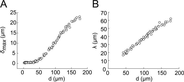
Figure 2—figure supplement 1. Biofilm diameter-dependence of δmax and λ. (A) Biofilm diameter-dependence of δmax. (B) Biofilm diameter-dependence of λ. Both δmax and λ linearly scale with the diameter d of the biofilm.
Figure 2—figure supplement 1. Biofilm diameter-dependence of δmax and λ. (A) Biofilm diameter-dependence of δmax. (B) Biofilm diameter-dependence of λ. Both δmax and λ linearly scale with the diameter d of the biofilm.CC BY · PubMed Central
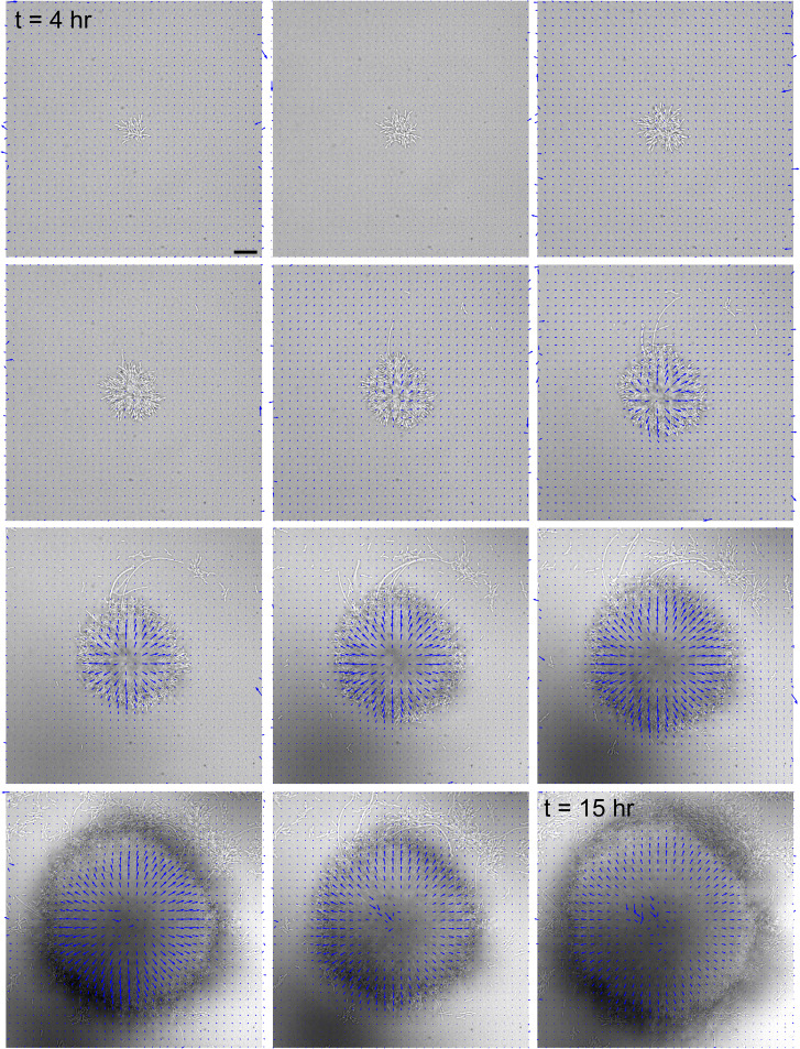
Figure 2—figure supplement 2. Hydrogel deformation field computed at different growth stages, superimposed with a brightfield image of the biofilm. Scale bar: 20 µm. The force field at each timestep is normalized by its maximum displacement, thereby showing relative deformations.
Figure 2—figure supplement 2. Hydrogel deformation field computed at different growth stages, superimposed with a brightfield image of the biofilm. Scale bar: 20 µm. The force field at each timestep is normalized by its maximum displacement, thereby showing relative deformations.CC BY · PubMed Central
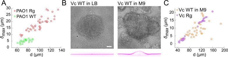
Figure 3. Wild-type and rugose biofilms deform soft-substrates. (A) Biofilm diameter-dependence of maximum deformation for rugose and smooth variants of P. aeruginosa. (B) Smooth variant of V. cholerae A1552 deforms hydrogels when growing in M9 medium, but not in LB. (C) Biofilm diameter-dependence of maximum deformation for rugose and smooth variants of V. cholerae. Data points correspond to biofilms grown in two microfluidic chambers for PAO1 Rg, PAO1 WT and Vc WT and to biofilms grown in one microfluidic chamber for Vc Rg. E = 38 kPa. Scale bars: 20 µm.
Figure 3. Wild-type and rugose biofilms deform soft-substrates. (A) Biofilm diameter-dependence of maximum deformation for rugose and smooth variants of P. aeruginosa. (B) Smooth variant of V. cholerae A1552 deforms hydrogels when growing in M9 medium, but not in LB. (C) Biofilm diameter-dependence of maximum deformation for rugose and smooth variants of V. cholerae. Data points correspond to biofilms grown in two microfluidic chambers for PAO1 Rg, PAO1 WT and Vc WT and to biofilms grown in one microfluidic chamber for Vc Rg. E = 38 kPa. Scale bars: 20 µm.CC BY · PubMed Central
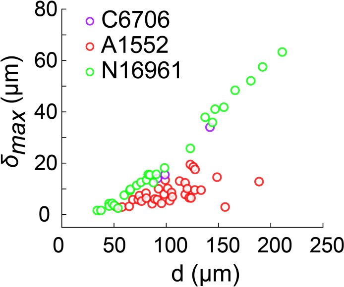
Figure 3—figure supplement 1. Biofilm diameter-dependence of maximum deformation for the smooth variant of different V. cholerae strains grown in M9. Data points correspond to different biofilms grown in two microfluidic chambers for A1552 and to biofilms grown in one microfluidic chamber for C6706 and N16961.
Figure 3—figure supplement 1. Biofilm diameter-dependence of maximum deformation for the smooth variant of different V. cholerae strains grown in M9. Data points correspond to different biofilms grown in two microfluidic chambers for A1552 and to biofilms grown in one microfluidic chamber for C6706 and N16961.CC BY · PubMed Central
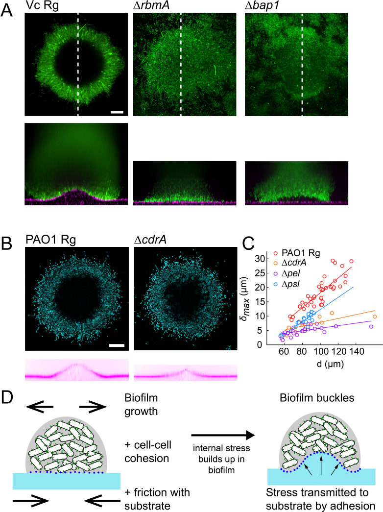
Figure 4. EPS composition drives biofilm and substrate deformations. (A) Deformations of hydrogel substrates by V. cholerae Rg, rbma- and bap1- biofilms. Biofilms formed by rbma- and bap1- fail to deform the substrate. bap1- biofilms delaminate from the hydrogel surface. (B) Comparison of hydrogel deformations by P. aeruginosa Rg and cdrA- biofilms. (C) Dependence of maximum deformations on P. aeruginosa Rg, cdrA-, pel- and psl- biofilm diameter. All matrix mutants tend to generate weaker deformations compared to Rg. Data points correspond to different biofilms grown in two microfluidic chambers. (D) A model for the mechanism of biofilm deformation of soft substrates. Buildup of mechanical stress in the biofilm induces buckling. Adhesion between the biofilm and the surface transmits buckling-generated stress to the hydrogel, inducing deformations. E = 38 kPa. Scale bars: 20 µm.
Figure 4. EPS composition drives biofilm and substrate deformations. (A) Deformations of hydrogel substrates by V. cholerae Rg, rbma- and bap1- biofilms. Biofilms formed by rbma- and bap1- fail to deform the substrate. bap1- biofilms delaminate from the hydrogel surface. (B) Comparison of hydrogel deformations by P. aeruginosa Rg and cdrA- biofilms. (C) Dependence of maximum deformations on P. aeruginosa Rg, cdrA-, pel- and psl- biofilm diameter. All matrix mutants tend to generate weaker deformations compared to Rg. Data points correspond to different biofilms grown in two microfluidic chambers. (D) A model for the mechanism of biofilm deformation of soft substrates. Buildup of mechanical stress in the biofilm induces buckling. Adhesion between the biofilm and the surface transmits buckling-generated stress to the hydrogel, inducing deformations. E = 38 kPa. Scale bars: 20 µm.CC BY · PubMed Central
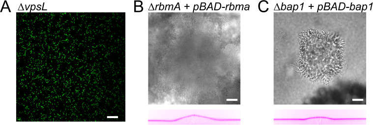
Figure 4—figure supplement 1. Deformation behaviour for vpsL deletion mutant and complementation strains. (A) VpsL deletion mutant can not form biofilms. Complementation of (B) V. cholerae rbmA and (C) bap1 deletion mutants (brightfield, top) restore the ability of the biofilm to deform the hydrogel (reslice, bottom). E = 38 kPa. Scale bars: 20 µm.
Figure 4—figure supplement 1. Deformation behaviour for vpsL deletion mutant and complementation strains. (A) VpsL deletion mutant can not form biofilms. Complementation of (B) V. cholerae rbmA and (C) bap1 deletion mutants (brightfield, top) restore the ability of the biofilm to deform the hydrogel (reslice, bottom). E = 38 kPa. Scale bars: 20 µm.CC BY · PubMed Central
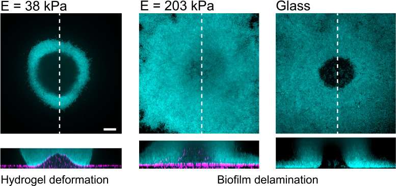
Figure 4—figure supplement 2. P. aeruginosa biofilms on substrates with different stiffness. Increasing hydrogel stiffness to 200 kPa induces delamination of biofilms, as observed on glass. Scale bars: 20 µm.
Figure 4—figure supplement 2. P. aeruginosa biofilms on substrates with different stiffness. Increasing hydrogel stiffness to 200 kPa induces delamination of biofilms, as observed on glass. Scale bars: 20 µm.CC BY · PubMed Central
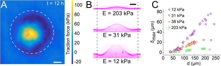
Figure 5. Biofilms generate large traction forces. (A) Traction force microscopy measurements at the hydrogel-biofilm interface. The dashed line shows the edge of the biofilm. Traction force is largest at the biofilm center, reaching 100 kPa. (B) Deformation profiles generated by V. cholerae Rg biofilms of equal diameters on three hydrogels with different stiffness. (C) Biofilm diameter-dependence of maximum deformation for four different hydrogel composition representing a typical range of tissue stiffnesses. The softest hydrogel can deform up to 80 µm for a biofilm diameter of 220 µm. Data points correspond to different biofilms grown in one microfluidic chamber. Scale bar: 20 µm.
Figure 5. Biofilms generate large traction forces. (A) Traction force microscopy measurements at the hydrogel-biofilm interface. The dashed line shows the edge of the biofilm. Traction force is largest at the biofilm center, reaching 100 kPa. (B) Deformation profiles generated by V. cholerae Rg biofilms of equal diameters on three hydrogels with different stiffness. (C) Biofilm diameter-dependence of maximum deformation for four different hydrogel composition representing a typical range of tissue stiffnesses. The softest hydrogel can deform up to 80 µm for a biofilm diameter of 220 µm. Data points correspond to different biofilms grown in one microfluidic chamber. Scale bar: 20 µm.CC BY · PubMed Central
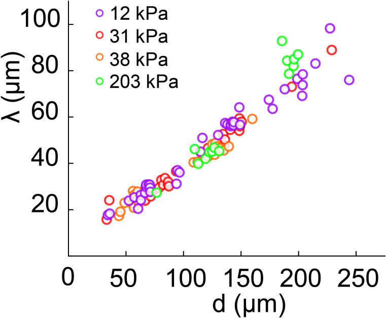
Figure 5—figure supplement 1. Biofilm diameter-dependence of λ for substrates with different moduli. λ scales linearly and it is not substrate-stiffness dependent.
Figure 5—figure supplement 1. Biofilm diameter-dependence of λ for substrates with different moduli. λ scales linearly and it is not substrate-stiffness dependent.CC BY · PubMed Central
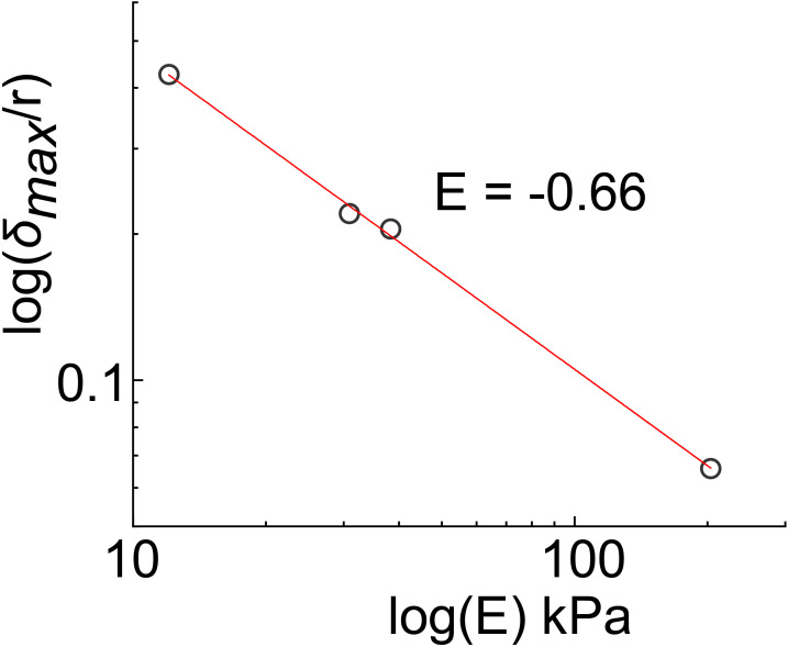
Figure 5—figure supplement 2. Power-law relationship between deformation δmax and substrate moduli (E). Values for δmax/r were extrapolated from linear regression of the data points in Figure 5C for r = 50 µm (d = 100 µm).
Figure 5—figure supplement 2. Power-law relationship between deformation δmax and substrate moduli (E). Values for δmax/r were extrapolated from linear regression of the data points in Figure 5C for r = 50 µm (d = 100 µm).CC BY · PubMed Central
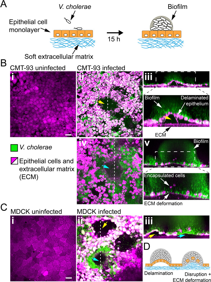
Figure 6. Biofilms deform and disrupt epithelial cell monolayer. (A) CMT-93 and MDCK cells grow at the surface of a soft ECM into a tight monolayer on which we seed a liquid inoculum of V. cholerae Rg. (B) Confocal images of uninfected (i) and infected (ii-v) monolayers of CMT-93 cells. Yellow arrow indicates gaps in the epithelial monolayer (ii and iii), blue arrow shows deformed ECM (iv). (C) Confocal images of uninfected (i) and infected (ii-iii) monolayers of MDCK cells, also showing delamination and rupture as illustrated in (D). Scale bars: 20 µm.
Figure 6. Biofilms deform and disrupt epithelial cell monolayer. (A) CMT-93 and MDCK cells grow at the surface of a soft ECM into a tight monolayer on which we seed a liquid inoculum of V. cholerae Rg. (B) Confocal images of uninfected (i) and infected (ii-v) monolayers of CMT-93 cells. Yellow arrow indicates gaps in the epithelial monolayer (ii and iii), blue arrow shows deformed ECM (iv). (C) Confocal images of uninfected (i) and infected (ii-iii) monolayers of MDCK cells, also showing delamination and rupture as illustrated in (D). Scale bars: 20 µm.CC BY · PubMed Central
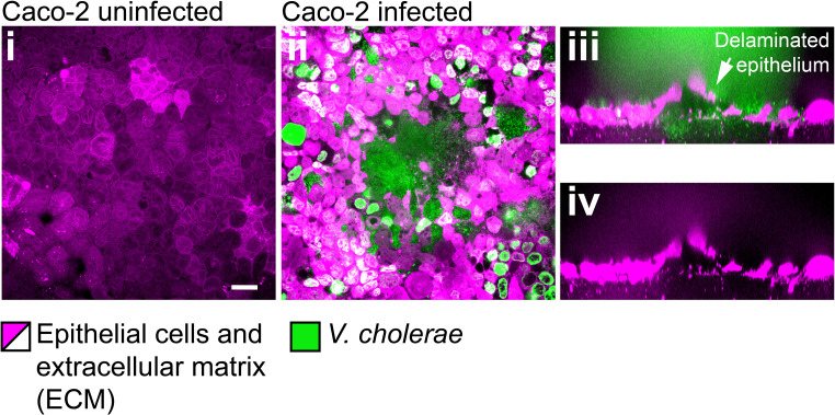
Figure 6—figure supplement 1. Confocal images of uninfected (i) and infected (ii-iv) monolayers of Caco-2 cells. Scale bars: 20 µm.
Figure 6—figure supplement 1. Confocal images of uninfected (i) and infected (ii-iv) monolayers of Caco-2 cells. Scale bars: 20 µm.CC BY · PubMed Central
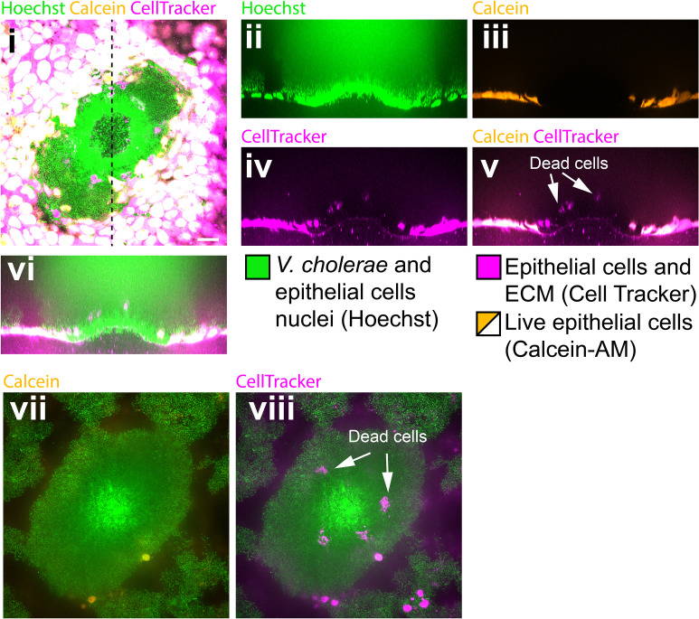
Figure 6—figure supplement 2. Biofilms perturb the viability of MDCK cell monalyers. (i) Confocal in plane visualization of a Vc Rg biofilm growing on top of an MDCK cell monolayer stained with CellTracker, Hoechst and Calcein-AM. Cross-section visualization of the infected MDCK monolayer stained with Hoechst (ii), Calcein-AM (iii), Cell Tracker (iv) and merged visualizations (v, vi). (vii, viii) Confocal in plane visualization of the biofilm on a focal plane above (i). Scale bars: 20 µm.
Figure 6—figure supplement 2. Biofilms perturb the viability of MDCK cell monalyers. (i) Confocal in plane visualization of a Vc Rg biofilm growing on top of an MDCK cell monolayer stained with CellTracker, Hoechst and Calcein-AM. Cross-section visualization of the infected MDCK monolayer stained with Hoechst (ii), Calcein-AM (iii), Cell Tracker (iv) and merged visualizations (v, vi). (vii, viii) Confocal in plane visualization of the biofilm on a focal plane above (i). Scale bars: 20 µm.CC BY · PubMed Central
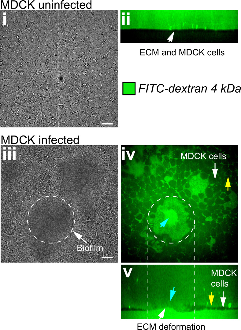
Figure 6—figure supplement 3. Biofilms increase the permeability of MDCK cell monolayers. (i) Bright-field in plane image of an uninfected MDCK cell monolayer grown at the surface of a soft ECM. (ii) Confocal cross-section shows that uninfected monolayers are impermeable to FITC labeled Dextran. (iii) Bright-field in plane visualization of an MDCK cell monolayer infected with Vc Rg. The dashed line shows the approximate edge of the biofilm. Confocal in plane (iv) and cross section (v) visualization of FITC-labeled Dextran permeability through the damaged epithelium. Blue arrows show Dextran permeability through the biofilm in direct contact with the ECM, yellow arrows indicate Dextran permeability through epithelial cells junctions. Scale bars: 20 µm.
Figure 6—figure supplement 3. Biofilms increase the permeability of MDCK cell monolayers. (i) Bright-field in plane image of an uninfected MDCK cell monolayer grown at the surface of a soft ECM. (ii) Confocal cross-section shows that uninfected monolayers are impermeable to FITC labeled Dextran. (iii) Bright-field in plane visualization of an MDCK cell monolayer infected with Vc Rg. The dashed line shows the approximate edge of the biofilm. Confocal in plane (iv) and cross section (v) visualization of FITC-labeled Dextran permeability through the damaged epithelium. Blue arrows show Dextran permeability through the biofilm in direct contact with the ECM, yellow arrows indicate Dextran permeability through epithelial cells junctions. Scale bars: 20 µm.CC BY · PubMed Central

Verification

Claims the paper makes

The claims and statistics of this paper haven’t been checked yet.

Run the check — free, no account needed. It reads the open-access text and checks the sources it cites.

A few free checks a day · papers already verified open instantly · results are public

Claims other papers make about this one

Each is checked against this paper’s own text: does it say what the citing paper says it does?

Self-check

Percentages, sums and reported differences re-computed from the paper’s own numbers.

The self-check needs the paper’s full text.

Citations

Cites

Research works printed but not linked: 58

  1. Ahmad, 2017, Gut permeability and mucosal inflammation: bad, good or context dependent, Mucosal Immunology, 10, 307, 10.1038/mi.2016.128 · DOI
  2. Asally, 2012, Localized cell death focuses mechanical forces during 3D patterning in a biofilm, PNAS, 109, 18891, 10.1073/pnas.1212429109 · DOI
  3. Bartlett, 2017, A periplasmic polymer curves Vibrio cholerae and promotes pathogenesis, Cell, 168, 172, 10.1016/j.cell.2016.12.019 · DOI
  4. Berk, 2012, Molecular architecture and assembly principles of Vibrio cholerae biofilms, Science, 337, 236, 10.1126/science.1222981 · DOI
  5. Beroz, 2018, Verticalization of bacterial biofilms, Nature Physics, 14, 954, 10.1038/s41567-018-0170-4 · DOI
  6. Bischoff, 2014, Intestinal permeability--a new target for disease prevention and therapy, BMC Gastroenterology, 14, 10.1186/s12876-014-0189-7 · DOI · Paper page
  7. Bjarnsholt, 2013, The in vivo biofilm, Trends in Microbiology, 21, 466, 10.1016/j.tim.2013.06.002 · DOI · Paper page
  8. Charras, 2018, Tensile forces and mechanotransduction at Cell-Cell junctions, Current Biology, 28, R445, 10.1016/j.cub.2018.02.003 · DOI
  9. Colvin, 2012, The pel and psl polysaccharides provide Pseudomonas aeruginosa structural redundancy within the biofilm matrix, Environmental Microbiology, 14, 1913, 10.1111/j.1462-2920.2011.02657.x · DOI
  10. De Weirdt, 2015, Micromanagement in the gut: microenvironmental factors govern Colon mucosal biofilm structure and functionality, Npj Biofilms and Microbiomes, 1, 10.1038/npjbiofilms.2015.26 · DOI
  11. Discher, 2005, Tissue cells feel and respond to the stiffness of their substrate, Science, 310, 1139, 10.1126/science.1116995 · DOI · Paper page
  12. Donlan, 2002, Biofilms: survival mechanisms of clinically relevant microorganisms, Clinical Microbiology Reviews, 15, 167, 10.1128/CMR.15.2.167-193.2002 · DOI · Paper page
  13. Douarche, 2015, Bacillus subtilis Bacteria generate an internal mechanical force within a biofilm, Biophysical Journal, 109, 2195, 10.1016/j.bpj.2015.10.004 · DOI
  14. Drescher, 2016, Architectural transitions in Vibrio cholerae biofilms at single-cell resolution, PNAS, 113, E2066, 10.1073/pnas.1601702113 · DOI
  15. Dufrêne, 2020, Mechanomicrobiology: how Bacteria sense and respond to forces, Nature Reviews Microbiology, 18, 227, 10.1038/s41579-019-0314-2 · DOI
  16. Duvernoy, 2018, Asymmetric adhesion of rod-shaped Bacteria controls microcolony morphogenesis, Nature Communications, 9, 25, 10.1038/s41467-018-03446-y · DOI
  17. Engel, 2015, The bacterium Frischella perrara causes scab formation in the gut of its honeybee host, mBio, 6, 10.1128/mBio.00193-15 · DOI
  18. Flemming, 2016, Biofilms: an emergent form of bacterial life, Nature Reviews Microbiology, 14, 563, 10.1038/nrmicro.2016.94 · DOI · Paper page
  19. Flemming, 2010, The biofilm matrix, Nature Reviews Microbiology, 8, 623, 10.1038/nrmicro2415 · DOI · Paper page
  20. Fong, 2006, Identification and characterization of RbmA, a novel protein required for the development of rugose colony morphology and biofilm structure in Vibrio cholerae, Journal of Bacteriology, 188, 1049, 10.1128/JB.188.3.1049-1059.2006 · DOI
  21. Fong, 2010, Role of Vibrio polysaccharide (vps) genes in VPS production, biofilm formation and Vibrio cholerae pathogenesis, Microbiology, 156, 2757, 10.1099/mic.0.040196-0 · DOI
  22. Fong, 2007, The rbmBCDEF gene cluster modulates development of rugose colony morphology and biofilm formation in Vibrio cholerae, Journal of Bacteriology, 189, 2319, 10.1128/JB.01569-06 · DOI
  23. Gloag, 2018, Viscoelastic properties of Pseudomonas aeruginosa variant biofilms, Scientific Reports, 8, 1, 10.1038/s41598-018-28009-5 · DOI
  24. Hardy, 2016, A fruitful alliance: the synergy between Atopobium vaginae and Gardnerella vaginalis in bacterial vaginosis-associated biofilm, Sexually Transmitted Infections, 92, 487, 10.1136/sextrans-2015-052475 · DOI
  25. Harris, 2012, Characterizing the mechanics of cultured cell monolayers, PNAS, 109, 16449, 10.1073/pnas.1213301109 · DOI
  26. Hartmann, 2019, Emergence of three-dimensional order and structure in growing biofilms, Nature Physics, 15, 251, 10.1038/s41567-018-0356-9 · DOI
  27. Hickman, 2005, A chemosensory system that regulates biofilm formation through modulation of cyclic diguanylate levels, PNAS, 102, 14422, 10.1073/pnas.0507170102 · DOI
  28. Jennings, 2015, Pel is a cationic exopolysaccharide that cross-links extracellular DNA in the Pseudomonas aeruginosa biofilm matrix, PNAS, 112, 11353, 10.1073/pnas.1503058112 · DOI
  29. Kolewe, 2015, Fewer Bacteria adhere to softer hydrogels, ACS Applied Materials & Interfaces, 7, 19562, 10.1021/acsami.5b04269 · DOI
  30. Kovach, 2017, Evolutionary adaptations of biofilms infecting cystic fibrosis lungs promote mechanical toughness by adjusting polysaccharide production, Npj Biofilms and Microbiomes, 3, 10.1038/s41522-016-0007-9 · DOI
  31. Lee, 2014, The effects of varying poly(ethylene glycol) hydrogel crosslinking density and the crosslinking mechanism on protein accumulation in three-dimensional hydrogels, Acta Biomaterialia, 10, 4167, 10.1016/j.actbio.2014.05.023 · DOI
  32. Lichter, 2008, Substrata mechanical stiffness can regulate adhesion of viable Bacteria, Biomacromolecules, 9, 1571, 10.1021/bm701430y · DOI
  33. Mah, 2001, Mechanisms of biofilm resistance to antimicrobial agents, Trends in Microbiology, 9, 34, 10.1016/S0966-842X(00)01913-2 · DOI · Paper page
  34. O'Toole, 2000, Biofilm formation as microbial development, Annual Review of Microbiology, 54, 49, 10.1146/annurev.micro.54.1.49 · DOI · Paper page
  35. Pestrak, 2018, Pseudomonas aeruginosa rugose small-colony variants evade host clearance, are hyper-inflammatory, and persist in multiple host environments, PLOS Pathogens, 14, 10.1371/journal.ppat.1006842 · DOI
  36. Rivera-Yoshida, 2018, Microbial multicellular development: mechanical forces in action, Current Opinion in Genetics & Development, 51, 37, 10.1016/j.gde.2018.05.006 · DOI
  37. Rojas, 2018, Regulation of microbial growth by turgor pressure, Current Opinion in Microbiology, 42, 62, 10.1016/j.mib.2017.10.015 · DOI
  38. Rybtke, 2012, Fluorescence-based reporter for gauging cyclic di-GMP levels in Pseudomonas aeruginosa, Applied and Environmental Microbiology, 78, 5060, 10.1128/AEM.00414-12 · DOI
  39. Rybtke, 2015, The LapG protein plays a role in Pseudomonas aeruginosa biofilm formation by controlling the presence of the CdrA adhesin on the cell surface, MicrobiologyOpen, 4, 917, 10.1002/mbo3.301 · DOI
  40. Serra, 2015, Vertical stratification of matrix production is essential for physical integrity and architecture of macrocolony biofilms of Escherichia coli, Environmental Microbiology, 17, 5073, 10.1111/1462-2920.12991 · DOI
  41. Song, 2014, Stiffness of cross-linked poly(dimethylsiloxane) affects bacterial adhesion and antibiotic susceptibility of attached cells, Langmuir, 30, 10354, 10.1021/la502029f · DOI
  42. Starkey, 2009, Pseudomonas aeruginosa rugose small-colony variants have adaptations that likely promote persistence in the cystic fibrosis lung, Journal of Bacteriology, 191, 3492, 10.1128/JB.00119-09 · DOI
  43. Swidsinski, 2013, Presence of a polymicrobial endometrial biofilm in patients with bacterial vaginosis, PLOS ONE, 8, 10.1371/journal.pone.0053997 · DOI
  44. Takigawa, 1996, Poisson's ratio of polyacrylamide (PAAm) gels, Polymer Gels and Networks, 4, 1, 10.1016/0966-7822(95)00013-5 · DOI
  45. Teschler, 2015, Living in the matrix: assembly and control of Vibrio cholerae biofilms, Nature Reviews Microbiology, 13, 255, 10.1038/nrmicro3433 · DOI
  46. Thelin, 1996, Toxin-coregulated pilus, but not mannose-sensitive hemagglutinin, is required for colonization by Vibrio cholerae O1 el Tor biotype and O139 strains, Infection and Immunity, 64, 2853, 10.1128/IAI.64.7.2853-2856.1996 · DOI · Paper page
  47. Timoshenko, 2019, Theory of Elastic Stability
  48. Tischler, 2005, Cyclic diguanylate regulates Vibrio cholerae virulence gene expression, Infection and Immunity, 73, 5873, 10.1128/IAI.73.9.5873-5882.2005 · DOI
  49. Toyjanova, 2014, High resolution, large deformation 3D traction force microscopy, PLOS ONE, 9, 10.1371/journal.pone.0090976 · DOI
  50. Trejo, 2013, Elasticity and wrinkled morphology of Bacillus subtilis pellicles, PNAS, 110, 2011, 10.1073/pnas.1217178110 · DOI
  51. Tremaroli, 2012, Functional interactions between the gut Microbiota and host metabolism, Nature, 489, 242, 10.1038/nature11552 · DOI · Paper page
  52. Wang, 2005, On the buckling of a circular plate on an elastic foundation, Journal of Applied Mechanics, 72, 795, 10.1115/1.1988347 · DOI
  53. Wang, 2015, A three-dimensional phase diagram of growth-induced surface instabilities, Scientific Reports, 5, 1, 10.1038/srep08887 · DOI
  54. Wilking, 2013, Liquid transport facilitated by channels in Bacillus subtilis biofilms, PNAS, 110, 848, 10.1073/pnas.1216376110 · DOI
  55. Yan, 2016, Vibrio cholerae biofilm growth program and architecture revealed by single-cell live imaging, PNAS, 113, E5337, 10.1073/pnas.1611494113 · DOI
  56. Yan, 2018, Bacterial biofilm material properties enable removal and transfer by capillary peeling, Advanced Materials, 30, 10.1002/adma.201804153 · DOI
  57. Yan, 2019, Mechanical instability and interfacial energy drive biofilm morphogenesis, eLife, 8, 10.7554/eLife.43920 · DOI
  58. Yildiz, 1999, Vibrio cholerae O1 el Tor: identification of a gene cluster required for the rugose colony type, exopolysaccharide production, chlorine resistance, and biofilm formation, PNAS, 96, 4028, 10.1073/pnas.96.7.4028 · DOI

Cited by

Sorted by citations

See all 70

Metadata

Authors
Cont, A., Rossy, T., Al-Mayyah, Z., & Persat, A.
Venue
eLife
Published
2020-10-07
Volume
9
ISSN
2050-084X
Cited by
70
References
1
Language
en
Funders
  • Swiss National Science Foundation
  • Cavaglieri Foundation
  • Fondation Beytout
  • Gebert Rüf Stiftung

About these checks

Registry, retraction, Cites and statistics results are computed, not judged, with one exception: a printed reference that no index lists is read once by a model before it counts as a citation error. Claims are extracted by a model and kept only when their quote appears verbatim in the text; each cited source is then checked, with a model’s help, for whether it says what the claim relies on. Those verdicts can be wrong — read the claim’s quote and the cited source before relying on one.

Statistics and arithmetic are recomputed, not judged.

Confidence bands are shown only for fields calibrated on our annotated test set. None is calibrated yet, so no band is shown.

What counts as a claim ›How we measure confidence ›How we check this ›

Are you an author, or do you see a problem with this page? Request a review