

The DOI matches its Crossref record; 48 of the 48 references it cites were found in a registry or an index.
Structural and functional properties of the transporter SLC26A6 reveal mechanism of coupled anion exchange
Open accessPublished version of a preprint
Abstract
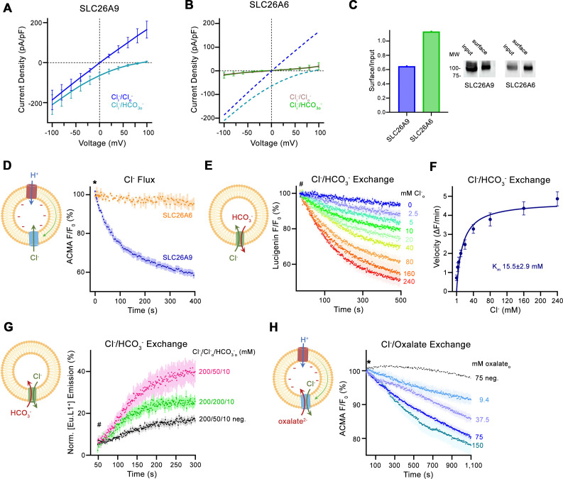
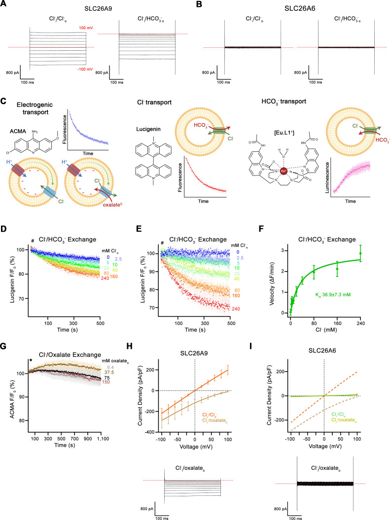
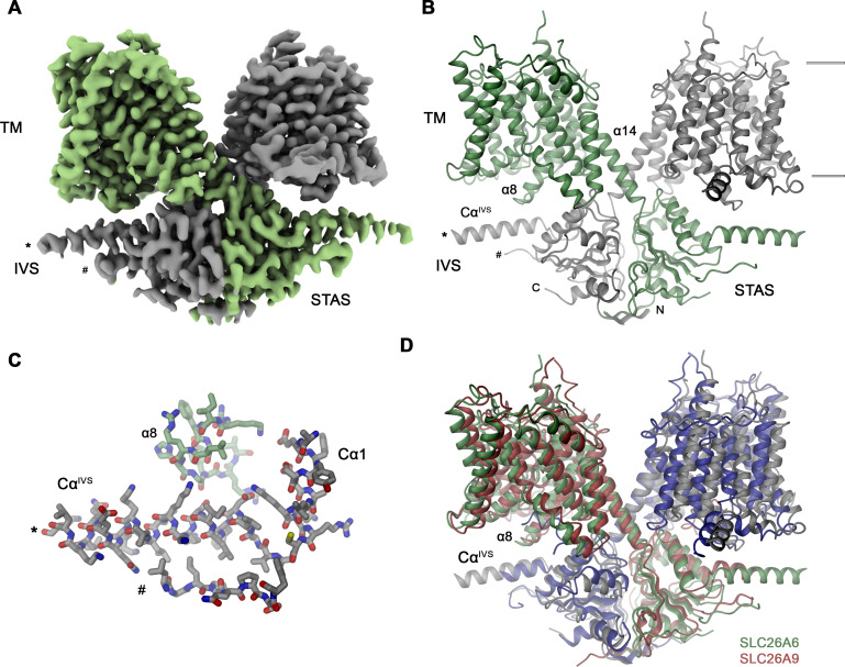
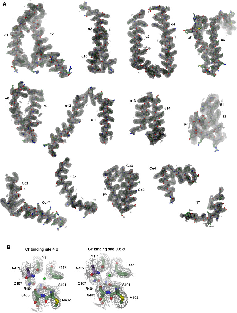
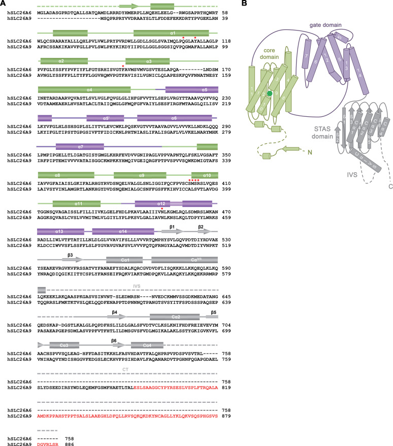
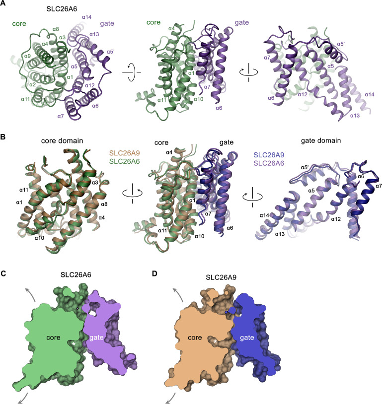
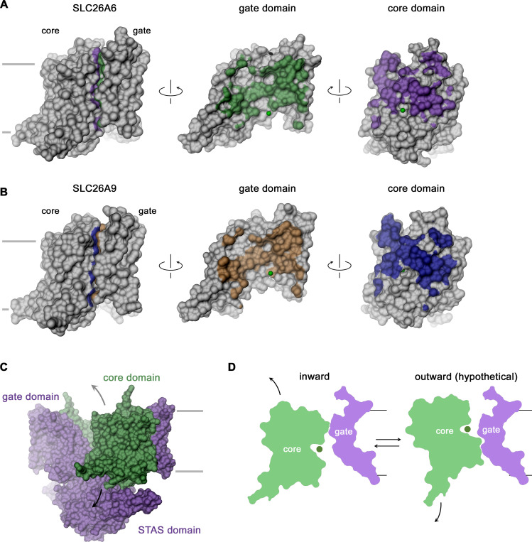
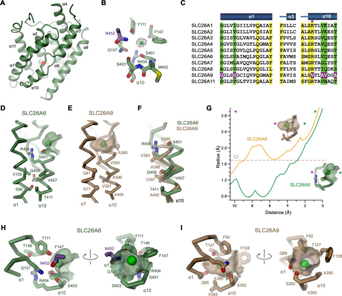
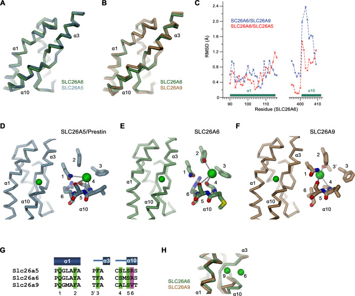
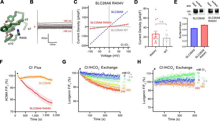
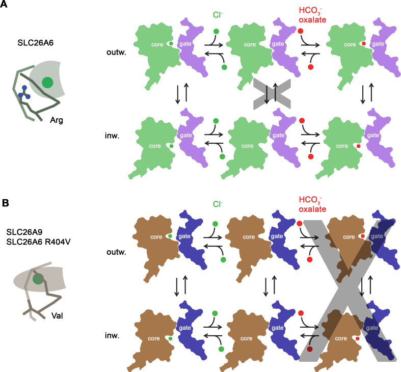
Members of the SLC26 family constitute a conserved class of anion transport proteins, which encompasses uncoupled transporters with channel-like properties, coupled exchangers and motor proteins. Among the 10 functional paralogs in humans, several participate in the secretion of bicarbonate in exchange with chloride and thus play an important role in maintaining pH homeostasis. Previously, we have elucidated the structure of murine SLC26A9 and defined its function as an uncoupled chloride transporter (Walter et al., 2019). Here we have determined the structure of the closely related human transporter SLC26A6 and characterized it as a coupled exchanger of chloride with bicarbonate and presumably also oxalate. The structure defines an inward-facing conformation of the protein that generally resembles known structures of SLC26A9. The altered anion selectivity between both paralogs is a consequence of a remodeled ion binding site located in the center of a mobile unit of the membrane-inserted domain, which also accounts for differences in the coupling mechanism.
Overview
The claims and statistics of this paper haven’t been checked yet.
Run the check — free, no account needed. It reads the open-access text and checks the sources it cites.
Integrity signals
Registry
Cites
Statistics
Figures
Verification
Claims the paper makes
The claims and statistics of this paper haven’t been checked yet.
Run the check — free, no account needed. It reads the open-access text and checks the sources it cites.
Self-check
Percentages, sums and reported differences re-computed from the paper’s own numbers.
The self-check needs the paper’s full text.
Citations
Cites
UCSF Chimera—A visualization system for exploratory research and analysis
Pettersen, E. F., Goddard, T. D., Huang, C. C., Couch, G. S., Greenblatt, D. M., Meng, E. C., & Ferrin, T. E. (2004) · Journal of Computational Chemistry · doi:10.1002/jcc.20084 · Cited by 45,828
Highly accurate protein structure prediction with AlphaFold
Jumper, J., Evans, R., Pritzel, A., Green, T., Figurnov, M., Ronneberger, O., Tunyasuvunakool, K., Bates, R., Žídek, A., Potapenko, A., et al. (2021) · Nature · doi:10.1038/s41586-021-03819-2 · Cited by 43,764
Coot : model-building tools for molecular graphics
Emsley, P., & Cowtan, K. (2004) · Acta Crystallographica Section D Biological Crystallography · doi:10.1107/s0907444904019158 · Cited by 30,571
Features and development of Coot
Emsley, P., Lohkamp, B., Scott, W. G., & Cowtan, K. (2010) · Acta Crystallographica Section D Biological Crystallography · doi:10.1107/s0907444910007493 · Cited by 29,915
cryoSPARC: algorithms for rapid unsupervised cryo-EM structure determination
Punjani, A., Rubinstein, J. L., Fleet, D. J., & Brubaker, M. A. (2017) · Nature Methods · doi:10.1038/nmeth.4169 · Cited by 11,626
Jalview Version 2—a multiple sequence alignment editor and analysis workbench
Waterhouse, A. M., Procter, J. B., Martin, D. M. A., Clamp, M. E., & Barton, G. J. (2009) · Bioinformatics · doi:10.1093/bioinformatics/btp033 · Cited by 10,349
UCSF ChimeraX : Structure visualization for researchers, educators, and developers
Pettersen, E. F., Goddard, T. D., Huang, C. C., Meng, E. C., Couch, G. S., Croll, T. I., Morris, J. H., & Ferrin, T. E. (2020) · Protein Science · doi:10.1002/pro.3943 · Cited by 9,762
Liebschner, D., Afonine, P. V., Baker, M. L., Bunkóczi, G., Chen, V. B., Croll, T. I., Hintze, B., Hung, L.-W., Jain, S., McCoy, A. J., et al. (2019) · Acta Crystallographica Section D Structural Biology · doi:10.1107/s2059798319011471 · Cited by 7,953
Varadi, M., Anyango, S., Deshpande, M., Nair, S., Natassia, C., Yordanova, G., Yuan, D., Stroe, O., Wood, G., Laydon, A., et al. (2022) · Nucleic Acids Research · doi:10.1093/nar/gkab1061 · Cited by 7,850
Real-space refinement in PHENIX for cryo-EM and crystallography
Afonine, P. V., Poon, B. K., Read, R. J., Sobolev, O. V., Terwilliger, T. C., Urzhumtsev, A., & Adams, P. D. (2018) · Acta Crystallographica Section D Structural Biology · doi:10.1107/s2059798318006551 · Cited by 3,255
HOLE: A program for the analysis of the pore dimensions of ion channel structural models
Smart, O. S., Neduvelil, J. G., Wang, X., Wallace, B. A., & Sansom, M. S. P. (1996) · Journal of Molecular Graphics · doi:10.1016/s0263-7855(97)00009-x · Cited by 1,803
Prestin is the motor protein of cochlear outer hair cells
Zheng, J., Shen, W., He, D. Z. Z., Long, K. B., Madison, L. D., & Dallos, P. (2000) · Nature · doi:10.1038/35012009 · Cited by 1,344
DeepEMhancer: a deep learning solution for cryo-EM volume post-processing
Sánchez-García, R., Gómez-Blanco, J., Cuervo, A., Carazo, J. M., Sorzano, C. Ó. S., & Vargas, J. (2021) · Communications Biology · doi:10.1038/s42003-021-02399-1 · Cited by 1,311
Addressing preferred specimen orientation in single-particle cryo-EM through tilting
Tan, Y. Z., Baldwin, P. R., Davis, J. H., Williamson, J. R., Potter, C. S., Carragher, B. O., & Lyumkis, D. (2017) · Nature Methods · doi:10.1038/nmeth.4347 · Cited by 895
Reduced surface: an efficient way to compute molecular surfaces.
Sanner, M. F., Olson, A. J., & Spehner, J.-C. (1996) · PubMed · doi:10.1002/(sici)1097-0282(199603)38:3%3c305::aid-bip4%3e3.0.co;2-y · Cited by 593
Shared Molecular Mechanisms of Membrane Transporters
Drew, D., & Boudker, O. (2016) · Annual Review of Biochemistry · doi:10.1146/annurev-biochem-060815-014520 · Cited by 517
The SLC26 gene family of anion transporters and channels
Alper, S. L., & Sharma, A. K. (2013) · Molecular Aspects of Medicine · doi:10.1016/j.mam.2012.07.009 · Cited by 411
Calcium oxalate urolithiasis in mice lacking anion transporter Slc26a6
Jiang, Z., Asplin, J. R., Evan, A. P., Rajendran, V. M., Velázquez, H., Nottoli, T., Binder, H. J., & Aronson, P. S. (2006) · Nature Genetics · doi:10.1038/ng1762 · Cited by 314
A Versatile and Efficient High-Throughput Cloning Tool for Structural Biology
Geertsma, E. R., & Dutzler, R. (2011) · Biochemistry · doi:10.1021/bi200178z · Cited by 248
Self-citationKnauf, F., Yang, C., Thomson, R. B., Mentone, S. A., Giebisch, G., & Aronson, P. S. (2001) · Proceedings of the National Academy of Sciences · doi:10.1073/pnas.141241098 · Cited by 244
Molecular characterization of the murine Slc26a6 anion exchanger: functional comparison with Slc26a1
Xie, Q., Welch, R., Mercado, A., Romero, M. F., & Mount, D. B. (2002) · American Journal of Physiology-Renal Physiology · doi:10.1152/ajprenal.00079.2002 · Cited by 223
Structure and mechanism of the uracil transporter UraA
Lu, F., Li, S., Jiang, Y., Jiang, J., He, F., Lu, G., Deng, D., Dang, S., Zhang, X., Wang, J., et al. (2011) · Nature · doi:10.1038/nature09885 · Cited by 205
Lohi, H., Kujala, M., Kerkelä, E., Saarialho‐Kere, U., Kestilä, M., & Kere, J. (2000) · Genomics · doi:10.1006/geno.2000.6355 · Cited by 205
The Solute Carrier 26 Family of Proteins in Epithelial Ion Transport
Dorwart, M. R., Shcheynikov, N., Yang, D., & Muallem, S. (2008) · Physiology · doi:10.1152/physiol.00037.2007 · Cited by 200
Coupling Modes and Stoichiometry of Cl−/HCO3− Exchange by slc26a3 and slc26a6
Shcheynikov, N., Wang, Y., Park, M., Ko, S. B. H., Dorwart, M. R., Naruse, S., Thomas, P., & Muallem, S. (2006) · The Journal of General Physiology · doi:10.1085/jgp.200509392 · Cited by 192
Structure of a prokaryotic fumarate transporter reveals the architecture of the SLC26 family
Geertsma, E. R., Chang, Y.-N., Shaik, F. R., Neldner, Y., Pardon, E., Steyaert, J., & Dutzler, R. (2015) · Nature Structural & Molecular Biology · doi:10.1038/nsmb.3091 · Cited by 179
Self-citationWang, Y., Soyombo, A. A., Shcheynikov, N., Zeng, W., Dorwart, M. R., Marino, C. R., Thomas, P., & Muallem, S. (2006) · The EMBO Journal · doi:10.1038/sj.emboj.7601387 · Cited by 166
Functional Comparison of Mouse slc26a6 Anion Exchanger with Human SLC26A6 Polypeptide Variants
Chernova, M. N., Jiang, L., Friedman, D. J., Darman, R. B., Lohi, H., Kere, J., Vandorpe, D. H., & Alper, S. L. (2004) · Journal of Biological Chemistry · doi:10.1074/jbc.m411703200 · Cited by 161
SLC26A9 is a Cl− channel regulated by the WNK kinases
Dorwart, M. R., Shcheynikov, N., Wang, Y., et al. (2007) · The Journal of Physiology · doi:10.1113/jphysiol.2007.135855 · Cited by 138
Diverse transport modes by the solute carrier 26 family of anion transporters
Ohana, E., Yang, D., Shcheynikov, N., & Muallem, S. (2008) · The Journal of Physiology · doi:10.1113/jphysiol.2008.164863 · Cited by 133
Walter, J. D., Sawicka, M., & Dutzler, R. (2019) · eLife · doi:10.7554/elife.46986 · Cited by 123
Self-citationStructural mechanism of the active bicarbonate transporter from cyanobacteria
Wang, C., Sun, B., Zhang, X., Huang, X., Zhang, M., Guo, H., Chen, X., Huang, F., Chen, T., Mi, H., et al. (2019) · Nature Plants · doi:10.1038/s41477-019-0538-1 · Cited by 104
The conformational cycle of prestin underlies outer-hair cell electromotility
Bavi, N., Clark, M. D., Contreras, G. F., Shen, R., Reddy, B. G., Milewski, W. M., & Perozo, E. (2021) · Nature · doi:10.1038/s41586-021-04152-4 · Cited by 100
Molecular mechanism of prestin electromotive signal amplification
Ge, J., Elferich, J., Dehghani‐Ghahnaviyeh, S., Zhao, Z., Meadows, M. A., von Gersdorff, H., Tajkhorshid, E., & Gouaux, E. (2021) · Cell · doi:10.1016/j.cell.2021.07.034 · Cited by 99
Bicarbonate transport in health and disease
Alka, K., & Casey, J. R. (2014) · IUBMB Life · doi:10.1002/iub.1315 · Cited by 94
SLC26A6 and NaDC-1 Transporters Interact to Regulate Oxalate and Citrate Homeostasis
Ohana, E., Shcheynikov, N., Moe, O. W., & Muallem, S. (2013) · Journal of the American Society of Nephrology · doi:10.1681/asn.2013010080 · Cited by 88
Structure and function of an Arabidopsis thaliana sulfate transporter
Wang, L., Chen, K., & Zhou, M. (2021) · Nature Communications · doi:10.1038/s41467-021-24778-2 · Cited by 82
Clark, J. S., Vandorpe, D. H., Chernova, M. N., Heneghan, J. F., Stewart, A. K., & Alper, S. L. (2008) · The Journal of Physiology · doi:10.1113/jphysiol.2007.143222 · Cited by 81
The CFTR trafficking mutation F508del inhibits the constitutive activity of SLC26A9
Bertrand, C. A., Mitra, S., Mishra, S., Wang, X., Zhao, Y., Pilewski, J. M., Madden, D. R., & Frizzell, R. A. (2017) · American Journal of Physiology-Lung Cellular and Molecular Physiology · doi:10.1152/ajplung.00178.2016 · Cited by 77
Single particle cryo-EM structure of the outer hair cell motor protein prestin
Butan, C., Song, Q., Bai, J.-P., Tan, W. J. T., Navaratnam, D. S., & Santos‐Sacchi, J. (2022) · Nature Communications · doi:10.1038/s41467-021-27915-z · Cited by 71
Structural insights into the gating mechanism of human SLC26A9 mediated by its C-terminal sequence
Chi, X., Jin, X., Chen, Y., Lu, X., Tu, X., Li, X., Zhang, Y., Lei, J., Huang, J., Huang, Z., et al. (2020) · Cell Discovery · doi:10.1038/s41421-020-00193-7 · Cited by 68
Regulatory Interaction between CFTR and the SLC26 Transporters
Shcheynikov, N., Ko, S. B. H., Zeng, W., Choi, J. Y., Dorwart, M. R., Thomas, P., & Muallem, S. (2006) · Novartis Foundation symposium · doi:10.1002/0470029579.ch12 · Cited by 67
Asymmetric pendrin homodimer reveals its molecular mechanism as anion exchanger
Liu, Q., Zhang, X., Huang, H., Chen, Y., Wang, F., Hao, A., Zhan, W., Mao, Q., Hu, Y., Han, L., et al. (2023) · Nature Communications · doi:10.1038/s41467-023-38303-0 · Cited by 46
Physiological and Pathological Functions of SLC26A6
Wang, J., Wang, W., Wang, H., & Tuo, B. (2021) · Frontiers in Medicine · doi:10.3389/fmed.2020.618256 · Cited by 45
Futamata, H., Fukuda, M., Umeda, R., Yamashita, K., Tomita, A., Takahashi, S., Shikakura, T., Hayashi, S., Kusakizako, T., Nishizawa, T., et al. (2022) · Nature Communications · doi:10.1038/s41467-022-34017-x · Cited by 44
Transmembrane Transport of Bicarbonate Unravelled**
Martínez‐Crespo, L., Hewitt, S. H., Simone, N. A. D., Šindelář, V., Davis, A. P., Butler, S. J., & Valkenier, H. (2021) · Chemistry - A European Journal · doi:10.1002/chem.202100491 · Cited by 40
Self-citationSlc26a6 functions as an electrogenic Cl−/HCO3− exchanger in cardiac myocytes
Kim, H. J., Myers, R., Sihn, C.-R., Rafizadeh, S., & Zhang, X.-D. (2013) · Cardiovascular Research · doi:10.1093/cvr/cvt195 · Cited by 25
Transmembrane Transport of Bicarbonate Unravelled
Martínez‐Crespo, L., Hewitt, S. H., Simone, N. A. D., Šindelář, V., Davis, A. P., Butler, S. J., & Valkenier, H. (2021) · Chemistry - A European Journal · doi:10.1002/chem.202101345 · Cited by 5
Self-citation
Metadata
- Authors
- Tippett, D. N., Breen, C., Butler, S. J., Sawicka, M., & Dutzler, R.
- Affiliations
- Loughborough University (GB)
- Corresponding author
- Dutzler, Raimund
- Venue
- eLife
- Publisher
- eLife Sciences Publications, Ltd
- Published
- 2023-06-23
- Volume
- 12
- ISSN
- 2050-084X (ISSN-L)
- PMC
- PMC10328499
- OpenAlex
- W4365514760
- Cited by
- 14
- References
- 48
- Citations by year
- 2026: 3 · 2025: 4 · 2024: 6 · 2023: 1
- FWCI
- 1.25
- Type
- journal article
- Language
- en
- Open access
- gold
- doi.org
- Licence
- Locations
- eLife (publishedVersion, cc-by)
- PubMed Central (publishedVersion, cc-by)
- PubMed (publishedVersion)
- DOAJ (DOAJ: Directory of Open Access Journals) (submittedVersion, cc-by-sa)
- Funders
- Schweizerischer Nationalfonds zur Förderung der Wissenschaftlichen Forschung (31003A_163421)
- Engineering and Physical Sciences Research Council (EP/S032339/1)
- Topics
- Life Sciences › Biochemistry, Genetics and Molecular Biology › Molecular Biology › Ion Transport and Channel Regulation
- Health Sciences › Medicine › Oncology › Drug Transport and Resistance Mechanisms
- Life Sciences › Biochemistry, Genetics and Molecular Biology › Molecular Biology › Ion channel regulation and function
- Keywords
- SLC26A6, SLC26 family, anion exchanger, chloride transport, bicarbonate secretion, oxalate transport, protein structure, anion selectivity, pH homeostasis, SLC26A9, inward-facing conformation
- MeSH terms
- Sulfate Transporters, Animals, Bicarbonates, Chlorides, Humans, Antiporters, Anion Transport Proteins, Chloride-Bicarbonate Antiporters, Mice
About these checks
Registry, retraction, Cites and statistics results are computed, not judged, with one exception: a printed reference that no index lists is read once by a model before it counts as a citation error. Claims are extracted by a model and kept only when their quote appears verbatim in the text; each cited source is then checked, with a model’s help, for whether it says what the claim relies on. Those verdicts can be wrong — read the claim’s quote and the cited source before relying on one.
Statistics and arithmetic are recomputed, not judged.
Confidence bands are shown only for fields calibrated on our annotated test set. None is calibrated yet, so no band is shown.
What counts as a claim ›How we measure confidence ›How we check this ›
Are you an author, or do you see a problem with this page? Request a review
Data sources
Bibliographic data: Semantic Scholar, OpenAlex, Crossref.
Contains information from the Semantic Scholar Academic Graph Datasets, made available under the ODC Attribution License (ODC-BY 1.0).
Semantic Scholar’s data is described in: Kinney, R., et al. (2023). The Semantic Scholar Open Data Platform. arXiv:2301.10140.
