Cited by 8Mitochondrial Function and PathologyGenomics and Chromatin DynamicsAutophagy in Disease and Therapy

The DOI matches its Crossref record; 2 of the 2 references it cites were found in a registry or an index.

A transcription network underlies the dual genomic coordination of mitochondrial biogenesis

Open access

Published version of a preprint

Abstract

Mitochondrial biogenesis requires the expression of genes encoded by both the nuclear and mitochondrial genomes. However, aside from a handful transcription factors regulating specific subsets of mitochondrial genes, the overall architecture of the transcriptional control of mitochondrial biogenesis remains to be elucidated. The mechanisms coordinating these two genomes are largely unknown. We performed a targeted RNAi screen in developing eyes with reduced mitochondrial DNA content, anticipating a synergistic disruption of tissue development due to impaired mitochondrial biogenesis and mitochondrial DNA (mtDNA) deficiency. Among 638 transcription factors annotated in the Drosophila genome, 77 were identified as potential regulators of mitochondrial biogenesis. Utilizing published ChIP-seq data of positive hits, we constructed a regulatory network revealing the logic of the transcription regulation of mitochondrial biogenesis. Multiple transcription factors in core layers had extensive connections, collectively governing the expression of nearly all mitochondrial genes, whereas factors sitting on the top layer may respond to cellular cues to modulate mitochondrial biogenesis through the underlying network. CG1603, a core component of the network, was found to be indispensable for the expression of most nuclear mitochondrial genes, including those required for mtDNA maintenance and gene expression, thus coordinating nuclear genome and mtDNA activities in mitochondrial biogenesis. Additional genetic analyses validated YL-1, a transcription factor upstream of CG1603 in the network, as a regulator controlling CG1603 expression and mitochondrial biogenesis.

Overview

The claims and statistics of this paper haven’t been checked yet.

Run the check — free, no account needed. It reads the open-access text and checks the sources it cites.

A few free checks a day · papers already verified open instantly · results are public

Integrity signals

Registry

Crossref · 10.7554/elife.96536.3

Cites

2 of 2 references verified0 citation errors · 0 not checkable

Statistics

Figures

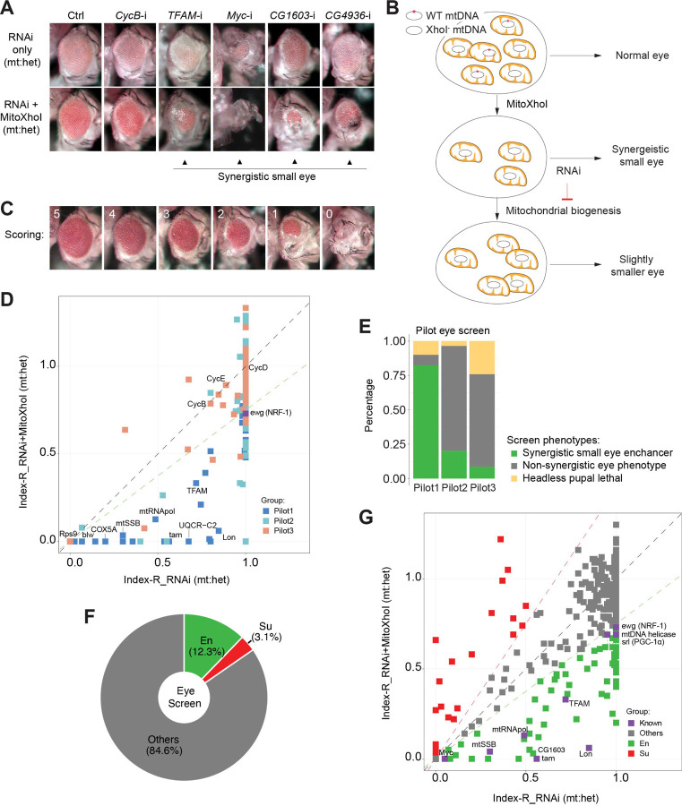
Figure 1. A genetic modifier screen identifying transcription factors regulating ETC biogenesis. (A) Representative images of adult eye for the control RNAi (Ctrl) and RNAi of selected genes tested in the eye screen, including CycB RNAi (CycB-i), TFAM RNAi (TFAM-i), Myc RNAi (Myc-i), CG1603 RNAi (CG1603-i) and CG4936 RNAi (CG4936-i). The upper panel shows eyes from RNAi-only offspring and lower panel displays eyes from RNAi+MitoXhoI offspring cultured at the same condition. Arrowheads indicate the synergistic small eye phenotype resulting from the combination of gene knockdown and the mtDNA deficiency caused by mitoXhoI in the background of heteroplasmic mtDNAs. (B) Schematic of the genetic modifier screen methodology (see text for details). (C) Representative images illustrating the scoring of eye size. (D) A plot illustrating the calling of positive hits in the pilot screen. Each datapoint represents the Index-R of RNAi (X values) or RNAi+MitoXhoI flies (Y values) for each gene belonging to the different groups (see Figure 1E and Supplementary file 1 for details). Genes with datapoints lie below the grey diagonal dash line exhibited a synergistic effect when combining their RNAi with mtDNA deficiency suggesting a potential role in regulating ETC biogenesis. The datapoint for ewg, the fly homolog of NRF-1, is labeled in purple. The green dashed line of slope 0.75 outlines the threshold for calling out positive hits based on ewg’s performance in the screen. (E) Graph summarizing the pilot screen of nuclear-encoded genes, demonstrating the efficacy of this screen in identifying genes involved in mitochondrial ETC biogenesis. Pilot group 1 (Pilot1) has 40 genes that are either nuclear-encoded ETC subunits or related to mtDNA maintenance and gene expression (Mito-EBR). Pilot2 has 84 genes involved in other mitochondrial processes. Pilot3 has 58 essential genes from other cellular components. (F) Graph summarizing the percentages of synergistic enhancers (En) and suppressors (Su) identified in the screen (see Figure 1G and Supplementary file 1 for details). (G) A plot illustrating the calling of positive hits in the screen of TF genes. Factors that are known to be involved in mitochondrial or ETC biogenesis are marked in purple (Known). The green dashed line outlines the threshold for calling out synergistic enhancers (En, green square). The red dashed line of slope 1.5 outlines the threshold for calling out suppressors (Su, red square).
Figure 1. A genetic modifier screen identifying transcription factors regulating ETC biogenesis. (A) Representative images of adult eye for the control RNAi (Ctrl) and RNAi of selected genes tested in the eye screen, including CycB RNAi (CycB-i), TFAM RNAi (TFAM-i), Myc RNAi (Myc-i), CG1603 RNAi (CG1603-i) and CG4936 RNAi (CG4936-i). The upper panel shows eyes from RNAi-only offspring and lower panel displays eyes from RNAi+MitoXhoI offspring cultured at the same condition. Arrowheads indicate the synergistic small eye phenotype resulting from the combination of gene knockdown and the mtDNA deficiency caused by mitoXhoI in the background of heteroplasmic mtDNAs. (B) Schematic of the genetic modifier screen methodology (see text for details). (C) Representative images illustrating the scoring of eye size. (D) A plot illustrating the calling of positive hits in the pilot screen. Each datapoint represents the Index-R of RNAi (X values) or RNAi+MitoXhoI flies (Y values) for each gene belonging to the different groups (see Figure 1E and Supplementary file 1 for details). Genes with datapoints lie below the grey diagonal dash line exhibited a synergistic effect when combining their RNAi with mtDNA deficiency suggesting a potential role in regulating ETC biogenesis. The datapoint for ewg, the fly homolog of NRF-1, is labeled in purple. The green dashed line of slope 0.75 outlines the threshold for calling out positive hits based on ewg’s performance in the screen. (E) Graph summarizing the pilot screen of nuclear-encoded genes, demonstrating the efficacy of this screen in identifying genes involved in mitochondrial ETC biogenesis. Pilot group 1 (Pilot1) has 40 genes that are either nuclear-encoded ETC subunits or related to mtDNA maintenance and gene expression (Mito-EBR). Pilot2 has 84 genes involved in other mitochondrial processes. Pilot3 has 58 essential genes from other cellular components. (F) Graph summarizing the percentages of synergistic enhancers (En) and suppressors (Su) identified in the screen (see Figure 1G and Supplementary file 1 for details). (G) A plot illustrating the calling of positive hits in the screen of TF genes. Factors that are known to be involved in mitochondrial or ETC biogenesis are marked in purple (Known). The green dashed line outlines the threshold for calling out synergistic enhancers (En, green square). The red dashed line of slope 1.5 outlines the threshold for calling out suppressors (Su, red square).CC BY-NC-ND · PubMed Central
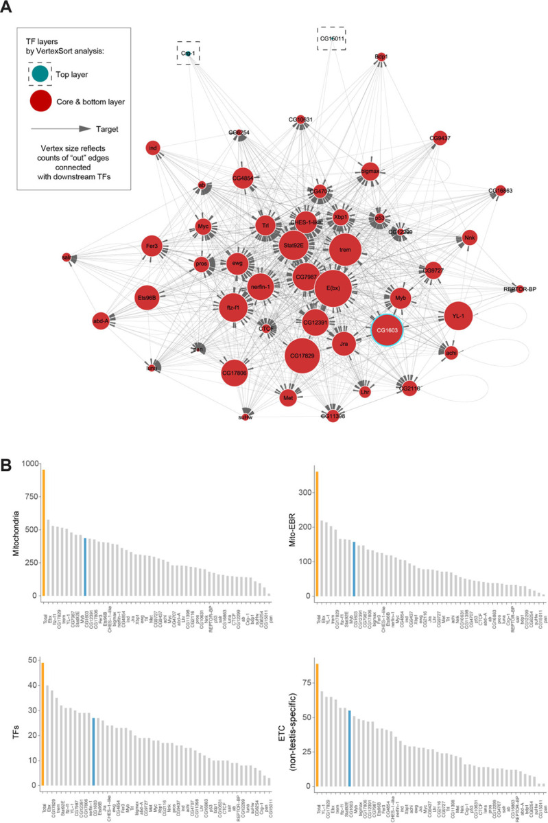
Figure 2. Regulatory network of mitochondrial biogenesis. (A) The potential transcriptional regulatory network of nuclear-encoded mitochondrial genes. (B) Bar graphs illustrating the promoter binding profiles of the 49 synergistic enhancer TFs within different groups of genes (nuclear-encoded mitochondrial genes, Mito-EBR genes, 49 synergistic enhancer TFs and ETC genes). Total gene number of the group (orange) and the number bound by CG1603 (cyan) were highlighted.
Figure 2. Regulatory network of mitochondrial biogenesis. (A) The potential transcriptional regulatory network of nuclear-encoded mitochondrial genes. (B) Bar graphs illustrating the promoter binding profiles of the 49 synergistic enhancer TFs within different groups of genes (nuclear-encoded mitochondrial genes, Mito-EBR genes, 49 synergistic enhancer TFs and ETC genes). Total gene number of the group (orange) and the number bound by CG1603 (cyan) were highlighted.CC BY-NC-ND · PubMed Central
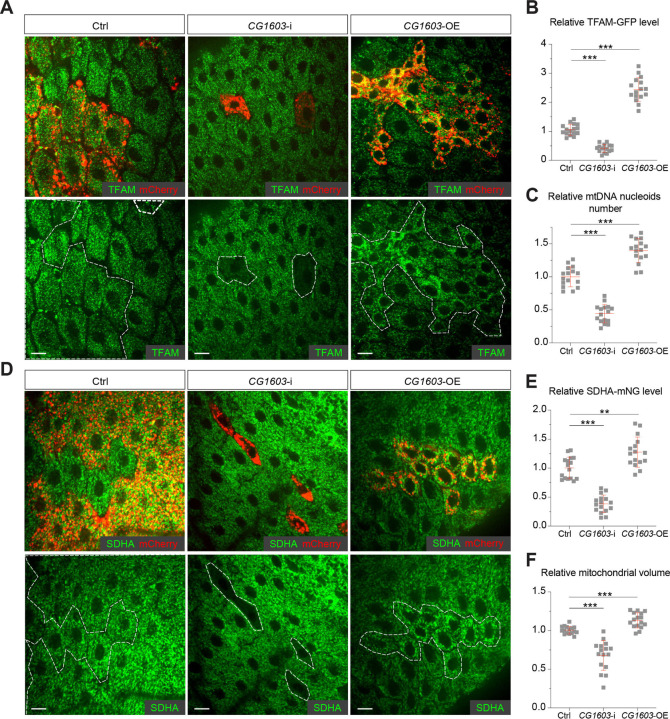
Figure 3. CG1603 promotes ETC gene expression and mitochondrial biogenesis. (A, D) Representative images of control RNAi (Ctrl), CG1603 RNAi (CG1603-i) and CG1603 overexpression (CG1603-OE) midgut EC clones with endogenously expressed TFAM-GFP (A) or SDHA-mNG (D) visualized in green. Clones were labeled by mCherry red and compared with wt neighbors. White dash lines aided in illustrating clones. Scale bars: 10 µm. (B, C, E, F) Quantification of the relative TFAM-GFP level (B), the relative mtDNA nucleoids number (C), the relative SDHA-mNG level (E), and the relative mitochondrial volume (F) in the EC clones to their wt neighbors. n=16 from 8 midguts for each group, error bar: SD. **: p<0.01, ***: p<0.001.
Figure 3. CG1603 promotes ETC gene expression and mitochondrial biogenesis. (A, D) Representative images of control RNAi (Ctrl), CG1603 RNAi (CG1603-i) and CG1603 overexpression (CG1603-OE) midgut EC clones with endogenously expressed TFAM-GFP (A) or SDHA-mNG (D) visualized in green. Clones were labeled by mCherry red and compared with wt neighbors. White dash lines aided in illustrating clones. Scale bars: 10 µm. (B, C, E, F) Quantification of the relative TFAM-GFP level (B), the relative mtDNA nucleoids number (C), the relative SDHA-mNG level (E), and the relative mitochondrial volume (F) in the EC clones to their wt neighbors. n=16 from 8 midguts for each group, error bar: SD. **: p<0.01, ***: p<0.001.CC BY-NC-ND · PubMed Central
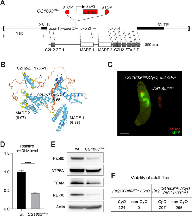
Figure 4. CG1603 gene model, product, mutant and the genomic DNA transgene. (A) Schematic representation of CG1603 genomic locus, showing the CG1603 transcript (5’ and 3’UTR in black bar and four exons in white), its protein product (586 amino acids in length, and characterized by seven C2H2-ZF and two MADF domains), the CG1603PBac mutant allele (with a piggyBac insertion in the second intron, which is marked by fluorescent DsRed driven by an eye-specific 3xP3 promoter and flanked by stop codons in all three reading frames terminating translation through downstream), and the genomic region (in square brackets, from 955 bp upstream of the CG1603 5’UTR to 656 bp downstream of CG1603 3’UTR) used for P[CG1603gDNA] transgene. (B) Predicted 3D structure of the CG1603 protein by AlphaFold. Labels indicate the N-and C-terminus, as well as the specific protein domains along with their predicted isoelectric point (pI). (C) Images of CG1603PBac / CyO, Act-GFP and homozygous CG1603PBac larvae cultured together at 25°C, day 4 after egg laying. Green: GFP; Red: DsRed. Scale bars: 1 mm. (D) Relative mtDNA levels in CG1603PBac mutant larvae to wild type (wt) control. n= 3, error bar: SD. ***: p<0.001. (E) Western blots of mitochondrial proteins in CG1603PBac mutant larvae to wild type (wt) control. (F) P[CG1603gDNA] restored viability of CG1603PBacflies. The number of progenies for each genotype is listed.
Figure 4. CG1603 gene model, product, mutant and the genomic DNA transgene. (A) Schematic representation of CG1603 genomic locus, showing the CG1603 transcript (5’ and 3’UTR in black bar and four exons in white), its protein product (586 amino acids in length, and characterized by seven C2H2-ZF and two MADF domains), the CG1603PBac mutant allele (with a piggyBac insertion in the second intron, which is marked by fluorescent DsRed driven by an eye-specific 3xP3 promoter and flanked by stop codons in all three reading frames terminating translation through downstream), and the genomic region (in square brackets, from 955 bp upstream of the CG1603 5’UTR to 656 bp downstream of CG1603 3’UTR) used for P[CG1603gDNA] transgene. (B) Predicted 3D structure of the CG1603 protein by AlphaFold. Labels indicate the N-and C-terminus, as well as the specific protein domains along with their predicted isoelectric point (pI). (C) Images of CG1603PBac / CyO, Act-GFP and homozygous CG1603PBac larvae cultured together at 25°C, day 4 after egg laying. Green: GFP; Red: DsRed. Scale bars: 1 mm. (D) Relative mtDNA levels in CG1603PBac mutant larvae to wild type (wt) control. n= 3, error bar: SD. ***: p<0.001. (E) Western blots of mitochondrial proteins in CG1603PBac mutant larvae to wild type (wt) control. (F) P[CG1603gDNA] restored viability of CG1603PBacflies. The number of progenies for each genotype is listed.CC BY-NC-ND · PubMed Central
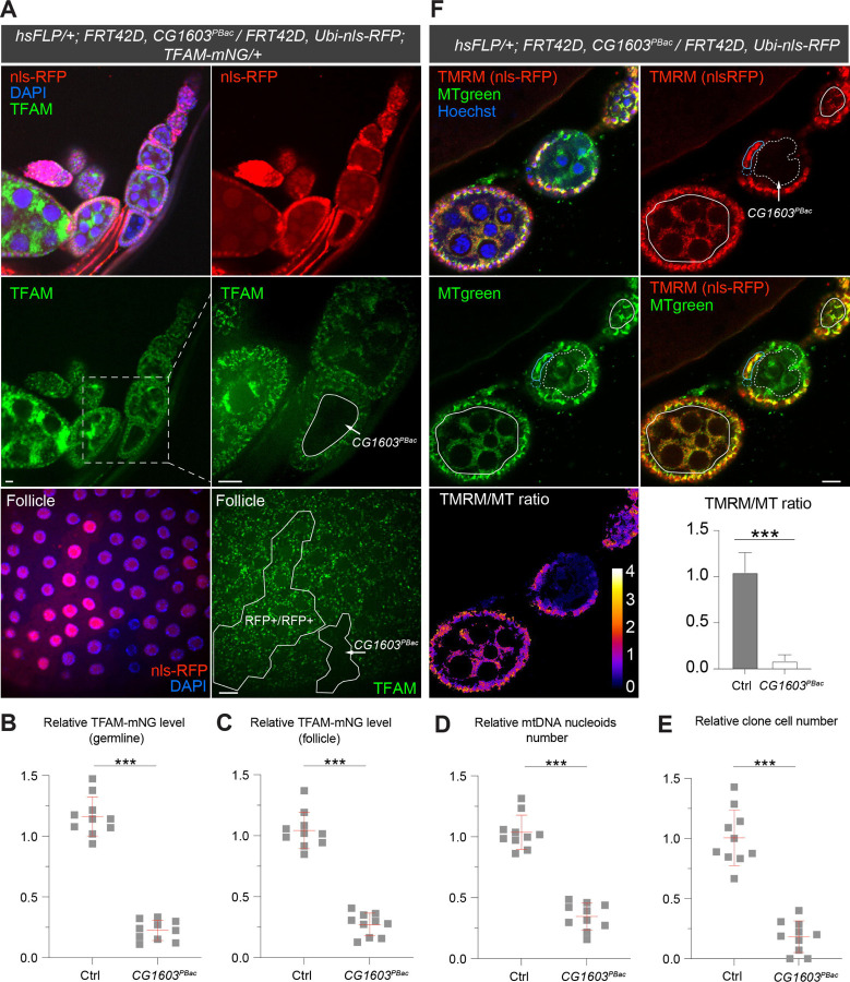
Figure 5. Clonal analyses confirmed CG1603’s role in mitochondrial biogenesis and activity. (A) Representative images of CG1603PBac mutant germline (top and middle panel) and follicle (bottom panel) clones in late-stage egg chambers of adult ovaries with endogenously expressed TFAM-mNG visualized in green. Homozygous mutant clones were marked by the absence of RFP and compared with either flanking RFP-positive cysts (germline) or homozygous wt twin (follicle). White dash lines aided in illustrating clones. The wt (RFP+/RFP+) follicle clone showed markedly higher RFP intensity than the heterozygous (RFP+/RFP-) cells, also see Figure 5—figure supplement B. Red: nls-RFP; Blue: DAPI. Scale bars: 10 μm. (B) Quantification of the relative TFAM-mNG level in the homozygous FRT42D control and CG1603PBac mutant germline clones in early-stage egg chamber to their anterior flanking RFP-positive cysts within the same ovariole. also see Figure 5—figure supplement A. n=10 for each group, error bar: SD. ***: p<0.001. (C-E) Quantification of the relative TFAM-mNG level (C), the relative mtDNA nucleoids number (D) and the relative clone cell number (E) in the homozygous FRT42D control and CG1603PBac mutant follicle clones to their wt twins. n=10 for each group, error bar: SD. ***: p<0.001. (F) TMRM / MitoTracker Green (MT) ratiometric live imaging and quantification of ovarioles containing homozygous CG1603PBac mutant germline clones (highlighted by white dash lines). Notably, in contrast to flanking control cysts (highlighted by white lines), Δψm was almost absent in mutant clones. Please note that compared to TMRM, nls-RFP signal was too low to be detected in ratiometric imaging. Nonetheless, the nls-RFP was readily detected in control cysts, but not in homozygous CG1603PBac clones, via visual observation, as depicted in Figure 5A and Figure 5—figure supplement. A twin pair of follicle clones in the same egg chamber were also highlighted (cyan line for control and cyan dash line for homozygous CG1603PBac mutant). The MT intensity was reduced in both the germline and follicle CG1603PBac clones, compared to germ cells in adjacent egg chambers and follicle cells in the same egg chamber, respectively. Quantification with background correction for MT intensity in germline clones is shown in Figure 5—figure supplement C. Blue: Hoechst. Scale bars: 10 µm. n= 8, error bar: SD. ***: p<0.001.
Figure 5. Clonal analyses confirmed CG1603’s role in mitochondrial biogenesis and activity. (A) Representative images of CG1603PBac mutant germline (top and middle panel) and follicle (bottom panel) clones in late-stage egg chambers of adult ovaries with endogenously expressed TFAM-mNG visualized in green. Homozygous mutant clones were marked by the absence of RFP and compared with either flanking RFP-positive cysts (germline) or homozygous wt twin (follicle). White dash lines aided in illustrating clones. The wt (RFP+/RFP+) follicle clone showed markedly higher RFP intensity than the heterozygous (RFP+/RFP-) cells, also see Figure 5—figure supplement B. Red: nls-RFP; Blue: DAPI. Scale bars: 10 μm. (B) Quantification of the relative TFAM-mNG level in the homozygous FRT42D control and CG1603PBac mutant germline clones in early-stage egg chamber to their anterior flanking RFP-positive cysts within the same ovariole. also see Figure 5—figure supplement A. n=10 for each group, error bar: SD. ***: p<0.001. (C-E) Quantification of the relative TFAM-mNG level (C), the relative mtDNA nucleoids number (D) and the relative clone cell number (E) in the homozygous FRT42D control and CG1603PBac mutant follicle clones to their wt twins. n=10 for each group, error bar: SD. ***: p<0.001. (F) TMRM / MitoTracker Green (MT) ratiometric live imaging and quantification of ovarioles containing homozygous CG1603PBac mutant germline clones (highlighted by white dash lines). Notably, in contrast to flanking control cysts (highlighted by white lines), Δψm was almost absent in mutant clones. Please note that compared to TMRM, nls-RFP signal was too low to be detected in ratiometric imaging. Nonetheless, the nls-RFP was readily detected in control cysts, but not in homozygous CG1603PBac clones, via visual observation, as depicted in Figure 5A and Figure 5—figure supplement. A twin pair of follicle clones in the same egg chamber were also highlighted (cyan line for control and cyan dash line for homozygous CG1603PBac mutant). The MT intensity was reduced in both the germline and follicle CG1603PBac clones, compared to germ cells in adjacent egg chambers and follicle cells in the same egg chamber, respectively. Quantification with background correction for MT intensity in germline clones is shown in Figure 5—figure supplement C. Blue: Hoechst. Scale bars: 10 µm. n= 8, error bar: SD. ***: p<0.001.CC BY-NC-ND · PubMed Central
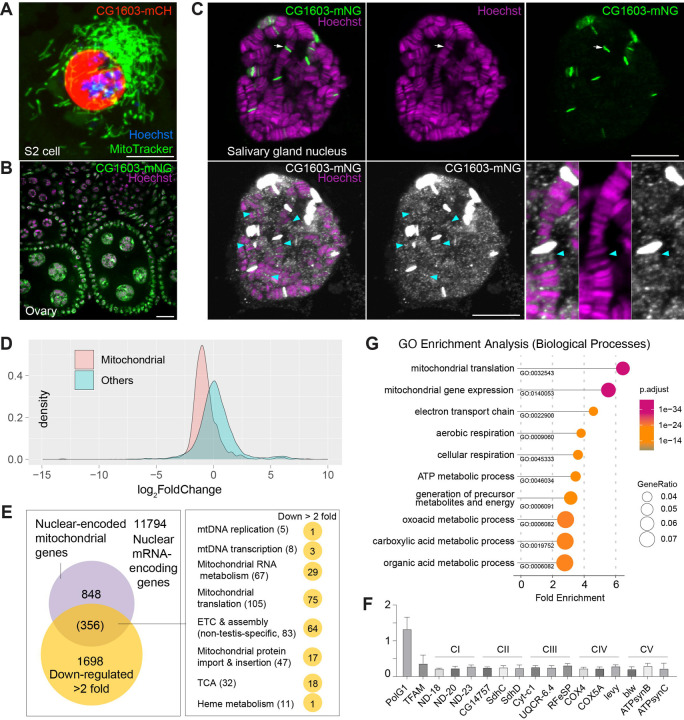
Figure 6. CG1603 localizes in the nucleus and is essential for regulating nuclear mitochondrial gene expression. (A-B) Representative images showing nuclear localization of CG1603 protein in cultured S2 cell (A) and adult ovary (B). Green: MitoTracker Green in S2 cell, and CG1603-mNG in tissues; Red: CG1603-mCH; Blue & Magenta: Hoechst. Scale bars: 10 μm. (C) Representative images showing bindings of endogenously expressed CG1603 proteins to less condensed euchromatin regions in the polytene chromosomes of salivary gland. High intensity CG1603-mNG bands were visualized in green in upper panel as indicated by arrow, and low intensity bands were visualized in white in lower panel as indicated by arrow heads with image B&C adjusted. Magenta: Hoechst. Scale bars: 10 μm. (D) Density plot illustrating the distribution of expression changes of the nuclear-encoded mitochondrial and non-mitochondrial genes in CG1603PBac mutant. (E) Graph illustrating the overlap between nuclear-encoded mitochondrial genes and differentially expressed genes (DEGs) that down-regulated > 2-fold, as well as the distribution of the overlapped genes in different mitochondrial function categories. (F) Relative mRNA levels of several ETC biogenesis-related genes in CG1603PBac mutant larvae to control, measured by real-time PCR. n= 3, error bar: SD. (G) Gene Ontology (GO) enrichment analyses of DEGs that down-regulated > 2-fold. The top 10 enriched biological processes are shown.
Figure 6. CG1603 localizes in the nucleus and is essential for regulating nuclear mitochondrial gene expression. (A-B) Representative images showing nuclear localization of CG1603 protein in cultured S2 cell (A) and adult ovary (B). Green: MitoTracker Green in S2 cell, and CG1603-mNG in tissues; Red: CG1603-mCH; Blue & Magenta: Hoechst. Scale bars: 10 μm. (C) Representative images showing bindings of endogenously expressed CG1603 proteins to less condensed euchromatin regions in the polytene chromosomes of salivary gland. High intensity CG1603-mNG bands were visualized in green in upper panel as indicated by arrow, and low intensity bands were visualized in white in lower panel as indicated by arrow heads with image B&C adjusted. Magenta: Hoechst. Scale bars: 10 μm. (D) Density plot illustrating the distribution of expression changes of the nuclear-encoded mitochondrial and non-mitochondrial genes in CG1603PBac mutant. (E) Graph illustrating the overlap between nuclear-encoded mitochondrial genes and differentially expressed genes (DEGs) that down-regulated > 2-fold, as well as the distribution of the overlapped genes in different mitochondrial function categories. (F) Relative mRNA levels of several ETC biogenesis-related genes in CG1603PBac mutant larvae to control, measured by real-time PCR. n= 3, error bar: SD. (G) Gene Ontology (GO) enrichment analyses of DEGs that down-regulated > 2-fold. The top 10 enriched biological processes are shown.CC BY-NC-ND · PubMed Central
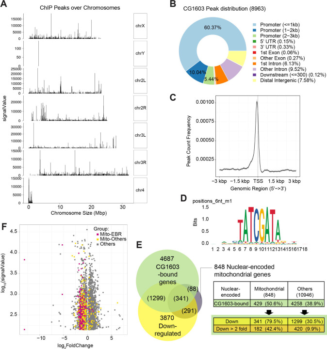
Figure 7. ChIP analysis identified nuclear mitochondrial genes that may be directly regulated by CG1603. (A) CG1603 ChIP peaks over all chromosomes. (B) Genomic distribution of CG1603 peaks. (C) Average profile of CG1603 peaks binding to transcription start site (TSS) regions. (D) Representative binding motif discovered with CG1603 ChIP peaks. (E) Summary of the number of nuclear-encoded mitochondrial and non-mitochondrial mRNA coding genes bound by CG1603, and the overlapping down-regulated differentially expressed genes (DEGs) in each group. (F) Scatterplot illustrating the signalValue of CG1603 ChIP peaks (y-axis) and log2 fold change in expression of DEGs between CG1603PBac mutant and control (x-axis). MitoES: the genes belong to the categories that are clearly essential to mitochondrial ETC biogenesis and maintenance, including ETC subunits and assembly factors, mtDNA replication and transcription, mitochondrial RNA metabolism and translation, as well as mitochondrial protein import and membrane insertion machinery.
Figure 7. ChIP analysis identified nuclear mitochondrial genes that may be directly regulated by CG1603. (A) CG1603 ChIP peaks over all chromosomes. (B) Genomic distribution of CG1603 peaks. (C) Average profile of CG1603 peaks binding to transcription start site (TSS) regions. (D) Representative binding motif discovered with CG1603 ChIP peaks. (E) Summary of the number of nuclear-encoded mitochondrial and non-mitochondrial mRNA coding genes bound by CG1603, and the overlapping down-regulated differentially expressed genes (DEGs) in each group. (F) Scatterplot illustrating the signalValue of CG1603 ChIP peaks (y-axis) and log2 fold change in expression of DEGs between CG1603PBac mutant and control (x-axis). MitoES: the genes belong to the categories that are clearly essential to mitochondrial ETC biogenesis and maintenance, including ETC subunits and assembly factors, mtDNA replication and transcription, mitochondrial RNA metabolism and translation, as well as mitochondrial protein import and membrane insertion machinery.CC BY-NC-ND · PubMed Central
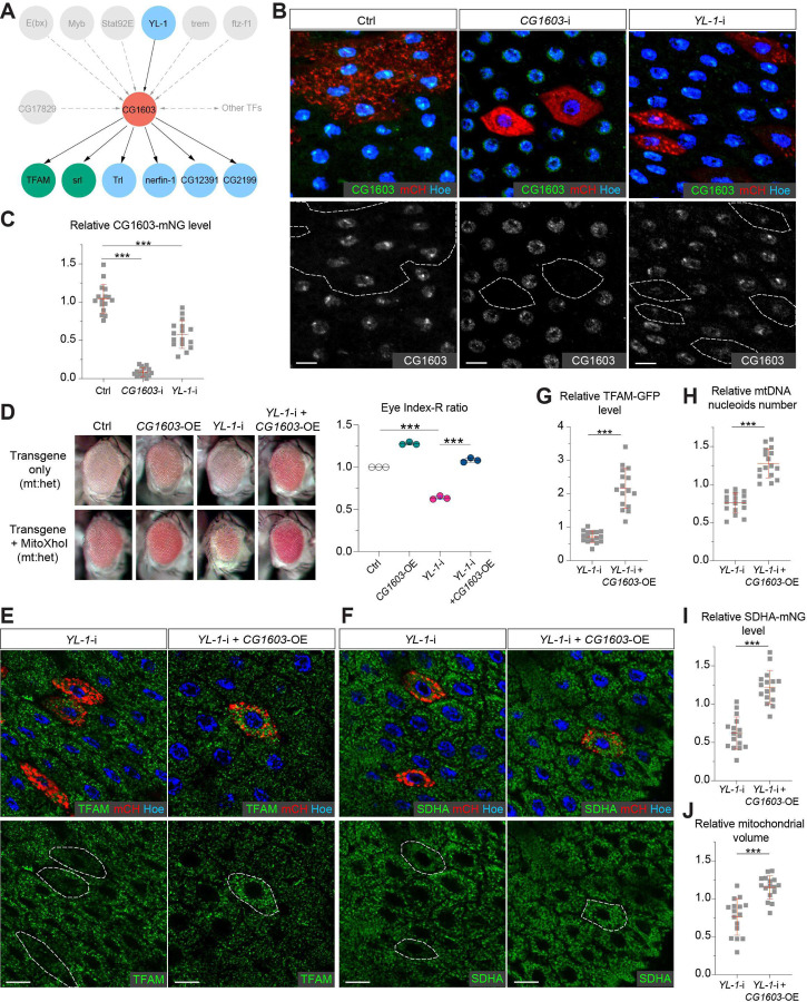
Figure 8. YL-1 is an upstream regulator of CG1603. (A) Schematic graph illustrating the CG1603 upstream and downstream (co-)TFs involved in regulating mitochondrial ETC biogenesis, inferred from ChIP-seq, RNAseq and genetics data. (B) Representative images of control RNAi (Ctrl), CG1603 RNAi (CG1603-i) and YL-1 RNAi (YL-1-i) midgut EC clones with endogenously expressed CG1603-mNG visualized in green or white. Clones were labeled by mCherry red and compared with wt neighbors. White dash lines aided in illustrating clones. Blue: Hoechst. Scale bars: 10 μm. (C) Quantification of the relative CG1603-mNG level in the EC clones to their wt neighbors. n=16 from 8 midguts for each group, error bar: SD. ***: p<0.001. (D) Representative eye image and Index-R ratio (RNAi + mitoXhoI / RNAi-only) of adult flies with indicated genotypes. Three biological repeats were performed for each group, error bar: SD. ***: p<0.001. (E-F) Representative images of YL-1 RNAi (YL-1-i) and YL-1 RNAi + CG1603 overexpression (YL-1-i + CG1603-OE) midgut EC clones with endogenously expressed TFAM-GFP (E) or SDHA-mNG (F) visualized in green. Clones were labeled by mCherry red and compared with wt neighbors. Blue: Hoechst. Scale bars: 10 µm. (G-J) Quantification of the relative TFAM-GFP level (G), the relative mtDNA nucleoids number (H), the relative SDHA-mNG level (I) and the relative mitochondrial volume (J) in the EC clones to their wt neighbors. n=16 from 8 midguts for each group, error bar: SD. ***: p<0.001.
Figure 8. YL-1 is an upstream regulator of CG1603. (A) Schematic graph illustrating the CG1603 upstream and downstream (co-)TFs involved in regulating mitochondrial ETC biogenesis, inferred from ChIP-seq, RNAseq and genetics data. (B) Representative images of control RNAi (Ctrl), CG1603 RNAi (CG1603-i) and YL-1 RNAi (YL-1-i) midgut EC clones with endogenously expressed CG1603-mNG visualized in green or white. Clones were labeled by mCherry red and compared with wt neighbors. White dash lines aided in illustrating clones. Blue: Hoechst. Scale bars: 10 μm. (C) Quantification of the relative CG1603-mNG level in the EC clones to their wt neighbors. n=16 from 8 midguts for each group, error bar: SD. ***: p<0.001. (D) Representative eye image and Index-R ratio (RNAi + mitoXhoI / RNAi-only) of adult flies with indicated genotypes. Three biological repeats were performed for each group, error bar: SD. ***: p<0.001. (E-F) Representative images of YL-1 RNAi (YL-1-i) and YL-1 RNAi + CG1603 overexpression (YL-1-i + CG1603-OE) midgut EC clones with endogenously expressed TFAM-GFP (E) or SDHA-mNG (F) visualized in green. Clones were labeled by mCherry red and compared with wt neighbors. Blue: Hoechst. Scale bars: 10 µm. (G-J) Quantification of the relative TFAM-GFP level (G), the relative mtDNA nucleoids number (H), the relative SDHA-mNG level (I) and the relative mitochondrial volume (J) in the EC clones to their wt neighbors. n=16 from 8 midguts for each group, error bar: SD. ***: p<0.001.CC BY-NC-ND · PubMed Central

Verification

Claims the paper makes

The claims and statistics of this paper haven’t been checked yet.

Run the check — free, no account needed. It reads the open-access text and checks the sources it cites.

A few free checks a day · papers already verified open instantly · results are public

Self-check

Percentages, sums and reported differences re-computed from the paper’s own numbers.

The self-check needs the paper’s full text.

Citations

Cites

Research works printed but not linked: 116

  1. Acosta-Alvear, 2007, XBP1 controls diverse cell type- and condition-specific transcriptional regulatory networks, Molecular Cell, 27, 53, 10.1016/j.molcel.2007.06.011 · DOI
  2. Alam, 2003, Human mitochondrial DNA is packaged with TFAM, Nucleic Acids Research, 31, 1640, 10.1093/nar/gkg251 · DOI
  3. Angelini, 2014, Understanding gene regulatory mechanisms by integrating ChIP-seq and RNA-seq data: statistical solutions to biological problems, Frontiers in Cell and Developmental Biology, 2, 10.3389/fcell.2014.00051 · DOI
  4. Berger, 2002, The mechanisms of action of PPARs, Annual Review of Medicine, 53, 409, 10.1146/annurev.med.53.082901.104018 · DOI · Paper page
  5. Boyer, 2004, The SANT domain: a unique histone-tail-binding module?, Nature Reviews. Molecular Cell Biology, 5, 158, 10.1038/nrm1314 · DOI
  6. Chen, 2015, Genetic mosaic analysis of a deleterious mitochondrial DNA mutation in Drosophila reveals novel aspects of mitochondrial regulation and function, Molecular Biology of the Cell, 26, 674, 10.1091/mbc.E14-11-1513 · DOI
  7. Chen, 2019, Human mitochondrial DNA diseases and Drosophila models, Journal of Genetics and Genomics = Yi Chuan Xue Bao, 46, 201, 10.1016/j.jgg.2019.03.009 · DOI
  8. Chen, 2020, Mitochondrial DNA segregation and replication restrict the transmission of detrimental mutation, The Journal of Cell Biology, 219, 10.1083/jcb.201905160 · DOI
  9. Couvillion, 2016, Synchronized mitochondrial and cytosolic translation programs, Nature, 533, 499, 10.1038/nature18015 · DOI
  10. Deblois, 2011, Functional and physiological genomics of estrogen-related receptors (ERRs) in health and disease, Biochimica et Biophysica Acta, 1812, 1032, 10.1016/j.bbadis.2010.12.009 · DOI
  11. Dobin, 2013, STAR: ultrafast universal RNA-seq aligner, Bioinformatics, 29, 15, 10.1093/bioinformatics/bts635 · DOI · Paper page
  12. Eisen, 1998, Cluster analysis and display of genome-wide expression patterns, PNAS, 95, 14863, 10.1073/pnas.95.25.14863 · DOI · Paper page
  13. Fedotova, 2017, C2H2 zinc finger proteins: the largest but poorly explored family of higher eukaryotic transcription factors, Acta Naturae, 9, 47, 10.32607/20758251-2017-9-2-47-58 · DOI
  14. Fernández-Vizarra, 2011, Tissue-specific differences in mitochondrial activity and biogenesis, Mitochondrion, 11, 207, 10.1016/j.mito.2010.09.011 · DOI
  15. Finck, 2007, Peroxisome proliferator-activated receptor gamma coactivator-1 (PGC-1) regulatory cascade in cardiac physiology and disease, Circulation, 115, 2540, 10.1161/CIRCULATIONAHA.107.670588 · DOI
  16. Gorman, 2016, Mitochondrial diseases, Nature Reviews. Disease Primers, 2, 10.1038/nrdp.2016.80 · DOI
  17. Hill, 2014, Selective propagation of functional mitochondrial DNA during oogenesis restricts the transmission of a deleterious mitochondrial variant, Nature Genetics, 46, 389, 10.1038/ng.2920 · DOI
  18. Hock, 2009, Transcriptional control of mitochondrial biogenesis and function, Annual Review of Physiology, 71, 177, 10.1146/annurev.physiol.010908.163119 · DOI
  19. Jiang, 2018, Integrating ChIP-seq with other functional genomics data, Briefings in Functional Genomics, 17, 104, 10.1093/bfgp/ely002 · DOI
  20. Jothi, 2009, Genomic analysis reveals a tight link between transcription factor dynamics and regulatory network architecture, Molecular Systems Biology, 5, 10.1038/msb.2009.52 · DOI
  21. Kudron, 2018, The ModERN Resource: Genome-wide binding profiles for hundreds of Drosophila and Caenorhabditis elegans transcription factors, Genetics, 208, 937, 10.1534/genetics.117.300657 · DOI
  22. Kurland, 2000, Origin and evolution of the mitochondrial proteome, Microbiology and Molecular Biology Reviews, 64, 786, 10.1128/MMBR.64.4.786-820.2000 · DOI
  23. Laws, 2015, Genetic mosaic analysis of stem cell lineages in the Drosophila Ovary, Methods in Molecular Biology, 1328, 57, 10.1007/978-1-4939-2851-4_4 · DOI
  24. Li, 2009, Fast and accurate short read alignment with Burrows-Wheeler transform, Bioinformatics, 25, 1754, 10.1093/bioinformatics/btp324 · DOI · Paper page
  25. Li, 2009, The sequence alignment/map format and samtools, Bioinformatics, 25, 2078, 10.1093/bioinformatics/btp352 · DOI · Paper page
  26. Liang, 2016, Structural basis of H2A.Z recognition by SRCAP chromatin-remodeling subunit YL1, Nature Structural & Molecular Biology, 23, 317, 10.1038/nsmb.3190 · DOI
  27. Liao, 2006, An efficient genetic screen in Drosophila to identify nuclear-encoded genes with mitochondrial function, Genetics, 174, 525, 10.1534/genetics.106.061705 · DOI
  28. Liu, 2022, The PPR domain of mitochondrial RNA polymerase is an exoribonuclease required for mtDNA replication in Drosophila melanogaster, Nature Cell Biology, 24, 757, 10.1038/s41556-022-00887-y · DOI
  29. Love, 2014, Moderated estimation of fold change and dispersion for RNA-seq data with DESeq2, Genome Biology, 15, 10.1186/s13059-014-0550-8 · DOI · Paper page
  30. Lowden, 2021, Homeostatic control of nuclear-encoded mitochondrial gene expression by the histone variant H2A.Z is essential for neuronal survival, Cell Reports, 36, 10.1016/j.celrep.2021.109704 · DOI
  31. Maheshwari, 2008, Recurrent positive selection of the Drosophila hybrid incompatibility gene Hmr, Molecular Biology and Evolution, 25, 2421, 10.1093/molbev/msn190 · DOI
  32. Mandal, 2005, Mitochondrial regulation of cell cycle progression during development as revealed by the tenured mutation in Drosophila, Developmental Cell, 9, 843, 10.1016/j.devcel.2005.11.006 · DOI
  33. mforde84. 2016. ENCODE_TF_ChIP_pipeline. Software Heritage. swh:1:rev:e2c7dc403d6d44a8eb667c20b9bf45e091d27134. https://archive.softwareheritage.org/swh:1:dir:5639bfed7102cc98642baf0f340782b309fcb56d;origin=https://github.com/mforde84/ENCODE_TF_ChIP_pipeline;visit=swh:1:snp:df73736416d2b812d0676475f5f6436bedad3f3d;anchor=swh:1:rev:e2c7dc403d6d44a8eb667c20b9bf45e091d27134.
  34. Owusu-Ansah, 2008, Distinct mitochondrial retrograde signals control the G1-S cell cycle checkpoint, Nature Genetics, 40, 356, 10.1038/ng.2007.50 · DOI
  35. Park, 2009, ChIP-seq: advantages and challenges of a maturing technology, Nature Reviews. Genetics, 10, 669, 10.1038/nrg2641 · DOI · Paper page
  36. Perez-Torrado, 2006, Born to bind: the BTB protein-protein interaction domain, BioEssays, 28, 1194, 10.1002/bies.20500 · DOI
  37. Persikov, 2015, A systematic survey of the Cys2His2 zinc finger DNA-binding landscape, Nucleic Acids Research, 43, 1965, 10.1093/nar/gku1395 · DOI
  38. Prober, 2000, Ras1 promotes cellular growth in the Drosophila wing, Cell, 100, 435, 10.1016/s0092-8674(00)80679-0 · DOI
  39. Putri, 2022, Analysing high-throughput sequencing data in Python with HTSeq 2.0, Bioinformatics, 38, 2943, 10.1093/bioinformatics/btac166 · DOI
  40. Rath, 2021, MitoCarta3.0: an updated mitochondrial proteome now with sub-organelle localization and pathway annotations, Nucleic Acids Research, 49, D1541, 10.1093/nar/gkaa1011 · DOI · Paper page
  41. Rouyer, 1997, A new gene encoding A putative transcription factor regulated by the Drosophila circadian clock, The EMBO Journal, 16, 3944, 10.1093/emboj/16.13.3944 · DOI
  42. Scarpulla, 2008, Transcriptional paradigms in mammalian mitochondrial biogenesis and function, Physiological Reviews, 88, 611, 10.1152/physrev.00025.2007 · DOI · Paper page
  43. Scarpulla, 2012, Transcriptional integration of mitochondrial biogenesis, Trends in Endocrinology and Metabolism, 23, 459, 10.1016/j.tem.2012.06.006 · DOI · Paper page
  44. Schuldiner, 2008, piggyBac-based mosaic screen identifies a postmitotic function for cohesin in regulating developmental axon pruning, Developmental Cell, 14, 227, 10.1016/j.devcel.2007.11.001 · DOI
  45. Schultz, 2000, SMART: a web-based tool for the study of genetically mobile domains, Nucleic Acids Research, 28, 231, 10.1093/nar/28.1.231 · DOI
  46. Shen, 2022, Mitochondria as cellular and organismal signaling hubs, Annual Review of Cell and Developmental Biology, 38, 179, 10.1146/annurev-cellbio-120420-015303 · DOI
  47. Sobocińska, 2021, KRAB-ZFP transcriptional regulators acting as oncogenes and tumor suppressors: an overview, International Journal of Molecular Sciences, 22, 10.3390/ijms22042212 · DOI
  48. Spinelli, 2018, The multifaceted contributions of mitochondria to cellular metabolism, Nature Cell Biology, 20, 745, 10.1038/s41556-018-0124-1 · DOI · Paper page
  49. Taylor, 2005, Mitochondrial DNA mutations in human disease, Nature Reviews. Genetics, 6, 389, 10.1038/nrg1606 · DOI
  50. Thomas-Chollier, 2012, A complete workflow for the analysis of full-size ChIP-seq (and similar) data sets using peak-motifs, Nature Protocols, 7, 1551, 10.1038/nprot.2012.088 · DOI
  51. Verheul, 2020, The Why of YY1: Mechanisms of transcriptional regulation by yin yang 1, Frontiers in Cell and Developmental Biology, 8, 10.3389/fcell.2020.592164 · DOI
  52. Wang, 2019, Electron transport chain biogenesis activated by a JNK-insulin-Myc relay primes mitochondrial inheritance in Drosophila, eLife, 8, 10.7554/eLife.49309 · DOI
  53. Wang, 2023, Mechanical stimulation from the surrounding tissue activates mitochondrial energy metabolism in Drosophila differentiating germ cells, Developmental Cell, 58, 2249, 10.1016/j.devcel.2023.08.007 · DOI
  54. Wiedemann, 2017, Mitochondrial machineries for protein import and assembly, Annual Review of Biochemistry, 86, 685, 10.1146/annurev-biochem-060815-014352 · DOI
  55. Wolfe, 2000, DNA recognition by Cys2His2 zinc finger proteins, Annual Review of Biophysics and Biomolecular Structure, 29, 183, 10.1146/annurev.biophys.29.1.183 · DOI · Paper page
  56. Wysocka, 2006, A PHD finger of NURF couples histone H3 lysine 4 trimethylation with chromatin remodelling, Nature, 442, 86, 10.1038/nature04815 · DOI
  57. Xu, 2008, Manipulating the metazoan mitochondrial genome with targeted restriction enzymes, Science, 321, 575, 10.1126/science.1160226 · DOI
  58. Xu, 2021, The SRCAP chromatin remodeling complex promotes oxidative metabolism during prenatal heart development, Development, 148, 10.1242/dev.199026 · DOI
  59. Yu, 2012, clusterProfiler: an R package for comparing biological themes among gene clusters, OMICS, 16, 284, 10.1089/omi.2011.0118 · DOI · Paper page
  60. Yu, 2015, ChIPseeker: an R/Bioconductor package for ChIP peak annotation, comparison and visualization, Bioinformatics, 31, 2382, 10.1093/bioinformatics/btv145 · DOI
  61. Zhang, 2008, Model-based analysis of chip-seq (MACS), Genome Biology, 9, 10.1186/gb-2008-9-9-r137 · DOI · Paper page
  62. Zhang, 2015, The cAMP phosphodiesterase Prune localizes to the mitochondrial matrix and promotes mtDNA replication by stabilizing TFAM, EMBO Reports, 16, 520, 10.15252/embr.201439636 · DOI
  63. Zhang, 2016, The mitochondrial outer membrane protein MDI promotes local protein synthesis and mtDNA replication, The EMBO Journal, 35, 1045, 10.15252/embj.201592994 · DOI
  64. Zhang, 2019, PINK1 inhibits local protein synthesis to limit transmission of deleterious mitochondrial DNA mutations, Molecular Cell, 73, 1127, 10.1016/j.molcel.2019.01.013 · DOI
  65. Zhang, 2020, Mitochondria regulate intestinal stem cell proliferation and epithelial homeostasis through FOXO, Molecular Biology of the Cell, 31, 1538, 10.1091/mbc.E19-10-0560 · DOI
  66. Zhou, 2016, Computational prediction of the global functional genomic landscape: applications, methods, and challenges, Human Heredity, 81, 88, 10.1159/000450827 · DOI
  67. Zhang F, Xu H. 2024. CG1603 regulation of nuclear-encoded mitochondrial gene expression. NCBI Gene Expression Omnibus. https://www.ncbi.nlm.nih.gov/geo/query/acc.cgi?acc=GSE282638. GSE282638.
  68. modENCODEProject. 2016. TF ChIP-seq_ab. ENCODE. https://www.encodeproject.org/experiments/ENCSR140RMO. ENCSR140RMO.
  69. modENCODEProject. 2019. TF ChIP-seq_abd-A. ENCODE. https://www.encodeproject.org/experiments/ENCSR609JDR. ENCSR609JDR.
  70. modENCODEProject. 2016. TF ChIP-seq_achi. ENCODE. https://www.encodeproject.org/experiments/ENCSR959SWC. ENCSR959SWC.
  71. modENCODEProject. 2016. TF ChIP-seq_bdp1. ENCODE. https://www.encodeproject.org/experiments/ENCSR504AYW. ENCSR504AYW.
  72. modENCODEProject. 2018. TF ChIP-seq_bigmax. ENCODE. https://www.encodeproject.org/experiments/ENCSR224YOT. ENCSR224YOT.
  73. modENCODEProject. 2016. TF ChIP-seq_CG10631. ENCODE. https://www.encodeproject.org/experiments/ENCSR751FSX. ENCSR751FSX.
  74. modENCODEProject. 2016. TF ChIP-seq_CG11398. ENCODE. https://www.encodeproject.org/experiments/ENCSR486MQI. ENCSR486MQI.
  75. modENCODEProject. 2018. TF ChIP-seq_CG12299. ENCODE. https://www.encodeproject.org/experiments/ENCSR028DHQ. ENCSR028DHQ.
  76. modENCODEProject. 2018. TF ChIP-seq_CG12391. ENCODE. https://www.encodeproject.org/experiments/ENCSR483VVZ. ENCSR483VVZ.
  77. modENCODEProject. 2018. TF ChIP-seq_CG15011. ENCODE. https://www.encodeproject.org/experiments/ENCSR608VFH. ENCSR608VFH.
  78. modENCODEProject. 2019. TF ChIP-seq_CG1603. ENCODE. https://www.encodeproject.org/experiments/ENCSR680NFF. ENCSR680NFF.
  79. modENCODEProject. 2016. TF ChIP-seq_CG16863. ENCODE. https://www.encodeproject.org/experiments/ENCSR848GYF. ENCSR848GYF.
  80. modENCODEProject. 2018. TF ChIP-seq_CG17806. ENCODE. https://www.encodeproject.org/experiments/ENCSR118TAH. ENCSR118TAH.
  81. modENCODEProject. 2019. TF ChIP-seq_CG17829. ENCODE. https://www.encodeproject.org/experiments/ENCSR810MLF. ENCSR810MLF.
  82. modENCODE. 2018. TF ChIP-seq_CG2116. ENCODE. https://www.encodeproject.org/experiments/ENCSR456UZW. ENCSR456UZW.
  83. modENCODEProject. 2018. TF ChIP-seq_CG4707. ENCODE. https://www.encodeproject.org/experiments/ENCSR302AQS. ENCSR302AQS.
  84. modENCODEProject. 2016. TF ChIP-seq_CG4854. ENCODE. https://www.encodeproject.org/experiments/ENCSR860FUA. ENCSR860FUA.
  85. modENCODEProject. 2017. TF ChIP-seq_CG6254. ENCODE. https://www.encodeproject.org/experiments/ENCSR233UXG. ENCSR233UXG.
  86. modENCODEProject. 2018. TF ChIP-seq_CG7987. ENCODE. https://www.encodeproject.org/experiments/ENCSR681RZY. ENCSR681RZY.
  87. modENCODEProject. 2018. TF ChIP-seq_CG9437. ENCODE. https://www.encodeproject.org/experiments/ENCSR965XSS. ENCSR965XSS.
  88. modENCODEProject. 2017. TF ChIP-seq_CG9727. ENCODE. https://www.encodeproject.org/experiments/ENCSR624PKN. ENCSR624PKN.
  89. modENCODEProject. 2016. TF ChIP-seq_CHES-1-like. ENCODE. https://www.encodeproject.org/experiments/ENCSR791TWT. ENCSR791TWT.
  90. modENCODEProject. 2016. TF ChIP-seq_Crg-1. ENCODE. https://www.encodeproject.org/experiments/ENCSR596YNC. ENCSR596YNC.
  91. modENCODEProject. 2017. TF ChIP-seq_CTCF. ENCODE. https://www.encodeproject.org/experiments/ENCSR661BEZ. ENCSR661BEZ.
  92. modENCODEProject. 2016. TF ChIP-seq_E(bx). ENCODE. https://www.encodeproject.org/experiments/ENCSR559AJG. ENCSR559AJG.
  93. modENCODEProject. 2019. TF ChIP-seq_Ets96B. ENCODE. https://www.encodeproject.org/experiments/ENCSR572JUN. ENCSR572JUN.
  94. modENCODEProject. 2017. TF ChIP-seq_ewg. ENCODE. https://www.encodeproject.org/experiments/ENCSR444JKK. ENCSR444JKK.
  95. modENCODEProject. 2018. TF ChIP-seq_Fer3. ENCODE. https://www.encodeproject.org/experiments/ENCSR367SBM. ENCSR367SBM.
  96. modENCODEProject. 2016. TF ChIP-seq_ftz-f1. ENCODE. https://www.encodeproject.org/experiments/ENCSR741KZZ. ENCSR741KZZ.
  97. modENCODEProject. 2016. TF ChIP-seq_ind. ENCODE. https://www.encodeproject.org/experiments/ENCSR486EPD. ENCSR486EPD.
  98. modENCODEProject. 2016. TF ChIP-seq_Jra. ENCODE. https://www.encodeproject.org/experiments/ENCSR471GSA. ENCSR471GSA.
  99. modENCODEProject. 2017. TF ChIP-seq_Lhr. ENCODE. https://www.encodeproject.org/experiments/ENCSR272DEA. ENCSR272DEA.
  100. modENCODEProject. 2016. TF ChIP-seq_luna. ENCODE. https://www.encodeproject.org/experiments/ENCSR770AUN. ENCSR770AUN.
  101. modENCODEProject. 2016. TF ChIP-seq_Met. ENCODE. https://www.encodeproject.org/experiments/ENCSR363SHZ. ENCSR363SHZ.
  102. modENCODEProject. 2016. TF ChIP-seq_Myb. ENCODE. https://www.encodeproject.org/experiments/ENCSR393AKW. ENCSR393AKW.
  103. modENCODEProject. 2017. TF ChIP-seq_Myc. ENCODE. https://www.encodeproject.org/experiments/ENCSR191VCQ. ENCSR191VCQ.
  104. modENCODEProject. 2019. TF ChIP-seq_nerfin-1. ENCODE. https://www.encodeproject.org/experiments/ENCSR335NNR. ENCSR335NNR.
  105. modENCODEProject. 2018. TF ChIP-seq_Nnk. ENCODE. https://www.encodeproject.org/experiments/ENCSR165KWP. ENCSR165KWP.
  106. modENCODEProject. 2016. TF ChIP-seq_p53. ENCODE. https://www.encodeproject.org/experiments/ENCSR808XNJ. ENCSR808XNJ.
  107. modENCODEProject. 2017. TF ChIP-seq_pan. ENCODE. https://www.encodeproject.org/experiments/ENCSR033IIP. ENCSR033IIP.
  108. modENCODEProject. 2019. TF ChIP-seq_pros. ENCODE. https://www.encodeproject.org/experiments/ENCSR682YQM. ENCSR682YQM.
  109. modENCODEProject. 2017. TF ChIP-seq_REPTOR-BP. ENCODE. https://www.encodeproject.org/experiments/ENCSR271ZJI. ENCSR271ZJI.
  110. modENCODEProject. 2016. TF ChIP-seq_salr. ENCODE. https://www.encodeproject.org/experiments/ENCSR042XCV. ENCSR042XCV.
  111. modENCODEProject. 2019. TF ChIP-seq_Stat92E. ENCODE. https://www.encodeproject.org/experiments/ENCSR290OJD. ENCSR290OJD.
  112. modENCODEProject. 2017. TF ChIP-seq_su(Hw). ENCODE. https://www.encodeproject.org/experiments/ENCSR761TCG. ENCSR761TCG.
  113. modENCODEProject. 2019. TF ChIP-seq_trem. ENCODE. https://www.encodeproject.org/experiments/ENCSR104XOO. ENCSR104XOO.
  114. modENCODEProject. 2016. TF ChIP-seq_Trl. ENCODE. https://www.encodeproject.org/experiments/ENCSR629WQT. ENCSR629WQT.
  115. modENCODEProject. 2016. TF ChIP-seq_Xbp1. ENCODE. https://www.encodeproject.org/experiments/ENCSR698IEF. ENCSR698IEF.
  116. modENCODEProject. 2017. TF ChIP-seq_YL-1. ENCODE. https://www.encodeproject.org/experiments/ENCSR396LMJ. ENCSR396LMJ.

Metadata

Authors
Zhang, F., Lee, A., Freitas, A. V., Herb, J. T., Wang, Z.-H., Gupta, S., Chen, Z., & Xu, H.
Affiliations
  • National Institutes of Health (US)
  • National Heart, Lung, and Blood Institute, National Institutes of Health
Corresponding author
Xu, Hong
Venue
eLife
Published
2024-12-27
Volume
13
ISSN
2050-084X (ISSN-L)
OpenAlex
W4405830359
Cited by
8
References
2
Citations by year
2026: 3 · 2025: 5
FWCI
1.09
Type
journal article
Language
en
Open access
Locations
  • eLife (publishedVersion, cc-by)
  • bioRxiv (Cold Spring Harbor Laboratory) (acceptedVersion, cc-by-nc-nd)
  • PubMed (publishedVersion)
  • PubMed Central (submittedVersion, cc-by-nc-nd)
Funders
National Heart, Lung, and Blood Institute
Topics
  • Life Sciences › Biochemistry, Genetics and Molecular Biology › Molecular Biology › Mitochondrial Function and Pathology
  • Life Sciences › Biochemistry, Genetics and Molecular Biology › Molecular Biology › Genomics and Chromatin Dynamics
  • Life Sciences › Biochemistry, Genetics and Molecular Biology › Cell Biology › Autophagy in Disease and Therapy
Keywords
mitochondrial biogenesis, transcriptional regulation, transcriptional regulatory networks, mitochondrial DNA maintenance, Drosophila eye development, RNAi screen, transcription factors

About these checks

Registry, retraction, Cites and statistics results are computed, not judged, with one exception: a printed reference that no index lists is read once by a model before it counts as a citation error. Claims are extracted by a model and kept only when their quote appears verbatim in the text; each cited source is then checked, with a model’s help, for whether it says what the claim relies on. Those verdicts can be wrong — read the claim’s quote and the cited source before relying on one.

Statistics and arithmetic are recomputed, not judged.

Confidence bands are shown only for fields calibrated on our annotated test set. None is calibrated yet, so no band is shown.

What counts as a claim ›How we measure confidence ›How we check this ›

Are you an author, or do you see a problem with this page? Request a review