The DOI matches its Crossref record; 117 of the 117 references it cites were found in a registry or an index.
Intergenerational transport of double-stranded RNA in C. elegans can limit heritable epigenetic changes
Open accessPublished version of a preprint
Abstract
RNAs in circulation carry sequence-specific regulatory information between cells in plant, animal, and host-pathogen systems. Such RNA can cross generational boundaries, as evidenced by somatic double-stranded RNA (dsRNA) in the nematode Caenorhabditis elegans silencing genes of matching sequence in progeny. Here we dissect the intergenerational path taken by dsRNA from parental circulation and discover that cytosolic import through the dsRNA importer SID-1 in the parental germline and/or developing progeny varies with developmental time and dsRNA substrates. Loss of SID-1 enhances initiation of heritable RNA silencing within the germline and causes changes in the expression of the s id-1 - d ependent g ene sdg-1 that last for more than 100 generations after restoration of SID-1. The SDG-1 protein is enriched in perinuclear germ granules required for heritable RNA silencing but is expressed from a retrotransposon targeted by such silencing. This auto-inhibitory loop suggests how retrotransposons could persist by hosting genes that regulate their own silencing.
Overview
The claims and statistics of this paper haven’t been checked yet.
Run the check — free, no account needed. It reads the open-access text and checks the sources it cites.
Integrity signals
Registry
Cites
Statistics
Figures
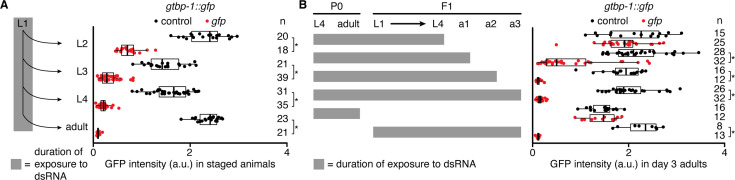
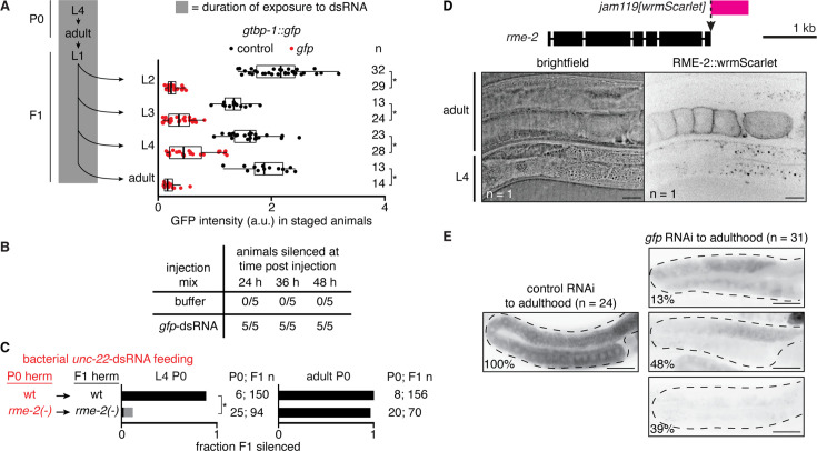
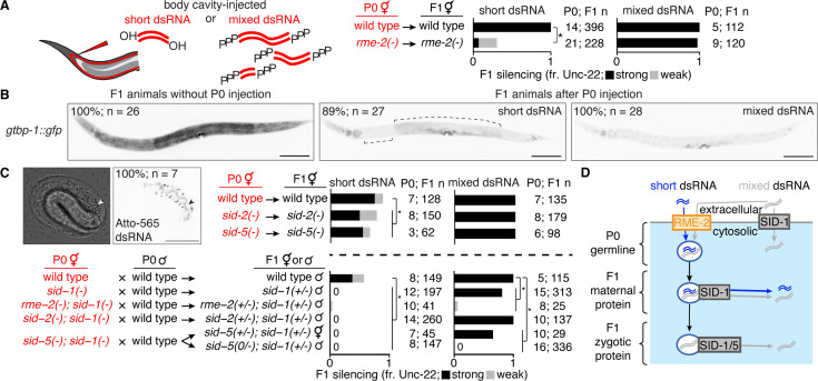
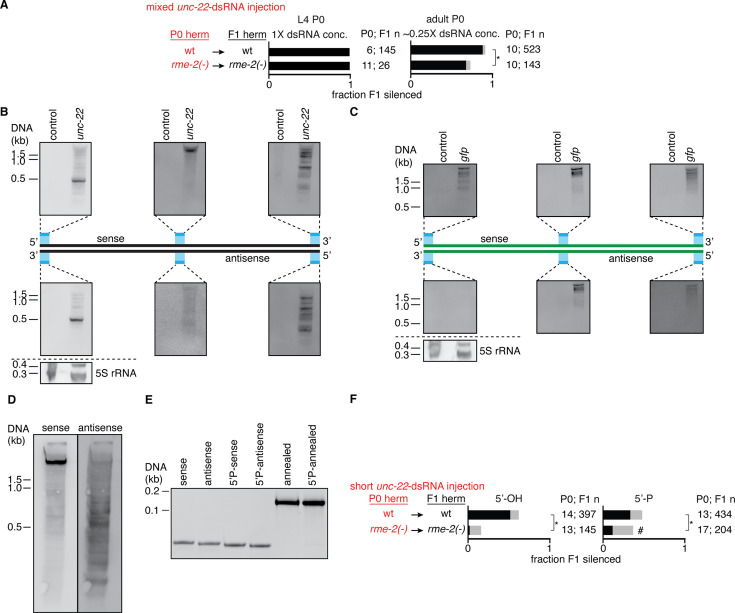
![Figure 1—figure supplement 1. Uptake of dsRNA into the proximal germline by RME-2 is required for silencing during early adulthood. (A) left Schematic depicting continuous exposure of gtbp-1::gfp P0 animals, starting at the L4 stage, and their F1 progeny to bacteria expressing dsRNA, followed by imaging of animals at the indicated stages. (right) Quantification of GTBP-1::GFP intensity (arbitrary units, a.u.) in representative germ cells (larvae) or embryos in utero (adults) of F1 animals at indicated stages after P0 and F1 exposure to control dsRNA (dark grey) or gfp-dsRNA (red). Numbers of animals scored at each stage (n) are indicated. Asterisks indicate P<0.05 with Bonferroni correction using Mann-Whitney U test for two-sided comparisons between animals exposed to control dsRNA or gfp-dsRNA. (B) Silencing of gtbp-1::gfp in germlines injected with duplex buffer (buffer) or in vitro transcribed gfp-dsRNA in duplex buffer during the first day of adulthood and scored for silencing 24, 36, and 48 h post injection. The numbers of animals out of 5 injected with each injection mix that exhibited silencing of both gonad arms are indicated for each time point. Animals injected with buffer never exhibited silencing in either gonad arm. (C) Hermaphrodite animals of the L4 stage (left) or young adult stage (24 hr post L4, right) of the indicated genotypes were fed unc-22-dsRNA expressed in bacteria for 24 hr (red). Hermaphrodite self-progeny of fed animals were scored for unc-22 silencing (strong, black; weak, grey). Numbers of fed P0 parents and scored F1 progeny (P0; F1 n) are as indicated. Asterisk indicates p<0.05 with Bonferroni correction using χ2 test. Previously generated rme-2(-) animals were used in this assay (DH1390). (D) Expression of RME-2. (top) Schematic showing insertion of wrmScarlet (rme-2(jam119[wrmScarlet])) at the rme-2 locus. Scale bar, 1 kb. (bottom) Brightfield and fluorescence images of a rme-2(jam119[wrmScarlet]) L4-stage and adult animal (n=1 confocal plane). Scale bars, 20 μm. (E) Representative fluorescence images of GTBP-1::GFP (black) in the germlines (dashed outline) of day 3 gtbp-1::gfp adult animals after P0 and F1 ingestion of control dsRNA (left) or gfp-dsRNA (right) up to the first day of adulthood. Numbers of animals imaged (n) and the percentages of animals exhibiting the depicted expression patterns are shown. Scale bars, 50 μm.](/paper-figures/13034b8a4c5842728dfd9c948eb1dbec/intergenerational-transport-of-double-stranded-rna-in-c-elegans-can-limit-figure-2.jpg)

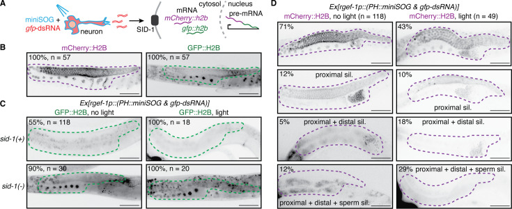
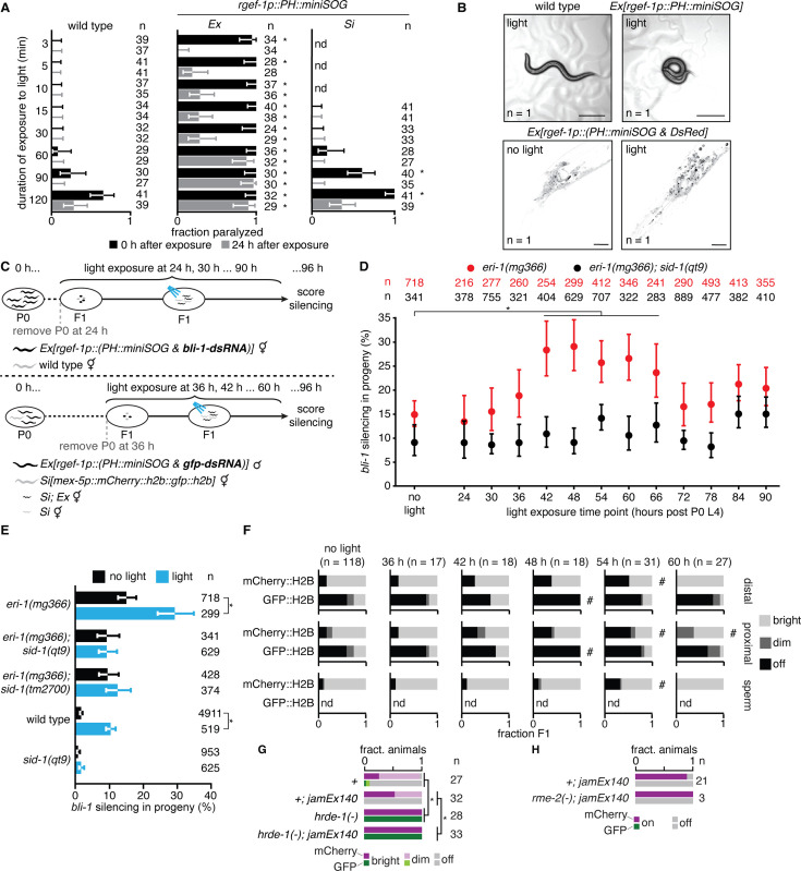
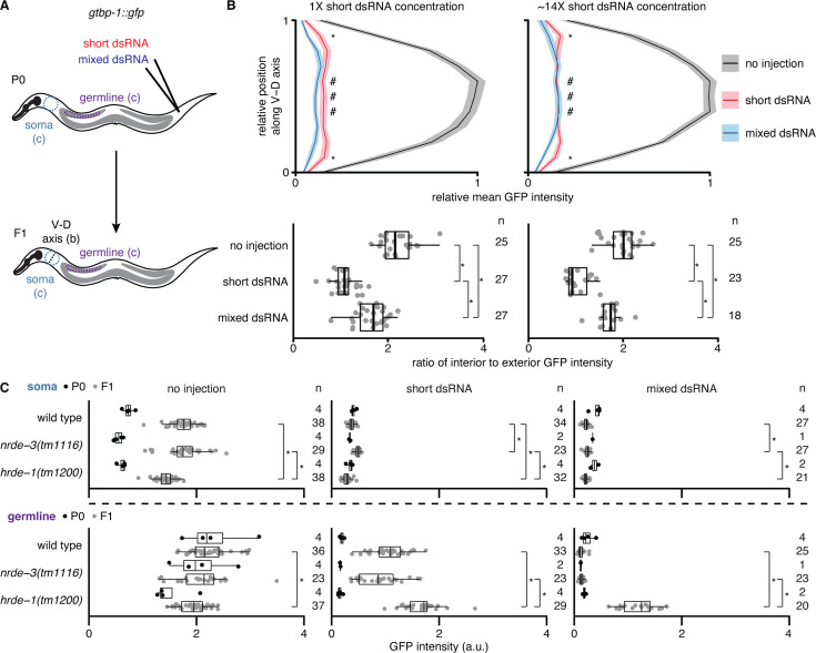
![Figure 2—figure supplement 1. Timed release of neuronal dsRNA by oxidative damage in neurons reveals period of enhanced gene silencing in the soma and germline. (A) Wild-type animals (left) and animals expressing membrane-tethered mini singlet oxygen generator protein (PH::miniSOG) from an extrachromosomal array (Ex, middle) or a single-copy transgene (Si, right) under a pan-neuronal promoter (rgef-1p) were exposed to blue light for different durations (minutes) and animals were scored for paralysis immediately after exposure (0 hr, black) and 24 hr later (24 hr, grey). (B) Functional and anatomical evidence for oxidative damage in neurons. (top) Widefield images of animals without (left) and with (right) Ex[rgef-1p::PH::miniSOG] after 5 min of blue light exposure. Animals paralyzed in (A) often appear coiled (right), likely indicative of a defect in neuronal signaling. Scale bar, 100 μm. (bottom) Confocal fluorescence images of neurons in the head region of animals with Ex[rgef-1p::(PH::miniSOG & DsRed)] without (left) and with (right) 30 minutes of blue light exposure showing light-induced changes (black, DsRed fluorescence). Scale bar, 20 μm. (C) Schematic of assay for measuring the impact of oxidative damage in neurons at different times during development on silencing by neuronal dsRNA. For measuring silencing in the hypodermis (top) or germline (bottom), cohorts of animals with Ex[rgef-1p::(PH::miniSOG & bli-1-dsRNA)] (top), or Ex[rgef-1p::(PH::miniSOG & gfp-dsRNA)] obtained by mating males with the array and hermaphrodites with Si[mex-5p::mCherry::h2b::gfp::h2b] (bottom) were exposed to blue light as indicated and scored for bli-1 silencing (top) or imaged (bottom) as stage-matched adults (at ~96 hr after the fourth larval stage of parent animals). (D) Percentages of eri-1(mg366) (red) or eri-1(mg366); sid-1(qt9) (black) animals silenced when assayed as described in (C, top). Silencing in the absence of exposure to blue light (no light) was also measured for comparison. (E) Percentages of stage-matched animals of the indicated genetic backgrounds with Ex[rgef-1p::(PH::miniSOG & bli-1-dsRNA)] that show bli-1 silencing without (black) or with (blue) a 1 hr exposure to blue light 48 hr after the fourth larval stage of parent animals. The 48 hr time point from (D) is replotted to facilitate comparison. (F) Fractions of animals exhibiting bright (light grey), dim (dark grey) or not detectable (black) mCherry::H2B or GFP::H2B fluorescence in the distal gonad (top), proximal gonad (middle) or sperm (bottom) when assayed as described in (C, bottom). Silencing in the absence of exposure to blue light (no light) was used as the reference. Numbers of animals scored (n), measurements that were not done (nd), significant differences using two-tailed Wilson’s estimates for single proportion compared to wild type (asterisks in A) or no light condition (asterisks in D and E) or χ2 test compared to no light condition (hashes in (F); p<0.05 with Bonferroni correction), and error bars (95% CI) are indicated. (G and H) Animals homozygous (G) or hemizygous (H) for the mex-5p::mCherry::h2b::gfp::h2b transgene (Figure 2) with or without neuronal gfp-dsRNA (jamEx140) were scored for expression of mCherry and GFP (bright, dim, off) in otherwise wild-type (+), hrde-1(-) (G) or rme-2(-) (H) backgrounds. Animals in (G) also have a dpy-2(e8) mutation linked to the mex-5p::mCherry::h2b::gfp::h2b transgene. Fraction silenced in wild-type animals (+) in (G) were calculated with n=31 for GFP and n=27 for mCherry. Asterisks indicates p<0.05 using χ2 test with Bonferroni correction.](/paper-figures/e9a919e151ef4034a91014af52c507d0/intergenerational-transport-of-double-stranded-rna-in-c-elegans-can-limit-figure-4.jpg)
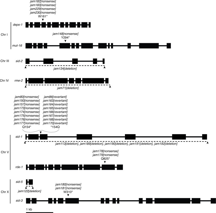
![Figure 2—figure supplement 2. Schematics depicting mutations generated in this study. Structures (boxes, exons; lines, introns) and chromosomal locations of genes with mutations generated using Cas9-mediated genome editing. Nonsense mutations (e.g. jam182[nonsense]) with associated amino acid changes (e.g. W161* for tryptophan at position 161 to stop) are indicated with black arrowheads and deletions of coding regions (e.g. jam134[deletion]) are indicated with a dashed line (deleted region) and flanking black arrowheads. Scale bar, 1 kb.](/paper-figures/cf1dc6b83aa542b19ff151fd4e959436/intergenerational-transport-of-double-stranded-rna-in-c-elegans-can-limit-figure-5.jpg)


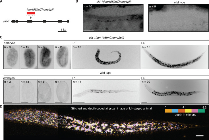
![Figure 4. The expression pattern of SID-1 varies during development. (A) Schematic depicting insertion of mCherry sequence that lacks piRNA binding sites (jam195[mCherry∆pi]; Zhang et al., 2018; Devanapally et al., 2021) into the sid-1 gene using Cas9-mediated genome editing. (B and C) Representative images showing fluorescence from SID-1::mCherry (black) in (B) the adult gonad arm, (C, left) developing embryos, (C, middle) L1-stage animals, or (C, right) L4-stage animals with sid-1(jam195[mCherry∆pi]) compared to autofluorescence in wild-type animals of the same stages. Numbers (n) of each stage imaged are indicated (100% of animals exhibited the depicted expression patterns). For animals imaged in (B), the distal germline was obstructed by the intestine in 1/10 sid-1(jam195[mCherry∆pi]) and 5/9 wild-type animals. (D) Airyscan image of an L1-staged animal assembled by stitching four different Z-stacks after depth-coding and taking maximum projections, illustrating the expression of SID-1::mCherry throughout the animal. Scale bar for adult gonad arms in (B) and embryos in (C), 20 μm; scale bar for larvae in (C), 50 μm and in (D), 10 μm. Also see Technical comments on ‘Making a sid-1 translational reporter’ in Materials and methods.](/paper-figures/fa912d801b30499f8995e85a14522946/intergenerational-transport-of-double-stranded-rna-in-c-elegans-can-limit-figure-4.jpg)
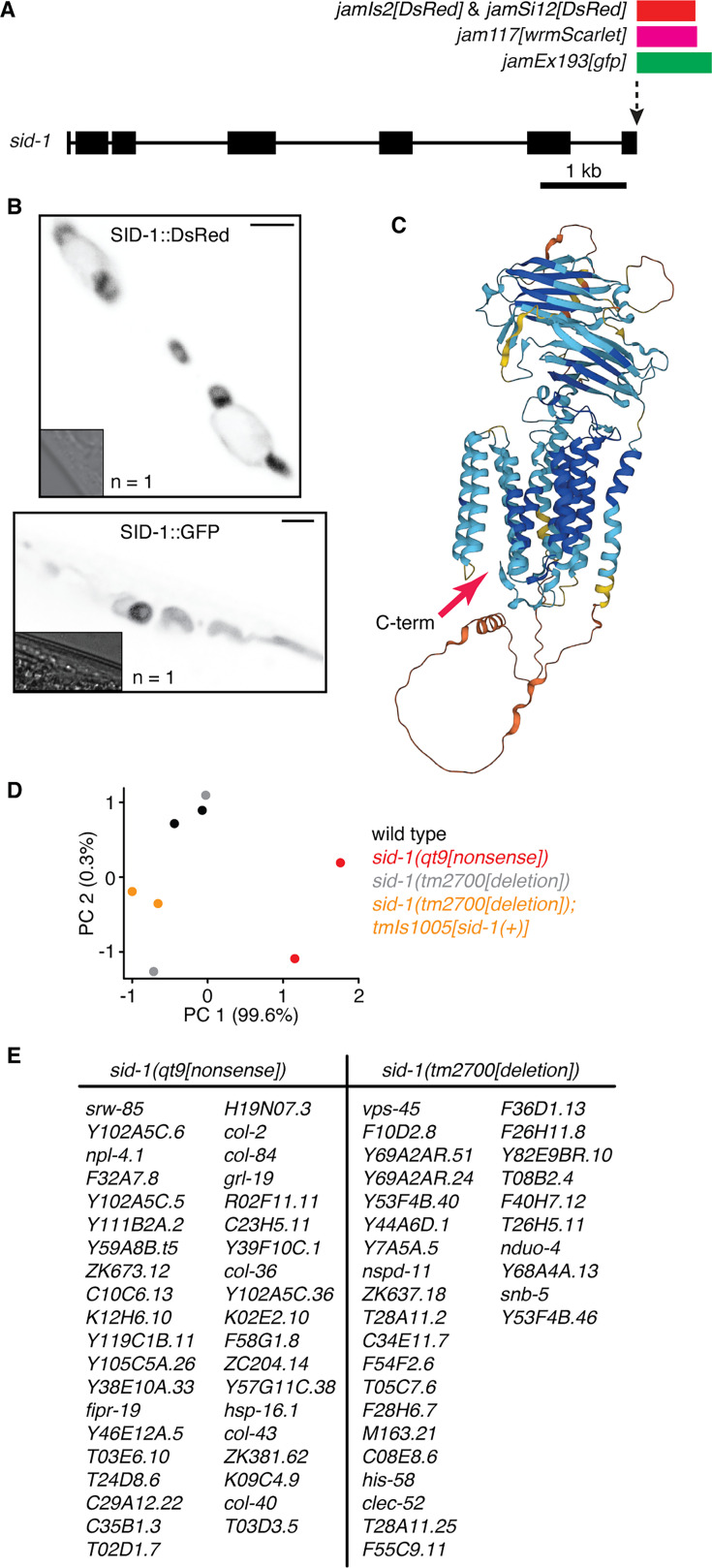
![Figure 4—figure supplement 1. Unsuccessful attempts to functionally tag SID-1 and to identify SID-1-dependent genes. (A) Schematic illustrating the tagging of sid-1 (box, exon; line, intron) at the 3’ end to generate fusion proteins with fluorophores (GFP, DsRed, or wrmScarlet) tagged at the C-terminus. (B) Images showing subcellularly localized fluorescence (black) from SID-1::DsRed (top) and SID-1::GFP (bottom) within muscle cells when expressed from multicopy transgenes. Scale bar = 10 µm and insets show brightfield images. (C) Structure of SID-1 predicted by AlphaFold shaded based on pLDDT scores (blue/cyan, high; yellow/orange, low). Red arrow indicates the C-terminus. (D) Principal component analysis of RNA-seq experiment comparing transcriptomes from wild-type, sid-1(qt9[nonsense]), sid-1(tm2700[deletion]), and sid-1(tm2700[deletion]); tmIs1005[sid-1(+)] animals. (E) List of sid-1-dependent genes identified by comparing polyA+ RNA from sid-1(qt9[nonsense]) animals with wild-type animals (left) and by comparing sid-1(tm2700[deletion]) animals with sid-1(tm2700[deletion]); tmIs1005[sid-1(+)] animals (right).](/paper-figures/dc7acce45fbb48d8afcb0b1cb3e606e2/intergenerational-transport-of-double-stranded-rna-in-c-elegans-can-limit-figure-10.jpg)
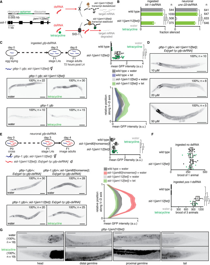
![Figure 4—figure supplement 2. Tetracycline-induced functional rescue of sid-1 expression is evident in somatic tissues but not within the germline. (A) Schematic illustrating a cell expressing sid-1 transcript with a tetracycline aptazyme (Wurmthaler et al., 2019) inserted into the sid-1 3’UTR (left) in the presence (bottom right) or absence (top right) of tetracycline. Tetracycline stabilizes sid-1 transcripts by inhibiting ribozyme-based cleavage in the 3’UTR and thereby allows for the expression of SID-1 protein and dsRNA import. (B) Fraction of wild-type or sid-1(jam112[tet]) animals silenced after ingestion of bli-1-dsRNA (left) or expression of neuronal unc-22-dsRNA (right) in the presence of water (grey bars) or 10 μM tetracycline (green bars). Numbers of animals scored for silencing (n) are depicted. (C) The extent of gfp silencing in gtbp-1::gfp; sid-1(jam112[tet]) day 3 adult animals after ingestion of gfp-dsRNA in the presence of water or 10 μM tetracycline. A schematic illustrating the experimental design (top left), representative images of animals from each condition with numbers of animals imaged (n) and percentages of animals represented (bottom left), and quantification of representative germline (top right) and somatic (bottom right) GTBP-1::GFP intensity (a.u.) are depicted. Mean germline GFP intensity was measured in representative regions of the posterior germline and somatic GFP intensity was measured along a dorsal to ventral axis in the tail region (shaded region represents 95% CI) to avoid increased autofluorescence in the intestines of animals exposed to tetracycline. Scale bars, 100 μm. (D) Representative images of gtbp-1::gfp; sid-1(jam112[tet]) F1 day 1 adult animals after P0 and F1 ingestion of gfp-dsRNA until day 1 of F1 adulthood in the presence of different concentrations of tetracycline (10 μM, 20 μM, 50 μM). Higher concentrations of tetracycline did not enhance silencing in gtbp-1::gfp; sid-1(jam112[tet]) animals. Scale bar, 100 μm. (E) The extent of gfp silencing in cross progeny of gtbp-1::gfp; sid-1(jam112[tet]) hermaphrodites injected with water or 10 μM tetracycline and sid-1(jam112[tet]); Ex[rgef-1p::gfp-dsRNA] males in the presence of water or 10 μM tetracycline. A schematic illustrating the experimental design including injection of gtbp-1::gfp; sid-1(jam112[tet]) hermaphrodites with water or 10 μM tetracycline (top left), representative images of animals with the Ex[rgef-1p::gfp-dsRNA] array from each condition with numbers of animals imaged (n) and percentages of animals represented (bottom left), and quantification of representative germline (top right) and somatic (bottom right) GFP intensity (a.u.) as in (C) are depicted. Scale bar, 100 μm. (F) Total brood of wild-type or sid-1(jam112[tet]) animals after culturing on OP50 E. coli or pos-1-dsRNA bacteria in the presence of water or 10 μM tetracycline. Silencing by pos-1-dsRNA typically results in inviable embryos (wild type, bottom), but culturing sid-1(jam112[tet]) with 10 μM tetracycline and pos-1-dsRNA only resulted in a minor decrease in brood size (sid-1(jam112[tet]), bottom). This decrease was not observed when sid-1(jam112[tet]) animals were cultured on 10 μM tetracycline plates in the absence of pos-1-dsRNA (top, brood of 1 animal; bottom, brood of 3 animals). (G) Representative fluorescence images of GTBP-1::GFP (black) in the heads, distal germlines, proximal germlines, and tails of gtbp-1::gfp animals with a tetracycline-aptazyme sequence inserted into the gtbp-1::gfp 3’UTR (gtbp-1(jam210[tet])) after culturing with water or 10 μM tetracycline. The numbers of animals imaged (n) and the percentages of animals with the represented expression patterns are depicted. An increase in GTBP-1::GFP intensity was observed in the soma and germline, but increased fluorescence in the intestine could not be distinguished from increased autofluorescence caused by culturing with 10 μM tetracycline. Scale bar, 50 μm.](/paper-figures/3f058ade20e643a29c36a8cadc40c29c/intergenerational-transport-of-double-stranded-rna-in-c-elegans-can-limit-figure-11.jpg)
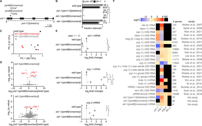
![Figure 5. Ancestral loss of SID-1 causes transgenerational changes in the mRNA levels of two germline genes that are subject to RNA regulation. (A) Schematic of modifications at the sid-1 gene generated using Cas9-mediated genome editing. Deletion of the entire coding sequence (jam113[deletion]), a nonsense mutation (jam80[nonsense]), and its reversion to wild-type sequence (jam86[revertant]) are depicted. (B) Fractions of animals with the indicated genotypes that show silencing in response to unc-22-dsRNA (grey) or bli-1-dsRNA (black). Numbers of animals scored (n), significant differences using two-tailed test with Wilson’s estimates for single proportions (asterisks, P<0.05 with Bonferroni correction) and 95% CI (error bars) are indicated. (C) Principal components explaining the variance between wild type (black), sid-1(jam80[nonsense]) (red), and sid-1(jam86[revertant]) (grey) polyA+ RNA samples. Almost all of the variance between samples is explained by PC 1. (D) Volcano plots of changes in the abundance of polyA+ RNA in sid-1(jam80[nonsense]) (top) and sid-1(jam86[revertant]) (bottom) animals compared with wild-type animals (black, q<0.05; red, both q<0.05 and change in the same direction in sid-1(jam80[nonsense]) and sid-1(jam113[deletion]); see Figure 5—figure supplement 1). While sid-1 transcript levels in sid-1(jam86[revertant]) are comparable to that in wild type (grey), sdg-1 (W09B7.2/F07B7.2) and sdg-2 (Y102A5C.36) transcript levels remain elevated in sid-1(jam86[revertant]) (red). (E) Levels of spliced sid-1 (top), sdg-1 (middle) and sdg-2 (bottom) transcripts measured using RT-qPCR. The median of three technical replicates is plotted for each of three biological replicates (bar indicates median) assayed before and after 1 year of passaging animals (year 1, dark grey; year 2, light grey). Asterisks indicate p<0.05 with Bonferroni correction using two-tailed Student’s t-test. (F) Heatmap showing changes in the levels of transcripts (total RNA or mRNA) or antisense small RNAs (22G RNA) from sid-1, sdg-1, sdg-2, and tbb-2 (abundant germline transcript for comparison). Fold changes (expressed as LogFC, indicating log2 for (m)RNA, log10 for piRNA binding, and log10 for 22G RNA) were deduced by integrating reports (study) of 21 experiments that identify subsets of genes as being subject to RNA-mediated regulation within the germline (# genes). These prior studies include comparisons of RNA or 22G RNA from wild-type animals with that from mutant animals (e.g. mut-16(-) 22G RNA), biochemical detection of piRNA binding to transcripts (piRNA-bound mRNA), and biochemical detection of 22G RNA binding to an Argonaute (HRDE-1-bound 22G RNA). ‘NS’ indicates cases where changes, if any, were not significant based on the criteria used in the study. A conservative value of two-fold is assigned to all genes reported as changing >two-fold in Ni et al., 2016.](/paper-figures/a334a266a6da4417bbf91d1c9e02da87/intergenerational-transport-of-double-stranded-rna-in-c-elegans-can-limit-figure-5.jpg)
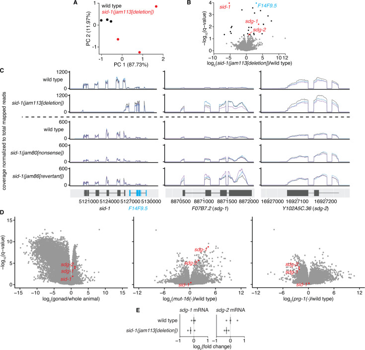
![Figure 5—figure supplement 1. Selective disruption of sid-1 followed by restoration to wild type reveals sid-1-dependent transcripts expressed in the germline that show heritable change. (A) Principal components explaining the variance between wild-type (black) and sid-1(jam113[deletion]) (red) animals. (B) Volcano plots of changes in the abundance of polyA+ RNA in sid-1(jam113[deletion]) animals compared with wild-type animals (black, q<0.05; red, q<0.05 and with change in the same direction in sid-1(jam80[nonsense]); see Figure 5D, top). (C) Read coverage in biological triplicate (black, blue and purple) at sid-1 and F14F9.5 (left), W09B7.2/F07B7.2 (sdg-1) (represented by F07B7.2 locus, middle) and Y102A5C.36 (sdg-2) (right) of polyA+ RNA in wild-type and sid-1(jam113[deletion]) animals (top), and in wild-type, sid-1(jam80[nonsense]), and sid-1(jam86[revertant]) animals (bottom) normalized to total mapped reads per sample. Deletion of sid-1 coding sequence caused accumulation of transcripts from F14F9.5 (blue), requiring point mutation (jam80[nonsense]) for selective disruption of sid-1 (see Figure 5). (D) Volcano plots of changes in the abundance of RNA in wild-type gonads vs. whole animals (left), mut-16(-) vs. wild-type animals (middle), and prg-1(-) vs. wild-type animals (right) using data from Reed et al., 2020. sdg-1, sdg-2 and sid-1 transcripts are highlighted (red). (E) Levels of spliced sdg-1 and sdg-2 transcripts in animals of the indicated genotypes measured using RT-qPCR. The median (line) of three technical replicates is plotted for each of three biological replicates. p>0.05 with Bonferroni correction using two-tailed Student’s t-test for wild type to mutant comparisons. Levels of sid-1 transcripts were not detectable in sid-1(jam113[deletion]) animals due to absence of sid-1 coding sequence (data not shown).](/paper-figures/9d7afd78d58543c9b2732ff4dd3336ca/intergenerational-transport-of-double-stranded-rna-in-c-elegans-can-limit-figure-13.jpg)
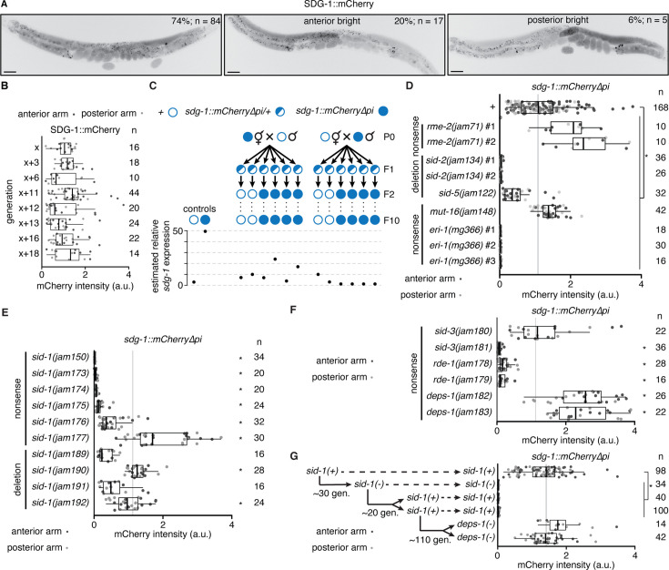
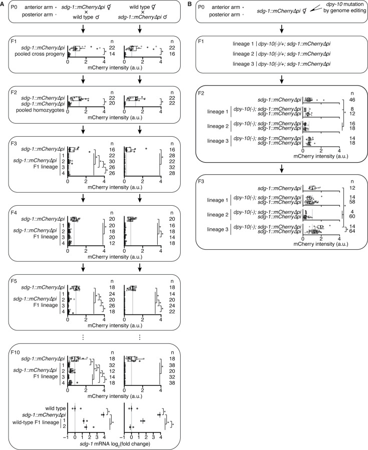
![Figure 6. The sdg-1 gene is prone to stochastic changes in gene expression that can become heritable. (A) Representative images showing fluorescence of SDG-1::mCherry (black) in a wild-type background. While most animals showed symmetric expression in the germline (left), animals with >two-fold difference in fluorescence between both gonad arms (bright anterior, middle and bright posterior, right) were also observed. Punctate fluorescence in the intestine likely represents autofluorescence. Scale bar, 50 μm. (B) Quantification of SDG-1::mCherry fluorescence intensity (arbitrary units, a.u.) in adult gonad arms (anterior arm, dark grey; posterior arm, light grey) of sdg-1(jam137[mCherry∆pi]) animals starting in one generation (x) and continuing in successive generations as indicated. Numbers of gonad arms quantified (n) is indicated. Expression in one generation was not significantly different when compared to that in the previous tested generation using Mann-Whitney U test for two-sided comparisons and Bonferroni correction. (C) Lineages and estimated relative sdg-1 expression 10 generations after mating wild-type (open circle) males with sdg-1::mCherry∆pi (filled circle) hermaphrodites and vice versa, and isolating sdg-1(+) or sdg-1::mCherry animals from F1 heterozygotes (half-filled circle). Expression of sdg-1 in the F10 generation was measured by RT-qPCR of sdg-1 mRNA purified from pooled wild-type animals of mixed stages or by quantification of SDG-1::mCherry fluorescence in gonad arms of adult sdg-1::mCherry∆pi animals. Relative levels of sdg-1 mRNA and SDG-1::mCherry fluorescence intensity were converted to units of estimated relative sdg-1 expression (see Materials and methods) for comparison. See Figure 6—figure supplement 2A for raw data. (D–F) Fluorescence intensity measurements (quantified as in B) in adult animals with sdg-1::mCherry∆pi (+) and additionally with mutations in genes introduced through genetic crosses (in regulators of dsRNA import rme-2, sid-2 or sid-5, or in regulators of RNA silencing mut-16 or eri-1) or through genome editing (in regulators of dsRNA import sid-1 or sid-3, or in regulators of RNA silencing rde-1 or deps-1). Asterisks indicate p<0.05 with Bonferroni correction using Mann-Whitney U test for two-sided comparisons between animals with sdg-1::mCherry∆pi (+) and animals with additional mutations. Nonsense mutations (nonsense) or deletion utations (deletion) introduced through genetic crosses (isolate numbers #1, #2, etc. in D) or genome editing (different alleles in E and F) and numbers of gonad arms (n) quantified for each isolate are indicated. Mutations in genes required for dsRNA import or subsequent silencing resulted in fewer animals with asymmetric fluorescence between gonad arms (a combined proportion of 21/197 for sid-1, sid-3, rde-1 and deps-1 mutants versus 22/84 for wild type, p=0.0009 using two-tailed test with Wilson’s estimates for single proportions). Animals with at least one gonad arm brighter than the dimmest wild-type gonad arm in (A) and with asymmetric gonad arms were found in different genotypes (anterior bright: sid-1(-) – 5/122, sid-3(-) – 1/29, rde-1(-) – 2/22, deps-1(-) – 4/24, and posterior bright: sid-1(-) – 6/122, rde-1(-) – 2/22, deps-1(-) – 1/24). (G) Fluorescence intensity measurements as in (B) of animals with sdg-1::mCherry∆pi that show loss of fluorescence when a nonsense mutation is introduced in sid-1 using genome editing ~30 generations (gen.) later remain changed despite reversion of sid-1 nonsense mutation to wild-type sequence after ~20 additional generations. Subsequent mutation of deps-1 after another ~110 generations restored SDG-1::mCherry fluorescence to wild-type levels. Also see Figure 6—figure supplements 1 and 2.](/paper-figures/c0a903afec1049559f14161591e7786c/intergenerational-transport-of-double-stranded-rna-in-c-elegans-can-limit-figure-6.jpg)
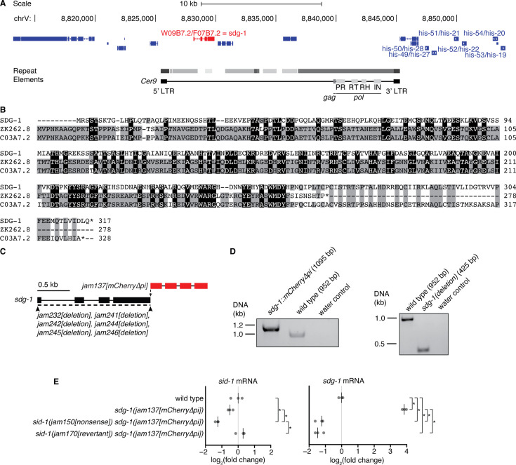
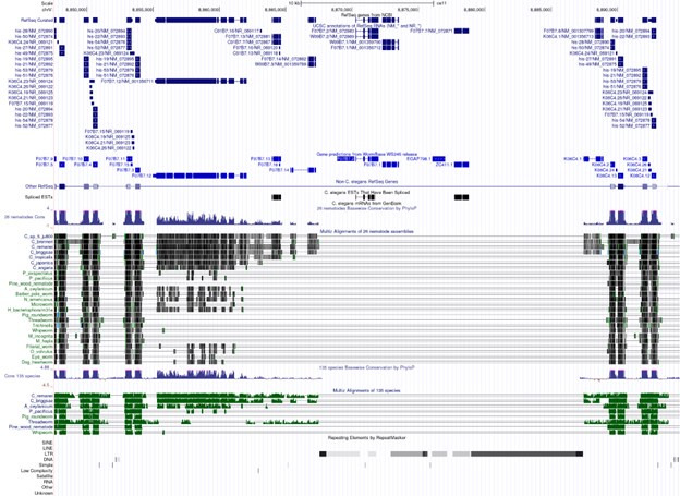
![Figure 6—figure supplement 1. The sid-1-dependent gene sdg-1 is expressed from two identical loci (W09B7.2/F07B7.2) and loss of its expression in sid-1(nonsense) animals fails to recover in sid-1(revertant) animals. (A) Schematic adapted from UCSC Genome Browser (Perez et al., 2025) depicting W09B7.2/F07B7.2 (red) located within a repeated ~40 kb locus on chromosome V (8813207–8854700 depicted; duplicate locus at 8855302–8896495) that includes many histone genes (dark blue; duplicate genes also depicted). W09B7.2/F07B7.2 are located within full-length Cer9 retrotransposons with repeated regions in grey (darker color indicates fewer repeat element-associated mismatches/insertions/deletions). Loci encoding gag and pol elements (PR: protease, RT: reverse transcriptase, RH: RNaseH, IN: integrase) within Cer9 are depicted. (B) Alignment of the SDG-1 protein sequence encoded by W09B7.2/F07B7.2 and the paralogs ZK262.8 and C03A7.2 with conserved residues between two (grey) or three (black) proteins highlighted. (C) Schematic depicting insertion of mCherry sequence that lacks piRNA binding sites (Zhang et al., 2018; Devanapally et al., 2021) at the 3’ end of sdg-1(jam137[mCherry∆pi]), as well as deletion of the sdg-1 coding sequence (jam232, jam241, jam242, jam244, jam245, and jam246). (D) Genotyping gels showing insertion of mCherry∆pi sequences (1095 bp) (left) or deletion of sdg-1 coding sequences (425 bp) (right) at both loci of sdg-1. Absence of wild-type bands in either case confirm genome editing of both copies. (E) Levels of spliced sid-1 (left) and sdg-1 (right) transcripts in wild-type animals and sdg-1(jam137[mCherry∆pi]) animals with a wild-type (+), sid-1(jam150[nonsense]) or sid-1(jam169[revertant]) background measured using RT-qPCR. The median of three technical replicates is plotted for each of three biological replicates (bar indicates median). Asterisks indicate p<0.05 with Bonferroni correction using two-tailed Student’s t-test.](/paper-figures/0132e81748e645ec9902379d041b9d68/intergenerational-transport-of-double-stranded-rna-in-c-elegans-can-limit-figure-15.jpg)
![Figure 6—figure supplement 2. Mating but not genome editing can initiate distinct heritable changes in sdg-1 expression. (A) (P0 to F10, top) Quantification of SDG-1::mCherry fluorescence intensity (a.u.) in adult gonad arms (anterior arm, dark grey; posterior arm, light grey) across generations after mating hermaphrodite and male sdg-1(jam137[mCherry∆pi]) animals with male and hermaphrodite wild-type animals, respectively. The generations assayed and numbers of gonad arms quantified (n) are indicated. In F1 and F2, fluorescence intensity values of animals with lineages that were not propagated to F10 but were heterozygous or homozygous sdg-1(jam137[mCherry∆pi]), respectively, were pooled with values of animals with lineages that were propagated to F10. In F3 to F10, top, animals from four different F1 lineages were scored. Fluorescence intensity of animals descending from the self-progeny of P0 sdg-1(jam137[mCherry∆pi]) animals was measured in each generation and is depicted, with the same data plotted for each mating direction for comparison. Asterisk indicates p<0.05 with Bonferroni correction using Mann-Whitney U test for two-sided comparisons. (F10, bottom) Levels of spliced sdg-1 mRNA transcripts in wild-type animals, sdg-1(jam137[mCherry∆pi]) animals and two lineages of wild-type F10 progeny from each cross direction, measured using RT-qPCR. The median of three technical replicates is plotted for each of three biological replicates (bar indicates median). Asterisks indicate p<0.05 with Bonferroni correction using two-tailed Student’s t-test. (B) (P0 and F1) Schematic illustrating mutation of dpy-10 in three P0 lineages of sdg-1(jam137[mCherry∆pi]) animals and subsequent segregation of the dpy-10 mutation. (F2 and F3) Both dpy-10(-) and dpy-10(+) F2 and F3 animals from each of the three P0 lineages were imaged and SDG-1::mCherry intensity was quantified (a.u.) in adult gonad arms (anterior arm, dark grey; posterior arm, light grey). Minor differences in SDG-1::mCherry expression were observed between mutants and nonmutants in some cases, as well as between lineages. The numbers of gonad arms quantified (n) are depicted. Asterisks indicates p<0.05 with Bonferroni correction using Mann-Whitney U test for two-sided comparisons.](/paper-figures/00f39ae4cabf43318e0c5b96619ab562/intergenerational-transport-of-double-stranded-rna-in-c-elegans-can-limit-figure-16.jpg)
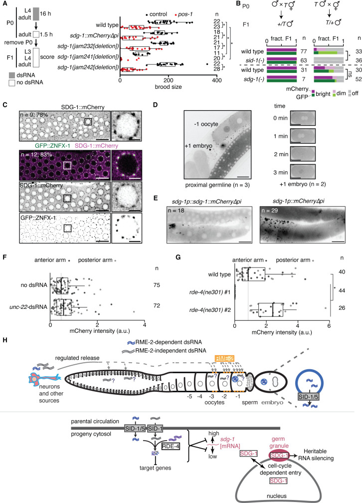
![Figure 7. SID-1 modifies RNA regulation within the germline, potentially through sdg-1 and other sid-1-dependent genes. (A) (left) Schematic of assay for sensitive detection of pos-1 silencing by ingested dsRNA. (right) Numbers of developed progeny (> 3rd larval stage) laid by wild-type animals, animals with a deletion (∆) in sdg-1 (jam232, jam241, jam242) or animals with overexpression (sdg-1::mCherry∆pi) of sdg-1 exposed to pos-1 dsRNA (red) or control dsRNA (black) for 16 hr are plotted. Asterisks indicate p<0.05 using Mann-Whitney U test for two-sided comparisons with Bonferroni correction. (B) Cross progeny males that inherited the mex-5p::mCherry::h2b::gfp::h2b transgene (T; Devanapally et al., 2021; also used in Figure 2) from maternal (left) or paternal (right) parents, both of wild-type, sid-1(-), or sdg-1(-) background, were scored for expression of mCherry and GFP (bright, dim, off). Wild-type data for top set (n=77 and n=33) are replotted from Devanapally et al., 2021 for comparison. Dashed line separates independent experiments. Asterisk indicates p<0.05 with Bonferroni correction using χ2 test; n.s. indicates not significant. (C) Representative AiryScan images of the distal germline (left; scale bar, 10 µm) or single germline nuclei (right; scale bar, 2 µm) showing SDG-1::mCherry alone (top) or with GFP::ZNFX-1 (bottom, merge and single channel images). The number of animals imaged (n) and the percentage that show enrichment of SDG-1::mCherry in perinuclear foci are indicated. Sites of SDG-1::mCherry enrichment coincide with GFP::ZNFX-1 localization. Boxes in left mark the nuclei shown in right. (D) Representative images showing entry of SDG-1::mCherry into the nucleus in –1 oocytes (left) and upon pronuclear fusion in early embryos during the time course indicated (right). Numbers of germlines and embryos imaged are indicated. Scale bars, 20 µm. Also see Videos 1–4. (E) Representative image of the hermaphrodite germline in animals with a translational (left) or transcriptional (right) reporter of sdg-1. Scale bar, 20 µm. Apparent extracellular punctae of SDG-1::mCherry and mCherry surrounding the proximal germline requires further study, but could be non-specific because similar localization is observed in animals with other promoters driving mCherry expression, but not GFP expression, in the germline (data not shown). The numbers of animals with the depicted fluorescence pattern are indicated. (F and G) Response of the transcriptional sdg-1 reporter (sdg-1p::mCherry∆pi[sdg-1(∆)]::sdg-1 3' UTR) to the addition of unc-22-dsRNA (F) or loss of rde-4 (G). Quantification and asterisk are as in Figure 6. (H) Models for dsRNA import into the germline (top) and subsequent RNA-mediated regulation of sdg-1 (bottom). See text for details.](/paper-figures/7f6c4a725f57436ca16febd35ff5ca26/intergenerational-transport-of-double-stranded-rna-in-c-elegans-can-limit-figure-7.jpg)

Verification
Claims the paper makes
The claims and statistics of this paper haven’t been checked yet.
Run the check — free, no account needed. It reads the open-access text and checks the sources it cites.
Self-check
Percentages, sums and reported differences re-computed from the paper’s own numbers.
The self-check needs the paper’s full text.
Citations
Cites
Fiji: an open-source platform for biological-image analysis
Schindelin, J., Arganda-Carreras, I., Frise, E., Kaynig, V., Longair, M., Pietzsch, T., Preibisch, S., Rueden, C., Saalfeld, S., Schmid, B., et al. (2012) · Nature Methods · doi:10.1038/nmeth.2019 · Cited by 66,890
The Sequence Alignment/Map format and SAMtools
Li, H., Handsaker, B., Wysoker, A., Fennell, T., Ruan, J., Homer, N., Marth, G., Abecasis, G., Durbin, R., & 1000 Genome Project Data Processing Subgroup (2009) · Bioinformatics · doi:10.1093/bioinformatics/btp352 · Cited by 62,041
Highly accurate protein structure prediction with AlphaFold
Jumper, J., Evans, R., Pritzel, A., Green, T., Figurnov, M., Ronneberger, O., Tunyasuvunakool, K., Bates, R., Žídek, A., Potapenko, A., et al. (2021) · Nature · doi:10.1038/s41586-021-03819-2 · Cited by 43,764
Cutadapt removes adapter sequences from high-throughput sequencing reads
Martin, M. (2011) · EMBnet.journal · doi:10.14806/ej.17.1.200 · Cited by 33,889
Potent and specific genetic interference by double-stranded RNA in Caenorhabditis elegans
Fire, A., Xu, S., Montgomery, M. K., Kostas, S. A., Driver, S. E., & Mello, C. C. (1998) · Nature · doi:10.1038/35888 · Cited by 15,429
THE GENETICS OF CAENORHABDITIS ELEGANS
Brenner, S. (1974) · Genetics · doi:10.1093/genetics/77.1.71 · Cited by 15,220
Graph-based genome alignment and genotyping with HISAT2 and HISAT-genotype
Kim, D., Paggi, J. M., Park, C., Bennett, C., & Salzberg, S. L. (2019) · Nature Biotechnology · doi:10.1038/s41587-019-0201-4 · Cited by 15,022
Salmon provides fast and bias-aware quantification of transcript expression
Patro, R., Duggal, G., Love, M. I., Irizarry, R. A., & Kingsford, C. (2017) · Nature Methods · doi:10.1038/nmeth.4197 · Cited by 12,700
A scaling normalization method for differential expression analysis of RNA-seq data
Robinson, M. D., & Oshlack, A. (2010) · Genome Biology · doi:10.1186/gb-2010-11-3-r25 · Cited by 7,876
Varadi, M., Anyango, S., Deshpande, M., Nair, S., Natassia, C., Yordanova, G., Yuan, D., Stroe, O., Wood, G., Laydon, A., et al. (2022) · Nucleic Acids Research · doi:10.1093/nar/gkab1061 · Cited by 7,850
voom: precision weights unlock linear model analysis tools for RNA-seq read counts
Law, C. W., Chen, Y., Shi, W., & Smyth, G. K. (2014) · Genome Biology · doi:10.1186/gb-2014-15-2-r29 · Cited by 6,155
The EMBL-EBI search and sequence analysis tools APIs in 2019
Madeira, F., Park, Y. M., Lee, J., Buso, N., Gur, T., Madhusoodanan, N., Basutkar, P., Tivey, A. R. N., Potter, S. C., Finn, R. D., et al. (2019) · Nucleic Acids Research · doi:10.1093/nar/gkz268 · Cited by 4,167
Differential analyses for RNA-seq: transcript-level estimates improve gene-level inferences
Soneson, C., Love, M. I., & Robinson, M. D. (2015) · F1000Research · doi:10.12688/f1000research.7563.1 · Cited by 3,929
Mello, C. C., Kramer, J. M., Stinchcomb, D. T., & Ambros, V. (1991) · The EMBO Journal · doi:10.1002/j.1460-2075.1991.tb04966.x · Cited by 3,095
Specific interference by ingested dsRNA
Timmons, L., & Fire, A. Z. (1998) · Nature · doi:10.1038/27579 · Cited by 1,938
Therapeutic siRNA: state of the art
Hu, B., Zhong, L., Weng, Y., Peng, L., Huang, Y., Zhao, Y., & Liang, X.-J. (2020) · Signal Transduction and Targeted Therapy · doi:10.1038/s41392-020-0207-x · Cited by 1,586
Adipose-derived circulating miRNAs regulate gene expression in other tissues
Thomou, T., Mori, M. A., Dreyfuss, J. M., Konishi, M., Sakaguchi, M., Wolfrum, C., Rao, T. N., Winnay, J. N., Garcia-Martin, R., Grinspoon, S. K., et al. (2017) · Nature · doi:10.1038/nature21365 · Cited by 1,539
Sperm tsRNAs contribute to intergenerational inheritance of an acquired metabolic disorder
Chen, Q., Yan, M., Cao, Z., Li, X., Zhang, Y., Shi, J., Feng, G.-H., Peng, H., Zhang, X., Zhang, Y., et al. (2016) · Science · doi:10.1126/science.aad7977 · Cited by 1,470
The current state and future directions of RNAi-based therapeutics
Setten, R. L., Rossi, J. J., & Han, S.-P. (2019) · Nature Reviews Drug Discovery · doi:10.1038/s41573-019-0017-4 · Cited by 1,443
The rde-1 Gene, RNA Interference, and Transposon Silencing in C. elegans
Tabara, H., Sarkissian, M., Kelly, W. G., Fleenor, J., Grishok, A., Timmons, L., Fire, A. Z., & Mello, C. C. (1999) · Cell · doi:10.1016/s0092-8674(00)81644-x · Cited by 1,307
Plants send small RNAs in extracellular vesicles to fungal pathogen to silence virulence genes
Cai, Q., Qiao, L., Wang, M., He, B., Lin, F., Palmquist, J., Huang, S.-D., & Jin, H. (2018) · Science · doi:10.1126/science.aar4142 · Cited by 1,289
Biogenesis and function of tRNA fragments during sperm maturation and fertilization in mammals
Sharma, U., Conine, C. C., Shea, J. M., Boskovic, A., Derr, A. G., Bing, X. Y., Belleannee, C., Kucukural, A., Serra, R. W., Sun, F., et al. (2016) · Science · doi:10.1126/science.aad6780 · Cited by 1,275
Systemic RNAi in C. elegans Requires the Putative Transmembrane Protein SID-1
Winston, W. M., Molodowitch, C., & Hunter, C. P. (2002) · Science · doi:10.1126/science.1068836 · Cited by 849
Arribere, J. A., Bell, R. T., Fu, B. X. H., Artiles, K. L., Hartman, P. S., & Fire, A. Z. (2014) · Genetics · doi:10.1534/genetics.114.169730 · Cited by 761
Paix, A., Folkmann, A. W., Rasoloson, D., & Seydoux, G. (2015) · Genetics · doi:10.1534/genetics.115.179382 · Cited by 664
The UCSC Genome Browser database: 2025 update
Perez, G., Barber, G. P., Benet‐Pagès, A., Casper, J., Clawson, H., Diekhans, M., Fischer, C., Gonzalez, J. N., Hinrichs, A. S., Lee, C. M., et al. (2024) · Nucleic Acids Research · doi:10.1093/nar/gkae974 · Cited by 623
Receptor-mediated Endocytosis in theCaenorhabditis elegansOocyte
Grant, B. D., & Hirsh, D. (1999) · Molecular Biology of the Cell · doi:10.1091/mbc.10.12.4311 · Cited by 603
Transport of dsRNA into Cells by the Transmembrane Protein SID-1
Feinberg, E. H., & Hunter, C. P. (2003) · Science · doi:10.1126/science.1087117 · Cited by 595
PRG-1 and 21U-RNAs Interact to Form the piRNA Complex Required for Fertility in C. elegans
Batista, P. J., Ruby, J. G., Claycomb, J. M., Chiang, R., Fahlgren, N., Kasschau, K. D., Chaves, D. A., Gu, W., Vasale, J. J., Duan, S., et al. (2008) · Molecular Cell · doi:10.1016/j.molcel.2008.06.002 · Cited by 592
Tabara, H., Yigit, E., Siomi, H., & Mello, C. C. (2002) · Cell · doi:10.1016/s0092-8674(02)00793-6 · Cited by 539
A nuclear Argonaute promotes multigenerational epigenetic inheritance and germline immortality
Buckley, B. A., Burkhart, K. B., Gu, S. G., Spracklin, G., Kershner, A. M., Fritz, H., Kimble, J. E., Fire, A. Z., & Kennedy, S. G. (2012) · Nature · doi:10.1038/nature11352 · Cited by 512
Retrovirus-like Gag Protein Arc1 Binds RNA and Traffics across Synaptic Boutons
Ashley, J., Cordy, B., Lucia, D., Fradkin, L. G., Budnik, V., & Thomson, T. (2018) · Cell · doi:10.1016/j.cell.2017.12.022 · Cited by 488
Kelly, W. G., Xu, S., Montgomery, M. K., & Fire, A. Z. (1997) · Genetics · doi:10.1093/genetics/146.1.227 · Cited by 463
C. elegans neurons jettison protein aggregates and mitochondria under neurotoxic stress
Melentijevic, I., Toth, M. L., Arnold, M. L., Guasp, R. J., Harinath, G., Nguyen, K. C., Taub, D., Parker, J. A., Neri, C., Gabel, C. V., et al. (2017) · Nature · doi:10.1038/nature21362 · Cited by 454
Genetic Requirements for Inheritance of RNAi in C. elegans
Grishok, A., Tabara, H., & Mello, C. C. (2000) · Science · doi:10.1126/science.287.5462.2494 · Cited by 449
Small RNAs Are Trafficked from the Epididymis to Developing Mammalian Sperm
Sharma, U., Sun, F., Conine, C. C., Reichholf, B., Kukreja, S., Herzog, V. A., Ameres, S. L., & Rando, O. J. (2018) · Developmental Cell · doi:10.1016/j.devcel.2018.06.023 · Cited by 446
Improved Mos1-mediated transgenesis in C. elegans
Frøkjær‐Jensen, C., Davis, M. W., Ailion, M., & Jørgensen, E. M. (2012) · Nature Methods · doi:10.1038/nmeth.1865 · Cited by 417
Sequence requirements for myosin gene expression and regulation in Caenorhabditis elegans.
Okkema, P. G., Harrison, S. M. W., Plunger, V., Aryana, A., & Fire, A. Z. (1993) · Genetics · doi:10.1093/genetics/135.2.385 · Cited by 416
Dokshin, G. A., Ghanta, K. S., Piscopo, K. M., & Mello, C. C. (2018) · Genetics · doi:10.1534/genetics.118.301532 · Cited by 416
C. elegans piRNAs Mediate the Genome-wide Surveillance of Germline Transcripts
Lee, H.-C., Gu, W., Shirayama, M., Youngman, E. M., Conte, D., & Mello, C. C. (2012) · Cell · doi:10.1016/j.cell.2012.06.016 · Cited by 372
Small RNAs Gained during Epididymal Transit of Sperm Are Essential for Embryonic Development in Mice
Conine, C. C., Sun, F., Song, L., Rivera‐Pérez, J. A., & Rando, O. J. (2018) · Developmental Cell · doi:10.1016/j.devcel.2018.06.024 · Cited by 370
An Argonaute Transports siRNAs from the Cytoplasm to the Nucleus
Guang, S., Bochner, A. F., Pavelec, D. M., Burkhart, K. B., Harding, S. J., Lachowiec, J., & Kennedy, S. G. (2008) · Science · doi:10.1126/science.1157647 · Cited by 351
Caenorhabditis elegans SID-2 is required for environmental RNA interference
Winston, W. M., Sutherlin, M. E., Wright, A. J., Feinberg, E. H., & Hunter, C. P. (2007) · Proceedings of the National Academy of Sciences · doi:10.1073/pnas.0611282104 · Cited by 329
Piwi/PRG-1 Argonaute and TGF-β Mediate Transgenerational Learned Pathogenic Avoidance
Moore, R. S., Kaletsky, R. L., & Murphy, C. T. (2019) · Cell · doi:10.1016/j.cell.2019.05.024 · Cited by 290
The piRNA targeting rules and the resistance to piRNA silencing in endogenous genes
Zhang, D., Tu, S., Stubna, M. W., Wu, W.-S., Huang, W.-C., Weng, Z., & Lee, H.-C. (2018) · Science · doi:10.1126/science.aao2840 · Cited by 240
C. elegans interprets bacterial non-coding RNAs to learn pathogenic avoidance
Kaletsky, R. L., Moore, R. S., Vrla, G. D., Parsons, L. R., Gitai, Z., & Murphy, C. T. (2020) · Nature · doi:10.1038/s41586-020-2699-5 · Cited by 240
Shen, E.-Z., Chen, H., Ozturk, A. R., Tu, S., Shirayama, M., Tang, W., Ding, Y.-H., Dai, S.-Y., Weng, Z., & Mello, C. C. (2018) · Cell · doi:10.1016/j.cell.2018.02.002 · Cited by 226
Das, S., Abdel‐Mageed, A. B., Adamidi, C., Adelson, P. D., Akat, K. M., Alsop, E. B., Ansel, K. M., Arango, J. I., D, N. A. M., Kilinc, S., et al. (2019) · Cell · doi:10.1016/j.cell.2019.03.023 · Cited by 223
Pastuzyn, E. D., Day, C. E., Kearns, R. B., Kyrke-Smith, M., Taibi, A. V., McCormick, J., Yoder, N., Belnap, D. M., Erlendsson, S., Morado, D. R., et al. (2018) · Cell · doi:10.1016/j.cell.2018.03.024 · Cited by 213
Spatiotemporal regulation of liquid-like condensates in epigenetic inheritance
Wan, G., Fields, B. D., Spracklin, G., Shukla, A., Phillips, C. M., & Kennedy, S. (2018) · Nature · doi:10.1038/s41586-018-0132-0 · Cited by 207
Uptake of Extracellular Double-Stranded RNA by SID-2
McEwan, D. L., Weisman, A. S., & Hunter, C. P. (2012) · Molecular Cell · doi:10.1016/j.molcel.2012.07.014 · Cited by 195
Neuronal Small RNAs Control Behavior Transgenerationally
Posner, R., Toker, I. A., Antonova, O., Star, E., Anava, S., Azmon, E., Hendricks, M., Bracha, S., Gingold, H., & Rechavi, O. (2019) · Cell · doi:10.1016/j.cell.2019.04.029 · Cited by 191
Chen, Q., Zhang, F., Dong, L., Wu, H., Xu, J., Li, H., Wang, J., Zhou, Z., Liu, C., Wang, Y., et al. (2020) · Cell Research · doi:10.1038/s41422-020-0389-3 · Cited by 160
SIDT2 Transports Extracellular dsRNA into the Cytoplasm for Innate Immune Recognition
Nguyen, T. A., Smith, B. R. C., Tate, M. D., Belz, G. T., Barrios, M. H., Elgass, K., Weisman, A. S., Baker, P. J., Preston, S., Whitehead, L. W., et al. (2017) · Immunity · doi:10.1016/j.immuni.2017.08.007 · Cited by 130
Devanapally, S., Ravikumar, S., & Jose, A. M. (2015) · Proceedings of the National Academy of Sciences · doi:10.1073/pnas.1423333112 · Cited by 127
Self-citationExtracellular RNA is transported from one generation to the next in Caenorhabditis elegans
Marré, J. A., Traver, E. C., & Jose, A. M. (2016) · Proceedings of the National Academy of Sciences · doi:10.1073/pnas.1608959113 · Cited by 117
Self-citationExport of RNA silencing from C. elegans tissues does not require the RNA channel SID-1
Jose, A. M., Smith, J. J., & Hunter, C. P. (2009) · Proceedings of the National Academy of Sciences · doi:10.1073/pnas.0809760106 · Cited by 116
Self-citationSID-5 Is an Endosome-Associated Protein Required for Efficient Systemic RNAi in C. elegans
Hinas, A., Wright, A. J., & Hunter, C. P. (2012) · Current Biology · doi:10.1016/j.cub.2012.08.020 · Cited by 116
ZNFX-1 Functions within Perinuclear Nuage to Balance Epigenetic Signals
Ishidate, T., Ozturk, A. R., Durning, D. J., Sharma, R., Shen, E.-Z., Chen, H., Seth, M., Shirayama, M., & Mello, C. C. (2018) · Molecular Cell · doi:10.1016/j.molcel.2018.04.009 · Cited by 106
P Granules Protect RNA Interference Genes from Silencing by piRNAs
Ouyang, J. P. T., Folkmann, A. W., Bernard, L., Lee, C.-Y., Seroussi, U., Charlesworth, A. G., Claycomb, J. M., & Seydoux, G. (2019) · Developmental Cell · doi:10.1016/j.devcel.2019.07.026 · Cited by 104
Genomic Analysis of Caenorhabditis elegans Reveals Ancient Families of Retroviral-like Elements
Bowen, N. J., & McDonald, J. F. (1999) · Genome Research · doi:10.1101/gr.9.10.924 · Cited by 102
Levy, A. D., Yang, J., & Kramer, J. M. (1993) · Molecular Biology of the Cell · doi:10.1091/mbc.4.8.803 · Cited by 101
Ni, J. Z., Kalinava, N., Chen, E., Huang, A., Trinh, T., & Gu, S. G. (2016) · Epigenetics & Chromatin · doi:10.1186/s13072-016-0052-x · Cited by 97
The SID-1 double-stranded RNA transporter is not selective for dsRNA length
Shih, J. D., Fitzgerald, M. C., Sutherlin, M. E., & Hunter, C. P. (2009) · RNA · doi:10.1261/rna.1286409 · Cited by 96
Yolk proteins of Caenorhabditis elegans
Sharrock, W. J. (1983) · Developmental Biology · doi:10.1016/0012-1606(83)90321-4 · Cited by 92
Highly efficient optogenetic cell ablation in C. elegans using membrane-targeted miniSOG
Xu, S., & Chisholm, A. D. (2016) · Scientific Reports · doi:10.1038/srep21271 · Cited by 90
A Cytoplasmic Argonaute Protein Promotes the Inheritance of RNAi
Xu, F., Feng, X., Chen, X., Weng, C., Yan, Q., Xu, T., Hong, M., & Guang, S. (2018) · Cell Reports · doi:10.1016/j.celrep.2018.04.072 · Cited by 87
Phase Separation in Germ Cells and Development
Dodson, A. E., & Kennedy, S. G. (2020) · Developmental Cell · doi:10.1016/j.devcel.2020.09.004 · Cited by 87
Conserved tyrosine kinase promotes the import of silencing RNA into Caenorhabditis elegans cells
Jose, A. M., Kim, Y. A., Leal-Ekman, J. S., & Hunter, C. P. (2012) · Proceedings of the National Academy of Sciences · doi:10.1073/pnas.1201153109 · Cited by 83
Self-citationDEPS-1 promotes P-granule assembly and RNA interference in C. elegans germ cells
Spike, C. A., Bader, J. R., Reinke, V., & Strome, S. (2008) · Development · doi:10.1242/dev.015552 · Cited by 80
The new 2D Superresolution mode for ZEISS Airyscan
Huff, J., Bergter, A., Birkenbeil, J., Kleppe, I. C., Engelmann, R., & Kržič, U. (2017) · Nature Methods · doi:10.1038/nmeth.f.404 · Cited by 75
Germ Granules Coordinate RNA-Based Epigenetic Inheritance Pathways
Dodson, A. E., & Kennedy, S. G. (2019) · Developmental Cell · doi:10.1016/j.devcel.2019.07.025 · Cited by 72
Reed, K. J., Svendsen, J. M., Brown, K. C., Montgomery, B. E., Marks, T. N., Vijayasarathy, T., Parker, D. M., Nishimura, E. O., Updike, D. L., & Montgomery, T. A. (2019) · Nucleic Acids Research · doi:10.1093/nar/gkz1178 · Cited by 72
Palominos, M. F., Verdugo, L., Gabaldón, C., Pollak, B., Ortíz-Severín, J., Varas, M. A., Chávez, F. P., & Calixto, A. (2017) · mBio · doi:10.1128/mbio.01234-17 · Cited by 69
Welker, N. C., Habig, J. W., & Bass, B. L. (2007) · RNA · doi:10.1261/rna.542107 · Cited by 63
Control of oocyte meiotic maturation in C. elegans
Huelgas‐Morales, G., & Greenstein, D. (2017) · Seminars in Cell and Developmental Biology · doi:10.1016/j.semcdb.2017.12.005 · Cited by 63
Two classes of silencing RNAs move between Caenorhabditis elegans tissues
Jose, A. M., Garcia, G. A., & Hunter, C. P. (2011) · Nature Structural & Molecular Biology · doi:10.1038/nsmb.2134 · Cited by 60
Self-citationConnecting the Dots: Linking Caenorhabditis elegans Small RNA Pathways and Germ Granules
Sundby, A. E., Molnar, R. I., & Claycomb, J. M. (2021) · Trends in Cell Biology · doi:10.1016/j.tcb.2020.12.012 · Cited by 59
Li, W., Koutmou, K. S., Leahy, D. J., & Li, M. (2015) · Journal of Biological Chemistry · doi:10.1074/jbc.m115.658864 · Cited by 57
Goh, W. S. S., Seah, J. W. E., Harrison, E., Chen, C., Hammell, C. M., & Hannon, G. J. (2014) · Genes & Development · doi:10.1101/gad.235622.113 · Cited by 56
An essential role for the piRNA pathway in regulating the ribosomal RNA pool in C. elegans
Wahba, L., Hansen, L. L., & Fire, A. Z. (2021) · Developmental Cell · doi:10.1016/j.devcel.2021.07.014 · Cited by 55
Charlesworth, A. G., Seroussi, U., Lehrbach, N. J., Renaud, M. S., Sundby, A. E., Molnar, R. I., Lao, R. X., Willis, A. R., Woock, J. R., Aber, M. J., et al. (2021) · Nucleic Acids Research · doi:10.1093/nar/gkab619 · Cited by 55
Wurmthaler, L. A., Sack, M., Gense, K., Hartig, J. S., & Gamerdinger, M. (2019) · Nature Communications · doi:10.1038/s41467-019-08412-w · Cited by 54
Efficient Generation of Endogenous Fluorescent Reporters by Nested CRISPR in Caenorhabditis elegans
Vicencio, J., Martínez-Fernández, C., Serrat, X., & Cerón, J. (2019) · Genetics · doi:10.1534/genetics.119.301965 · Cited by 54
The conserved helicase ZNFX-1 memorializes silenced RNAs in perinuclear condensates
Ouyang, J. P. T., Zhang, W. L., & Seydoux, G. (2022) · Nature Cell Biology · doi:10.1038/s41556-022-00940-w · Cited by 52
SID-1 Functions in Multiple Roles To Support Parental RNAi in Caenorhabditis elegans
Wang, E., & Hunter, C. P. (2017) · Genetics · doi:10.1534/genetics.117.300067 · Cited by 51
Choi, Y. S., Edwards, L., DiBello, A., & Jose, A. M. (2017) · Nucleic Acids Research · doi:10.1093/nar/gkx091 · Cited by 51
Self-citationGenome-wide profiling of the C. elegans dsRNAome
Whipple, J. M., Youssef, O. A., Aruscavage, P. J., Nix, D. A., Hong, C., Johnson, W. E., & Bass, B. (2015) · RNA · doi:10.1261/rna.048801.114 · Cited by 50
SIDT1 Localizes to Endolysosomes and Mediates Double-Stranded RNA Transport into the Cytoplasm
Nguyen, T. A., Smith, B. R. C., Elgass, K., Creed, S. J., Cheung, S., Tate, M. D., Belz, G. T., Wicks, I. P., Masters, S. L., & Pang, K. C. (2019) · The Journal of Immunology · doi:10.4049/jimmunol.1801369 · Cited by 50
piRNAs Coordinate Poly(UG) Tailing to Prevent Aberrant and Permanent Gene Silencing
Shukla, A., Perales, R., & Kennedy, S. G. (2021) · Current Biology · doi:10.1016/j.cub.2021.07.076 · Cited by 47
A Conditional Knockout Toolkit for Caenorhabditis elegans Based on the Cre/loxP Recombination
Kage‐Nakadai, E., Imae, R., Suehiro, Y., Yoshina, S., Hori, S., & Mitani, S. (2014) · PLoS ONE · doi:10.1371/journal.pone.0114680 · Cited by 46
Long, O. S., Benson, J. A., Kwak, J. H., Luke, C. J., Gosai, S. J., O’Reilly, L. P., Wāng, Y., Li, J., Vetica, A. C., Miedel, M. T., et al. (2014) · Human Molecular Genetics · doi:10.1093/hmg/ddu235 · Cited by 45
The Caenorhabditis elegans Argonautes ALG-1 and ALG-2: Almost Identical yet DifferentPreprint
Tops, B. B. J., Plasterk, R. H. A., & Ketting, R. F. (2006) · Cold Spring Harbor Symposia on Quantitative Biology · doi:10.1101/sqb.2006.71.035 · Cited by 43
A novel family of mammalian transmembrane proteins involved in cholesterol transport
Méndez-Acevedo, K. M., Valdés, V. J., Asanov, A. N., & Vaca, L. A. (2017) · Scientific Reports · doi:10.1038/s41598-017-07077-z · Cited by 42
Extracellular RNA in Central Nervous System Pathologies
Tielking, K., Fischer, S., Preissner, K. T., Vajkoczy, P., & Xu, R. (2019) · Frontiers in Molecular Neuroscience · doi:10.3389/fnmol.2019.00254 · Cited by 41
Mating can initiate stable RNA silencing that overcomes epigenetic recovery
Devanapally, S., Raman, P., Chey, M. S., Allgood, S. C., Ettefa, F., Diop, M., Lin, Y., Cho, Y. E., & Jose, A. M. (2021) · Nature Communications · doi:10.1038/s41467-021-24053-4 · Cited by 41
Self-citationCrane, M. M., Sands, B., Battaglia, C., Johnson, B., Yun, S. R., Kaeberlein, M. R., Brent, R., & Mendenhall, A. (2019) · Scientific Reports · doi:10.1038/s41598-019-45517-0 · Cited by 40
Price, I. F., Hertz, H. L., Pastore, B., Wagner, J., & Tang, W. (2021) · eLife · doi:10.7554/elife.72276 · Cited by 40
The use of fluorescent marker dyes for studying intercellular communication in nematode embryos
Bossinger, O., & Schierenberg, E. (1996) · The International Journal of Developmental Biology · doi:10.1387/ijdb.8735958 · Cited by 37
AMPK regulates germline stem cell quiescence and integrity through an endogenous small RNA pathway
Kadekar, P., & Roy, R. (2019) · PLoS Biology · doi:10.1371/journal.pbio.3000309 · Cited by 35
FDA approves fifth RNAi drug — Alnylam’s next-gen hATTR treatment
Mullard, A. (2022) · Nature Reviews Drug Discovery · doi:10.1038/d41573-022-00118-x · Cited by 35
Ow, M. C., Borziak, K., Nichitean, A. M., Dorus, S., & Hall, S. E. (2018) · PLoS Genetics · doi:10.1371/journal.pgen.1007219 · Cited by 34
Transcription polymerase–catalyzed emergence of novel RNA replicons
Jain, N., Blauch, L. R., Szymański, M. R., Das, R., Tang, S. K. Y., Yin, Y. W., & Fire, A. Z. (2020) · Science · doi:10.1126/science.aay0688 · Cited by 32
Harris, C. J., Molnár, A., Müller, S., & Baulcombe, D. C. (2015) · Nucleic Acids Research · doi:10.1093/nar/gkv604 · Cited by 30
Raman, P., Zaghab, S. M., Traver, E. C., & Jose, A. M. (2017) · Nucleic Acids Research · doi:10.1093/nar/gkx484 · Cited by 27
Self-citationPlacentino, M., Domingues, A. M. D. J., Schreier, J., Dietz, S., Hellmann, S., de Albuquerque, B. F. M., Butter, F., & Ketting, R. F. (2020) · The EMBO Journal · doi:10.15252/embj.2020105280 · Cited by 27
HDAC1 SUMOylation promotes Argonaute-directed transcriptional silencing in C. elegans
Kim, H., Ding, Y., Zhang, G., Yan, Y., Conte, D., Dong, M., & Mello, C. C. (2021) · eLife · doi:10.7554/elife.63299 · Cited by 27
Suen, K. M., Braukmann, F., Butler, R., Bensaddek, D., Akay, A., Lin, C., Milonaitytė, D., Doshi, N., Sapetschnig, A., Lamond, A. I., et al. (2020) · Nature Communications · doi:10.1038/s41467-020-18089-1 · Cited by 27
Wan, G., Bajaj, L., Fields, B. D., Dodson, A. E., Pagano, D. J., Fei, Y., & Kennedy, S. G. (2021) · The EMBO Journal · doi:10.15252/embj.2020105612 · Cited by 26
Zhao, C., Cai, S., Shi, R., Li, X., Deng, B., Li, R., Yang, S., Huang, J., Liang, Y., Lu, P., et al. (2024) · Nature Cell Biology · doi:10.1038/s41556-024-01514-8 · Cited by 25
Heritable epigenetic changes at single genes: challenges and opportunities in Caenorhabditis elegans
Chey, M. S., & Jose, A. M. (2021) · Trends in Genetics · doi:10.1016/j.tig.2021.08.011 · Cited by 17
Self-citationJose, A. M. (2020) · BioEssays · doi:10.1002/bies.201900254 · Cited by 16
Self-citationGaeta, A. L., Nourse, J. B., Willicott, K., McKay, L. E., Keogh, C. M., Peter, K., Russell, S. N., Hamamichi, S., Berkowitz, L. A., Caldwell, K. A., et al. (2022) · PLoS Genetics · doi:10.1371/journal.pgen.1010115 · Cited by 15
Compartmentalized localization of perinuclear proteins within germ granules in C. elegansPreprint
Huang, X., Feng, X., Yan, Y.-H., Xu, D., Wang, K., Zhu, C., Dong, M., Huang, X., Guang, S., & Chen, X. (2024) · bioRxiv (Cold Spring Harbor Laboratory) · doi:10.1101/2024.03.25.586584 · Cited by 15
Small RNAs couple embryonic developmental programs to gut microbes
Ohno, H., & Bao, Z. (2022) · Science Advances · doi:10.1126/sciadv.abl7663 · Cited by 13
Ravikumar, S., Devanapally, S., & Jose, A. M. (2019) · Nucleic Acids Research · doi:10.1093/nar/gkz748 · Cited by 11
Self-citationTissue homogeneity requires inhibition of unequal gene silencing during development
Lê, H. M. N., Looney, M. M., Strauss, B., Bloodgood, M., & Jose, A. M. (2016) · The Journal of Cell Biology · doi:10.1083/jcb.201601050 · Cited by 9
Self-citation
Research works printed but not linked: 1
- Shugarts N, Yi A, Chan W, Marré JA, Sathya A, Jose AM. 2025. Intergenerational transport of double-stranded RNA in C. elegans can limit heritable epigenetic changes. NCBI Gene Expression Omnibus. https://www.ncbi.nlm.nih.gov/geo/query/acc.cgi?acc=GSE185385. GSE185385.
Cited by
Sorted by citations
Intraspecific variation in the duration of epigenetic inheritance in wild isolates of C. elegans
Frejacques, F., Saglio, M., Aljohani, M. D., Chervova, A. A., Bourdon, L., Pelletier, K., Cecere, G., Frøkjær‐Jensen, C., Frézal, L., & Félix, M. (2026) · Current Biology · doi:10.1016/j.cub.2026.02.040 · Cited by 5
Lalit, F., & Jose, A. M. (2025) · Nucleic Acids Research · doi:10.1093/nar/gkae1246 · Cited by 3
Self-citationImplications for OLE RNA as a natural integral membrane RNA
Lyon, S. E., & Breaker, R. R. (2025) · RNA · doi:10.1261/rna.080489.125 · Cited by 3
Beyond Binary Thinking, a Philosophy of Epigenetics and RNA
Rabin, J. S. (2026) · SpringerBriefs in psychology · doi:10.1007/978-3-032-09952-5_7
Two SID-1-dependent genes sensitive to heritable epigenetic changes can also impact reproductionPreprint
Sathya, A., Shugarts, N., Yi, A., & Jose, A. M. (2026) · bioRxiv (Cold Spring Harbor Laboratory) · doi:10.64898/2026.06.08.730950
Self-citation
Metadata
- Authors
- Shugarts Devanapally, N. M., Sathya, A., Yi, A. L., Chan, W. M., Marre, J. A., & Jose, A. M.
- Affiliations
- Department of Cell Biology and Molecular Genetics, University of Maryland
- University of Maryland, College Park (US)
- Corresponding author
- Jose, Antony M
- Venue
- eLife
- Publisher
- eLife Sciences Publications, Ltd
- Published
- 2025-02-04
- Volume
- 13
- ISSN
- 2050-084X (ISSN-L)
- PMC
- PMC11793870
- OpenAlex
- W4407139509
- Cited by
- 6
- References
- 117
- Citations by year
- 2026: 4 · 2025: 1
- Type
- posted content
- Language
- en
- Open access
- gold
- doi.org
- Licence
- Locations
- eLife (publishedVersion, cc-by)
- PubMed Central (publishedVersion, cc-by)
- PubMed (publishedVersion)
- DOAJ (DOAJ: Directory of Open Access Journals) (submittedVersion, cc-by-sa)
- Funders
- National Institute of General Medical Sciences (R01GM111457, R01GM124356)
- University of Maryland (UMD CMNS Dean's Matching Award for "Training Program in Cell and Molecular Biology" T32GM080201)
- National Science Foundation (2120895)
- Topics
- Life Sciences › Biochemistry, Genetics and Molecular Biology › Molecular Biology › RNA Interference and Gene Delivery
- Life Sciences › Biochemistry, Genetics and Molecular Biology › Genetics › Chromosomal and Genetic Variations
- Life Sciences › Biochemistry, Genetics and Molecular Biology › Molecular Biology › Epigenetics and DNA Methylation
- Keywords
- double-stranded RNA, SID-1, SDG 1, germ granules, retrotransposons, epigenetic inheritance, germline, Caenorhabditis elegans
- MeSH terms
- Animals, Membrane Proteins, RNA, Double-Stranded, Caenorhabditis elegans, Retroelements, Caenorhabditis elegans Proteins, RNA Interference, Epigenesis, Genetic
About these checks
Registry, retraction, Cites and statistics results are computed, not judged, with one exception: a printed reference that no index lists is read once by a model before it counts as a citation error. Claims are extracted by a model and kept only when their quote appears verbatim in the text; each cited source is then checked, with a model’s help, for whether it says what the claim relies on. Those verdicts can be wrong — read the claim’s quote and the cited source before relying on one.
Statistics and arithmetic are recomputed, not judged.
Confidence bands are shown only for fields calibrated on our annotated test set. None is calibrated yet, so no band is shown.
What counts as a claim ›How we measure confidence ›How we check this ›
Are you an author, or do you see a problem with this page? Request a review