The DOI matches its Crossref record; 27 of the 27 references it cites were found in a registry or an index.
Unraveling the Role of the Tumor Extracellular Matrix to Inform Nanoparticle Design for Nanomedicine
Abstract
The extracellular matrix (ECM)—and its mechanobiology—regulates key cellular functions that drive tumor growth and development. Accordingly, mechanotherapy is emerging as an effective approach to treat fibrotic diseases such as cancer. Through restoring the ECM to healthy‐like conditions, this treatment aims to improve tissue perfusion, facilitating the delivery of chemotherapies. In particular, the manipulation of ECM is gaining interest as a valuable strategy for developing innovative treatments based on nanoparticles (NPs). However, further progress is required; for instance, it is known that the presence of a dense ECM, which hampers the penetration of NPs, primarily impacts the efficacy of nanomedicines. Furthermore, most 2D in vitro studies fail to recapitulate the physiological deposition of matrix components. To address these issues, a comprehensive understanding of the interactions between the ECM and NPs is needed. This review focuses on the main features of the ECM and its complex interplay with NPs. Recent advances in mechanotherapy are discussed and insights are offered into how its combination with nanomedicine can help improve nanomaterials design and advance their clinical translation.
Overview
The claims and statistics of this paper haven’t been checked yet.
Run the check — free, no account needed. It reads the open-access text and checks the sources it cites.
Integrity signals
Registry
Cites
Statistics
Figures
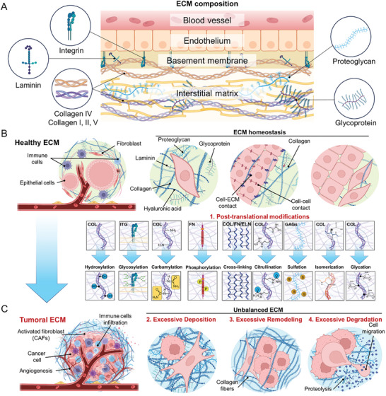
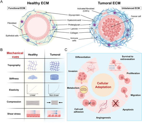
![Figure 1 Structural and compositional desmoplastic ECM alterations occurring during cancer development. A) The ECM is generally divided into an interstitial matrix and a basement membrane, which is specific to different tissues and organs. The basement membrane, which is rich in laminins and collagen IV, provides structural and organizational stability to the upper cell layer. The interstitial matrix lies below the basement membrane and is rich in collagens, proteoglycans (such as hyaluronan), and glycoproteins (such as fibronectin).[ 12 ] B) In a healthy microenvironment, epithelial cells interact with neighboring cells, including immune cells and resident fibroblasts, to maintain ECM homeostasis. C) However, during carcinogenesis, the ECM undergoes significant alterations, supporting a TME that sustains cancer development. These changes involve the activation of fibroblasts into cancer‐associated fibroblasts (CAFs) and the development of a fibrotic, stiff environment through: (1) posttranslational modifications of ECM components (collagen (COL), integrins (ITG), fibronectin (FN), elastin (ELN), glycosaminoglycans (GAGs); (2) excessive ECM deposition of collagen, laminin, hyaluronic acid, proteoglycans, and glycoproteins; (3) excessive ECM remodeling; and (4) excessive ECM degradation. These modifications support angiogenesis and the growth, survival, and invasion of tumor cells.](/paper-figures/605eec42fd1b45ddbe174516a0acfaf7/unraveling-the-role-of-the-tumor-extracellular-matrix-to-inform-nanoparticle-figure-1.jpg)

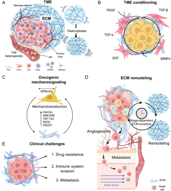
![Figure 3 ECM composition, organization, and remodeling sustain the evolution of the TME and promote cancer progression and development. A) The TME is a complex system composed of various cellular and acellular components. The tumor ECM is significantly different from that in healthy tissues in composition and mechanical properties. An increased and abundant deposition of collagens and stiffening of the surrounding stroma is characteristic of pathology and, together with fiber reorganization, establish the hallmarks of desmoplasia.[ 42 ] B) The irregular secretion of different factors (such as TGF‐β, PDGF, and EGF) and the increased activity of MMPs in the preneoplastic niche foster tumorigenicity and perpetuate the effects of desmoplasia. These occurrences facilitate the creation of a TME that supports the survival of cancer cells and the progression of the tumor. C) Mechanosensing pathways are commonly dysregulated in cancer cells, orchestrating the transcription of genes associated with cell proliferation, survival, and migration. A reciprocal loop forms between these pathways and ECM remodeling, mutually reinforcing one another and thereby promoting cancer progression. D) The presence of desmoplasia and the occurrence of ECM remodeling play a role in prompting cancer cells to adopt a more aggressive phenotype. This includes heightened migratory and invasive capabilities, which allow cancer cells to extravasate from the tumoral niche and enter the bloodstream, taking advantage of the newly formed vessels resulting from angiogenesis. During the process, some cells may evade clearance mechanisms and survive shear stress‐induced death, potentially leading to metastasis. E) The presence of tumor‐associated ECM presents several clinical challenges in cancer treatment. It acts as a physical barrier, limiting drug penetration and reducing the effectiveness of chemotherapy and immunotherapy, while releasing biochemical signals that promote immune evasion and foster drug resistance. It also enhances tumor cell migration and supports metastasis, which is responsible for most cancer‐related deaths. ECs, endothelial cells; CAFs, cancer‐associated fibroblasts; CCs, cancer cells.](/paper-figures/64efa02c968c495f9b7a462b4f8d4342/unraveling-the-role-of-the-tumor-extracellular-matrix-to-inform-nanoparticle-figure-3.jpg)
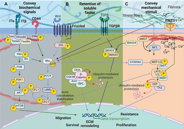
![Figure 4 The aberrant tumorigenic ECM modulates several intracellular signaling pathways involved in tumor development and progression. A) The cell membrane is the main hub that conveys changes occurring at the ECM level into the cell; receptors on the cell membrane such as integrins, hyaluronan receptor (CD44), G protein‐coupled receptor (e.g., Frizzled receptor), and TGF‐β receptor play a major role in the mechanosignaling cascade. B) Importantly, the ECM directly interacts with these receptors and promotes the retention of secreted soluble factors (e.g., Wnt and TGF‐βB) at the TME, which can consequently be activated during disease development and fuel tumor progression by initiating a signaling cascade that can inhibit growth suppressors and pro‐apoptotic proteins (e.g., via FAK‐Src signaling), promote cell proliferation and differentiation (e.g., via Wnt/β‐catenin pathway), and enhance migration and invasiveness through EMT (e.g., via TGF‐β pathway).[ 1 ] C) ECM stiffness also regulates the activity of Piezo1 ion channel, leading to the influx of Ca2+ ions and subsequent activation of downstream signaling pathways that modulate cell behavior. Mechanical cues also control the activity of angiomotin (AMOT) proteins and neurofibromin 2 (NF2, also known as Moesin‐Ezrin‐Radixin‐Like (MERLIN) tumor suppressor), which are negative regulators of yes‐associated protein (YAP). Following increased ECM stiffness, the assembly of actin results in the sequestration of AMOT, which prevents its interaction with NF2. This leads to the inhibition of the Hippo signaling pathway, resulting in the shuttling of YAP to the nucleus, where it promotes the transcription of genes encoding proteins that participate in focal adhesion assembly,[ 44 ] immune evasion,[ 45 ] resistance to therapy,[ 46 ] and ECM remodeling.](/paper-figures/6615500b3ec94d509a6c40d06f86b216/unraveling-the-role-of-the-tumor-extracellular-matrix-to-inform-nanoparticle-figure-4.jpg)
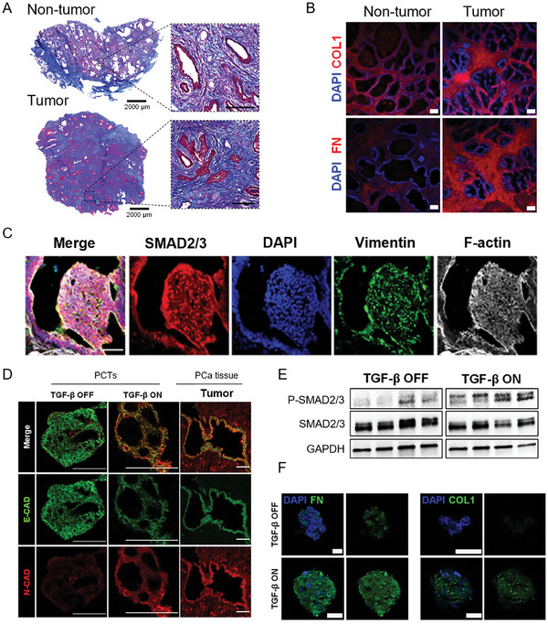
![Figure 5 TGF‐β promotes prostate cancer (PCa) development by inducing desmoplasia, EMT, cancer cell invasion, and migration. A) Representative images of Masson's trichrome staining of tumor and adjacent non‐tumor prostate tissues. Collagen is stained in blue. Scale bars: 2000 and 200 µm (magnified area). B) Representative confocal microscopy images of the indicated ECM markers, such as collagen I (COL1) and fibronectin (FN), in tumor and adjacent non‐tumor prostate tissues. Scale bars: 200 µm. C) Representative confocal images of tumor tissues stained for 4′,6‐diamidino‐2‐phenylindole (DAPI) (blue), SMAD 2/3 (red), vimentin (green), and f‐actin (white). Scale bar in all images: 50 µm. D) Representative confocal images of the PCTs showing the expression levels of epithelial‐cadherin (E‐CAD, green) and N‐cadherin (N‐CAD, red)—markers of benign epithelium and EMT, respectively—in the presence or absence of TGF‐β pathway activation. The tumoroids were fixed with 4% paraformaldehyde and then embedded in optical cutting temperature compound for slicing, prior to immunofluorescence staining. Scale bars: 50 µm. E) Protein quantification by western blot of SMAD2/3 and phospho‐SMAD2 (p‐SMAD2), which is phosphorylated and activated downstream of the TGF‐β pathway. F) Representative confocal images depicting the expression of collagen I and fibronectin in PCTs in the presence or absence of TGF‐β pathway activation. Scale bars: 50 µm.[ 58 ] Reproduced with permission.[ 58 ] Copyright 2023, Elsevier B.V.](/paper-figures/c9764128b4e04f0aa1b06ad6c77e5b03/unraveling-the-role-of-the-tumor-extracellular-matrix-to-inform-nanoparticle-figure-5.jpg)
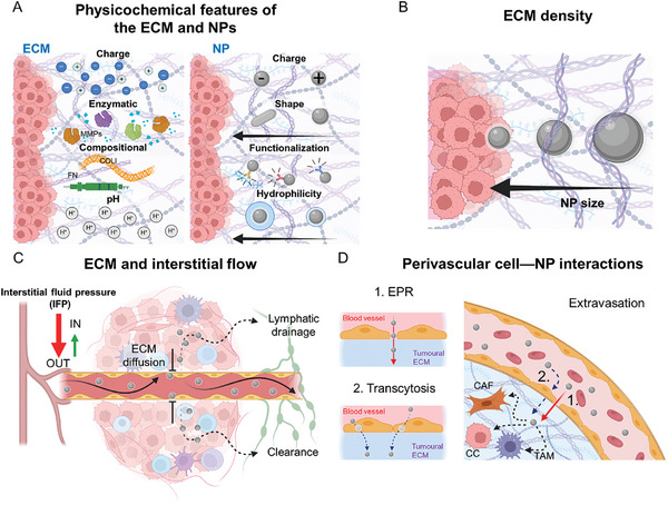
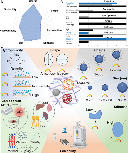
![Figure 6 The different features of the ECM contribute to restricting the accumulation of NPs at the tumor site, thereby reducing the effectiveness of nanomedicines. A) The biochemical features of the ECM affect its interaction with NPs, which consequently influences the optimal physicochemical properties of the NPs—i.e., surface charge, shape, hydrophilicity, and surface functionalization. Considering the physicochemical properties of both the ECM and NPs, it is essential to evaluate the outcome of NP–ECM interactions. B) Concurrently, ECM density impairs the diffusion of NPs via steric hindrance. Consequently, smaller NPs (<50 nm) are better able to penetrate the ECM than larger NPs. C) Likewise, IFP limits the diffusion of drugs and NPs at the TME. Abnormal angiogenesis, increased matrix deposition, and fibrosis can alter the hydration of the stroma surrounding the tumor, creating an interstitial fluidic gradient that hinders the extravasation of small molecules or favors their clearance via lymphatic vessels.[ 65 ] D) Two main mechanisms drive the accumulation of NPs in tumors: 1) EPR effect and 2) transcytosis. EPR relies on the leakiness of the fast‐growing vessels typical of the angiogenesis of tumors. Transcytosis involves the active internalization of the NPs by ECs of the blood vessels at the apical level and their subsequent exocytosis at the basal level. Although EPR has been demonstrated to be heterogeneous and inconsistent between animal and human tumors, and even among different human cancers, to date, transcytosis has only been observed in animal models. Once extravasated, NPs face interactions with and possible clearance by different cells residing at the TME, mainly by fibroblasts (CAFs) and macrophages (TAMs). Thus, targeting these cells may enhance the accumulation of NPs and drugs at the TME.](/paper-figures/1d8005b1a6184e559ff6110013a42c7c/unraveling-the-role-of-the-tumor-extracellular-matrix-to-inform-nanoparticle-figure-6.jpg)
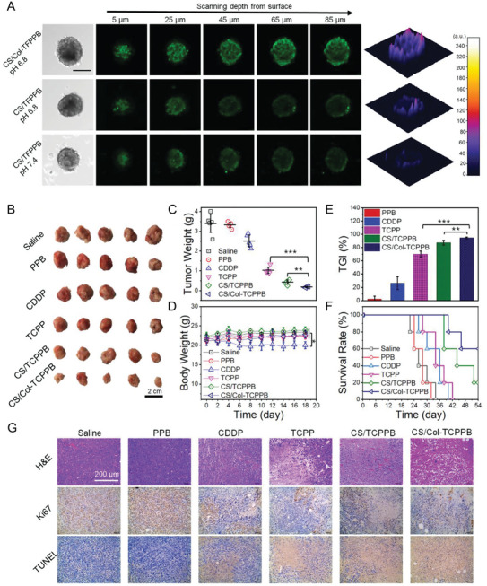
![Figure 7 The use of biocompatible coatings to protect NPs and their cargo can increase the delivery efficacy at the TME of drugs and enzymes that are suitable for hampering the deposition and stiffening of ECM, thus increasing the anti‐tumoral efficacy of the nanoformulation. A) Representative confocal images showing the in vitro penetration of NPs in breast cancer spheroids; the NPs were coated with chondroitin sulfate, labeled with fluorescein isothiocyanate, and encapsulated collagenase enzyme (CS/col‐TFPPB); the CS/TFPPB formulation does not contain collagenase enzyme. Scale bar for all images: 200 µm. B) Efficiency of the nanoformulation was corroborated in vivo in 4T1 breast tumor‐bearing mice. Comparison of the images of tumors obtained from mice treated with control groups (saline, poly(ethylene glycol)‐block‐poly(β‐amino ester) (PPB), cis‐diamminedichloroplatinum (CDDP, cisplatinum), and succinic anhydride‐modified cisplatin‐conjugated poly(ε‐caprolactone)‐block‐poly(ethylene oxide)‐triphenylphosphonium (TCPP)) and the nanoformulations CS‐Col‐TCPPB and CS/TCPPB revealed a significant reduction in tumor volume for the group treated with CS‐Col‐TCPPB NPs. Scale bar: 2 cm. C) Comparison of the weight of the tumors excised from the different treated mice confirmed that CS‐Col‐TCPPB was more effective in halting tumor progression than that of other formulations. D) This efficacy was not associated with systemic toxicity, as only the free drug (CDDP) treatment was associated with a reduction in mouse body weight. E) Tumor growth inhibition (TGI) for the nanoformulations coated with chondroitin sulfate was significantly higher than that of the uncoated NPs, with the presence of collagenase further increasing TGI. F) The data were further confirmed by assessing the survival rate of the 4T1 tumor‐bearing mice treated with various regimens. G) Hematoxylin and eosin (H&E) staining, terminal deoxynucleotidyl transferase‐mediated dUTP nick‐end labeling (TUNEL), and immunohistochemical analyses (Ki67) were used to further evaluate the antitumor effect. In H&E staining, nuclei are stained blue and the cytoplasm is stained red. Ki67‐positive proliferating cells and TUNEL‐positive apoptotic cells are stained brown. Scale bar for all images: 200 µm.[ 71b ] Reproduced with permission.[ 71b ] Copyright 2020, WILEY‐VCH Verlag GmbH & Co. KGaA, Weinheim.](/paper-figures/815378bd063e4302802c838826bc7b0f/unraveling-the-role-of-the-tumor-extracellular-matrix-to-inform-nanoparticle-figure-7.jpg)
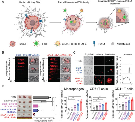
![Figure 8 The combination of ECM‐targeting therapy and immunotherapy can enhance NP penetration into tumors and improve cancer therapy. A) Schematic of the mechanism of the delivery of LNPs encapsulating FAK siRNA, Cas9 mRNA, and targeted sgRNAs to the tumor. This approach aims to enhance NP penetration by targeting the mechanical properties of cancer cells while simultaneously improving immunotherapy by knocking down PD‐L1 via gene editing.[ 25a ] B) Representative images of the penetration of NPs into the IGROV1 tumor spheroids treated with LNPs carrying a scrambled siRNA (siCtrl + Cy5‐mRNA‐LNPs) or an siRNA against FAK (siFAK + Cy5‐mRNA‐LNPs) after 48 h incubation. Scale bar for all images: 50 µm. C) mCherry expression (left) and deep distribution (right) in tumor spheroids treated with phosphate‐buffered saline (PBS), siCtrl + Cy5‐mRNA‐LNPs, siFAK + Cy5‐mRNA‐LNPs, carrying mRNA for expressing the red fluorescent mCherry protein. Scale bars: 100 µm. D) Excised tumors (left) and tumor size (right) show the in vivo therapeutic efficacy of the LNPs carrying siFAK and CRISPR/Cas9 construct for PD‐L1 silencing in cancer cells (siFAK + CRISPR‐PD‐L1‐LNPs) and the other control groups in mice bearing ID8‐Luc xenograft tumors. Scale bar for all images: 1 cm. E) Flow cytometry analyses of macrophages, CD8+ T cells, and CD4+ T cells in the tumors from mice treated with PBS, cargo‐free (Empty) LNPs, siCtrl + CRISPR‐PD‐L1‐LNPs, siFAK + CRISPR‐Ctrl‐LNPs, or siFAK+CRISPR‐PD‐L1‐LNPs after 30‐day therapy. Reproduced with permission.[ 25a ] Copyright 2022, Nature.](/paper-figures/7d81530fe2784868b305a4ad18e3cf56/unraveling-the-role-of-the-tumor-extracellular-matrix-to-inform-nanoparticle-figure-8.jpg)
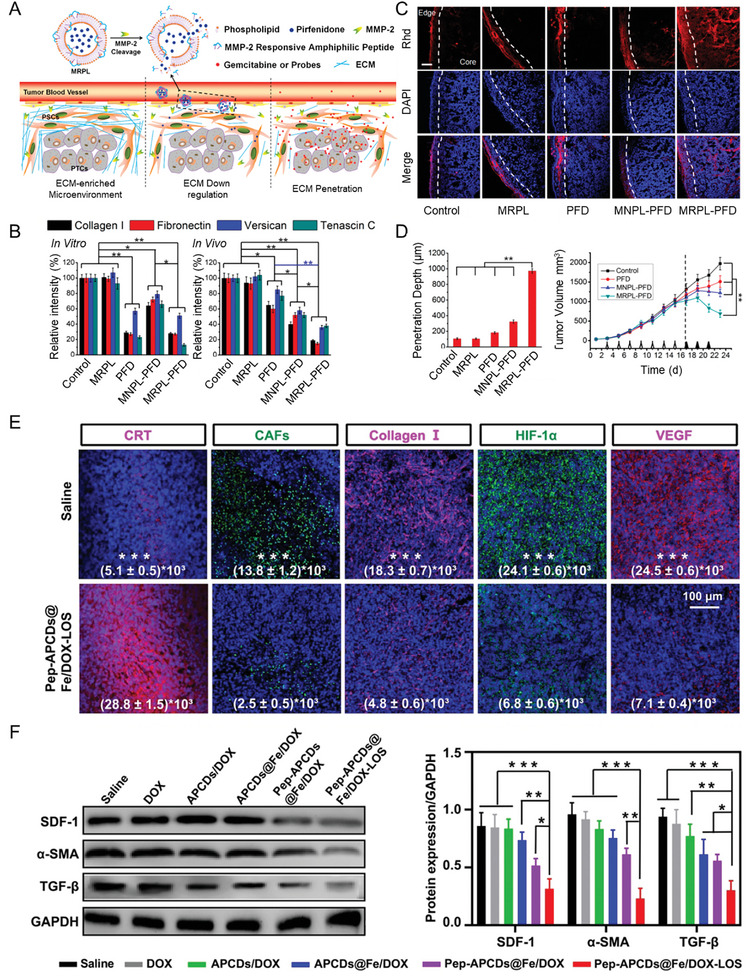
![Figure 9 The use of drugs acting on the pathways involved in ECM deposition and organization can reduce IFP and improve the perfusion of nanomedicines and small molecules into tumors, thus improving their antitumor efficacy. A) Schematic of the delivery strategy of NPs loaded with PFD in the ECM‐enriched environment typical of pancreatic tumor. The NPs can achieve a tumor‐specific release of PFD in the tumor stroma via a process mediated by MMP‐2 activity. The subsequent release of PFD inhibits ECM deposition and increases the perfusion of small molecules in the tumor tissue. B) Expression levels of multiple ECM components (collagen I, fibronectin, versican, and tenascin C) from immunohistochemistry measurements derived from in vitro (left) and in vivo (right) treatments of pancreatic stellate cells and a murine pancreatic tumor model, respectively, with various formulations: MMP‐2‐responsive liposomes (MRPL) alone or encapsulating PFD (MRPL‐PFD) and MMP‐2‐nonresponsive liposomes (MNPL) alone or encapsulating PFD (MNPL‐PFD). C) Rhodamine (Rhd) penetration and distribution in a murine model of pancreatic tumor after 2 weeks of treatment with the different PFD formulations. Frozen tumor sections were stained with DAPI (blue) to label nuclei. Rhd is visible in red. Scale bar for all images: 100 µm. D) Quantification of the depth of Rhd penetration in tumors treated with the PFD formulations (left). Growth curves of pancreatic tumors in mice treated with the different PFD formulations and subsequently injected with gemcitabine (right).[ 90b ] E) Immunofluorescence analysis of calreticulin (CRT; red), CAFs (green), collagen I (red), hypoxia‐inducible factor‐1α (HIF‐1α) (green), and VEGF (red) at the endpoint after treatment with CD nanoassemblies carrying DOX and LOS (Pep‐APCDs@Fe/DOX‐LOS) compared to control saline treatment. Scale bar for all images: 100 µm. F) Western blot analysis showing the protein levels of SDF‐1, α‐SMA, and TGF‐β found in 4T1 tumor‐bearing mice after treatment with different nanoformulations (left) and the relative quantification (right).[ 93a ] (A–D) Reproduced with permission.[ 90b ] Copyright 2017, American Chemical Society. (E, F) Reproduced with permission.[ 93a ] Copyright 2020, Wiley‐VCH GmbH.](/paper-figures/4870cef7c1044225864b3b00e9192998/unraveling-the-role-of-the-tumor-extracellular-matrix-to-inform-nanoparticle-figure-9.jpg)
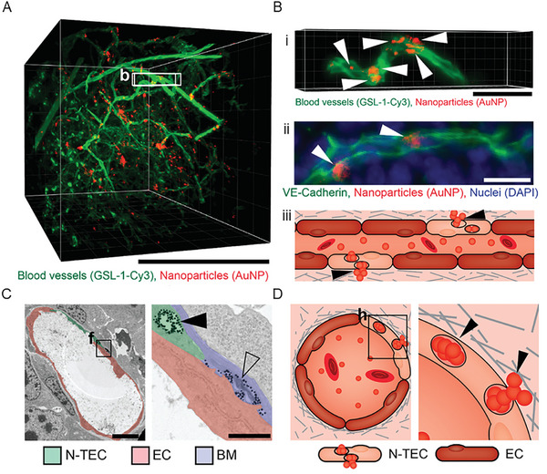
![Figure 10 The accumulation of NPs at the tumor can be mediated by processes beyond the classical EPR effect, such as transcytosis by a specific subset of ECs present in the tumor vasculature. A) 3D microscopy image of murine tumor derived from 4T1 cells showing blood vessels in green and 50 nm gold NPs in red. Scale bar: 500 µm. B) Magnified region of (A) (b‐squared selection) showing an individual vessel with clusters of NPs, indicated by white arrows (i). 2D section of a representative blood vessel stained with vascular endothelial (VE)‐cadherin to highlight EC boundaries; clusters of NPs are highlighted with white arrows (ii). Schematic showing NP transport mediated by specific ECs; black arrows indicate clusters of NPs (iii). Scale bar: 50 µm. C) Transmission electron microscopy image of a representative tumor blood vessel (left) and a close‐up image of a region of the vessel where NPs accumulated (right). The solid black arrow shows NPs packaged in a vacuole of an endothelial cell (N‐TEC). The open arrow shows NPs in the extravascular space surrounded by the basement membrane. The N‐TEC is highlighted in green, cells without NP uptake are in red, and the basement membrane is shown in blue. Scale bars: 5 µm (left), 1 µm (right). D) Schematic of the mechanism of NP transcytosis by N‐TECs at the interface between blood vessels and the extracellular space. Clusters of NPs are indicated by black arrows.[ 101 ] Reproduced with permission.[ 101 ] Copyright 2021, American Chemical Society.](/paper-figures/9c5e5e3212744558946ff11b9c020d9c/unraveling-the-role-of-the-tumor-extracellular-matrix-to-inform-nanoparticle-figure-10.jpg)
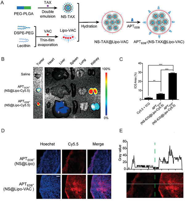
![Figure 11 The modulation of the TME via the inhibition of pro‐fibrotic cells and the reduction of ECM deposition can increase the accumulation of nanodrugs at tumors. A) Schematic of the synthesis of NPs (specifically nanospheres (NS)) encapsulating vactosertib (VAC) and paclitaxel (TAX) (NS‐TAX@Lipo‐VAC) and functionalized with a peptide specific for the fibronectin EDB (APTEDB) (APTEDB‐NS‐TAX@Lipo‐VAC). PLGA, poly(lactic‐co‐glycolic acid). B) NPs functionalized with APTEDB or a scrambled peptide (APTSCR) and labeled with Cy5.5 dye were injected intravenously into mice with orthotopic pancreatic cancer. Ex vivo Cy5.5 fluorescence images of major organs and tumors show the prevalent accumulation of the NPs functionalized with APTEDB at the tumor. C) The results were further confirmed with photoacoustic imaging, by encapsulating ICG into the nanoformulation, which revealed the higher accumulation of the APTEDB‐functionalized NPs at the tumor compared to the NPs functionalized with APTSCR. D) By adding vactosertib to the nanoformulation, the NPs functionalized with APTEDB showed higher accumulation at the tumor, and E) could penetrate deeper into the tumoral mass compared to the NPs without the drug.[ 102a ] Reproduced with permission.[ 102a ] Copyright 2021, American Chemical Society.](/paper-figures/d463747ddcd04a2299ddbc52c3da9c73/unraveling-the-role-of-the-tumor-extracellular-matrix-to-inform-nanoparticle-figure-11.jpg)


Verification
Claims the paper makes
The claims and statistics of this paper haven’t been checked yet.
Run the check — free, no account needed. It reads the open-access text and checks the sources it cites.
Claims other papers make about this one
Each is checked against this paper’s own text: does it say what the citing paper says it does?
In the citing paper
“Tumor vasculature, genetic anomalies, and varying quantities of extracellular matrix (ECM) components can all make it harder for MNPs to accurately target certain tumor locations [79].”
From Advances in green-synthesized magnetic nanoparticles for targeted cancer therapy: mechanisms, applications, and future perspectives (2026) · cites this paper as [79]
Self-check
Percentages, sums and reported differences re-computed from the paper’s own numbers.
The self-check needs the paper’s full text.
Citations
Cites
A framework for advancing our understanding of cancer-associated fibroblasts
Sahai, E., Astsaturov, I., Cukierman, E., DeNardo, D. G., Egeblad, M., Evans, R. M., Fearon, D., Greten, F. R., Hingorani, S. R., Hunter, T., et al. (2020) · Nature Reviews Cancer · doi:10.1038/s41568-019-0238-1 · Cited by 3,863
Accumulation of sub-100 nm polymeric micelles in poorly permeable tumours depends on size
Cabral, H., Matsumoto, Y., Mizuno, K., Chen, Q., Murakami, M., Kimura, M., Terada, Y., Kano, M. R., Miyazono, K., Uesaka, M., et al. (2011) · Nature Nanotechnology · doi:10.1038/nnano.2011.166 · Cited by 2,429
Cheng, Q., Wei, T., Farbiak, L., Johnson, L. T., Dilliard, S. A., & Siegwart, D. J. (2020) · Nature Nanotechnology · doi:10.1038/s41565-020-0669-6 · Cited by 2,383
High Interstitial Fluid Pressure - an Obstacle in Cancer Therapy
Heldin, C.-H., Rubin, K., Pietras, K., & Östman, A. (2004) · Nature reviews. Cancer · doi:10.1038/nrc1456 · Cited by 2,222
Drost, J., & Clevers, H. (2018) · Nature Reviews Cancer · doi:10.1038/s41568-018-0007-6 · Cited by 1,790
The entry of nanoparticles into solid tumours
Sindhwani, S., Syed, A. M., Ngai, J., Kingston, B. R., Maiorino, L., Rothschild, J., MacMillan, P., Zhang, Y., Rajesh, N. U., Hoang, T., et al. (2020) · Nature Materials · doi:10.1038/s41563-019-0566-2 · Cited by 1,732
Calvo, F., Ege, N., Grande-Garcia, A., Hooper, S., Jenkins, R. P., Chaudhry, S. I., Harrington, K., Williamson, P., Moeendarbary, E., Charras, G., et al. (2013) · Nature Cell Biology · doi:10.1038/ncb2756 · Cited by 1,478
The evolution of commercial drug delivery technologies
Vargason, A. M., Anselmo, A. C., & Mitragotri, S. (2021) · Nature Biomedical Engineering · doi:10.1038/s41551-021-00698-w · Cited by 1,361
Tumor targeting via EPR: Strategies to enhance patient responses
Golombek, S. K., May, J.-N., Theek, B., Appold, L., Drude, N., Kiessling, F., & Lammers, T. (2018) · Advanced Drug Delivery Reviews · doi:10.1016/j.addr.2018.07.007 · Cited by 1,262
Chauhan, V. P., Martin, J. D., Liu, H., Lacorre, D. A., Jain, S. R., Kozin, S. V., Stylianopoulos, T., Mousa, A. S., Han, X., Adstamongkonkul, P., et al. (2013) · Nature Communications · doi:10.1038/ncomms3516 · Cited by 1,031
Research works printed but not linked: 192
- 10.15252/embr.201439246 · DOI
- 10.1242/jcs.023820 · DOI · Paper page
- 10.1038/s41467-020-18794-x · DOI · Paper page
- 10.1038/s41563-020-00894-0 · DOI
- 10.1002/ar.24086 · DOI
- 10.1136/jmg.2005.039669 · DOI
- 10.1111/j.0022-202X.2004.23406.x · DOI · Paper page
- 10.1016/j.molmed.2005.04.006 · DOI
- 10.1126/scitranslmed.adg3840 · DOI
- 10.1038/s41568-020-00329-7 · DOI
- 10.1186/s13045-019-0770-1 · DOI · Paper page
- 10.1038/d41587-019-00019-2 · DOI
- 10.1038/nrclinonc.2010.139 · DOI
- Netti P. A., 2000, Cancer Res., 60, 2497
- 10.3389/fonc.2021.650453 · DOI
- 10.1021/acsami.5b07116 · DOI
- 10.1136/gutjnl-2016-312473 · DOI
- 10.1016/j.bbcan.2020.188435 · DOI
- 10.1634/theoncologist.2016-0479 · DOI
- 10.1634/theoncologist.2017-0024 · DOI
- 10.1016/j.tips.2017.10.009 · DOI
- S.Hua M. B. C.deMatos J. M.Metselaar G.Storm 2018 9. · DOI
- 10.1080/17425247.2017.1262346 · DOI
- 10.1038/s41565-022-01122-3 · DOI · Paper page
- 10.1002/advs.202302965 · DOI
- 10.1016/j.devcel.2021.05.011 · DOI
- 10.1038/nrc2544 · DOI
- 10.1039/D4TB00328D · DOI
- 10.1242/dmm.004077 · DOI · Paper page
- 10.1016/j.tcb.2010.08.015 · DOI
- 10.1038/s41416-020-01150-7 · DOI
- 10.1038/s41580-020-00306-w · DOI
- 10.1038/s41392-021-00544-0 · DOI · Paper page
- 10.1038/s41467-022-32255-7 · DOI
- 10.1038/s41563-020-0615-x · DOI
- 10.1016/j.devcel.2019.03.026 · DOI
- 10.1152/ajpcell.00238.2015 · DOI
- Yankaskas C. L., 2021, Sci. Adv., 7
- 10.3389/fmolb.2019.00160 · DOI · Paper page
- 10.4161/onci.20239 · DOI
- 10.1038/s41587-022-01294-2 · DOI · Paper page
- 10.1002/advs.202305769 · DOI
- 10.1038/ncomms15321 · DOI
- 10.1158/1541-7786.MCR-19-0375 · DOI
- 10.1016/j.trecan.2019.02.010 · DOI · Paper page
- J.Seong N.Wang Y.Wang 2013 17 597. · DOI
- 10.1038/nrm1549 · DOI
- 10.1083/jcb.201501025 · DOI
- 10.1038/nature10137 · DOI · Paper page
- 10.1038/nrc3876 · DOI
- 10.1016/j.biomaterials.2019.03.009 · DOI
- 10.1016/j.trecan.2023.07.006 · DOI
- 10.1038/ncomms8223 · DOI · Paper page
- 10.1016/j.celrep.2016.10.033 · DOI
- 10.1016/j.biopha.2021.111692 · DOI
- 10.1038/nature25501 · DOI · Paper page
- 10.1016/j.cell.2008.07.001 · DOI · Paper page
- 10.1016/j.matbio.2023.11.001 · DOI
- 10.1038/natrevmats.2016.14 · DOI · Paper page
- 10.1038/s41563-020-0755-z · DOI
- 10.1016/j.cis.2024.103124 · DOI
- 10.1016/j.mtbio.2022.100364 · DOI
- 10.1002/adhm.202101428 · DOI
- 10.1152/physiol.00026.2017 · DOI
- 10.1186/s13287-022-03070-0 · DOI
- 10.1016/j.stem.2020.10.018 · DOI
- 10.1016/j.bpj.2010.06.016 · DOI
- 10.1021/am506112u · DOI
- 10.1177/1759091415592389 · DOI
- 10.1039/C9MH00835G · DOI
- 10.1002/adhm.201500441 · DOI · Paper page
- 10.1021/acs.langmuir.0c01624 · DOI
- 10.1002/anie.201104449 · DOI
- 10.1021/acsami.7b09458 · DOI
- 10.1002/adma.201906745 · DOI
- 10.1021/acsami.0c03022 · DOI
- 10.1016/j.jcis.2018.04.084 · DOI
- 10.1073/pnas.1522080113 · DOI
- 10.1016/j.biomaterials.2015.05.006 · DOI
- b)C.Wong T.Stylianopoulos J.Cui J.Martin V. P.Chauhan W.Jiang Z.Popović R. K.Jain M. G.Bawendi D.Fukumura 2011 108 2426. · DOI
- 10.2174/1381612826666201125111625 · DOI
- 10.1016/j.jconrel.2020.04.019 · DOI
- 10.1186/s12989-017-0199-z · DOI
- 10.1039/C5NR08463F · DOI
- 10.1158/0008-5472.CAN-12-2071 · DOI
- 10.2217/17435889.2.3.265 · DOI
- 10.1016/j.cis.2018.03.006 · DOI
- 10.1039/D0MH00870B · DOI
- 10.1039/C7CS00522A · DOI
- 10.1039/C9CS00129H · DOI
- 10.1016/j.jconrel.2017.02.035 · DOI
- 10.1016/j.biomaterials.2018.04.045 · DOI
- 10.1186/s40580-017-0124-z · DOI
- 10.1016/j.nano.2020.102183 · DOI
- 10.3390/nano11020438 · DOI
- 10.1021/acs.nanolett.6b00068 · DOI
- c)J.Li C.Xie J.Huang Y.Jiang Q.Miao K.Pu 2018 57 3995. · DOI
- 10.1021/acsnano.1c10590 · DOI
- 10.1002/adma.202106390 · DOI
- 10.1016/j.biomaterials.2012.12.012 · DOI
- 10.1021/acsami.7b05119 · DOI
- 10.1021/acsnano.9b03978 · DOI
- 10.1042/CS20200039 · DOI
- 10.1038/s41467-018-05906-x · DOI · Paper page
- 10.1016/j.biomaterials.2020.119772 · DOI
- 10.1021/acsnano.7b01026 · DOI
- 10.1021/acsami.0c10416 · DOI
- Chen Y., 2019, ACS Nano, 13, 1751
- 10.1002/anie.202014397 · DOI
- 10.1186/s12967-020-02376-y · DOI
- 10.1001/jamaoncol.2019.0892 · DOI
- 10.1021/acsami.9b13957 · DOI
- 10.1016/j.biomaterials.2018.01.014 · DOI
- 10.1016/j.jconrel.2016.11.015 · DOI · Paper page
- 10.2174/0929867327666200625151134 · DOI
- 10.1038/s41565-019-0485-z · DOI
- 10.1021/acsnano.1c04510 · DOI
- 10.1021/acsnano.0c08860 · DOI
- 10.1038/s41467-020-14425-7 · DOI
- 10.4161/cbt.5.12.3354 · DOI
- 10.1021/acsnano.6b02776 · DOI
- Zhou S., 2021, Adv. Funct. Mater., 31
- 10.1016/j.ebiom.2021.103664 · DOI
- 10.1073/pnas.1917424117 · DOI
- 10.1021/acsnano.8b03900 · DOI
- 10.1016/j.carbpol.2020.116887 · DOI
- 10.1021/acs.chemmater.7b03128 · DOI
- H.Wang X.Han Z.Dong J.Xu J.Wang Z.Liu 2019 29 1902440. · DOI
- 10.1039/D0BM00916D · DOI
- 10.1016/j.ebiom.2022.103873 · DOI
- 10.1016/j.smim.2017.09.004 · DOI
- 10.3390/pharmaceutics13101670 · DOI
- 10.1146/annurev.cellbio.22.010305.104315 · DOI
- 10.1016/j.matbio.2022.05.003 · DOI
- 10.1021/acs.bioconjchem.0c00270 · DOI
- A. E.Gilchrist S.Lee Y.Hu B. A. C.Harley 2019 8 1900751. · DOI
- 10.1002/EXP.20210093 · DOI
- 10.1016/j.addr.2021.04.001 · DOI
- 10.1038/s41568-022-00514-w · DOI
- 10.1126/sciadv.abi9119 · DOI
- 10.1371/journal.pone.0118090 · DOI
- 10.1016/j.actbio.2022.11.038 · DOI · Paper page
- 10.1016/j.bioactmat.2021.12.005 · DOI
- A. R.Perestrelo A. C.Silva J. O.‐D. L.Cruz F.Martino V.Horváth G.Caluori O.Polanský V.Vinarský G.Azzato G. d.Marco V.Žampachová P.Skládal S.Pagliari A.Rainer P.Pinto‐do‐Ó A.Caravella K.Koci D. S.Nascimento G.Forte 2021 128 24. · DOI
- 10.1038/nm.4352 · DOI
- 10.1021/acs.jproteome.7b00191 · DOI
- 10.1186/s42047-021-00089-0 · DOI
- 10.1038/s41467-022-29279-4 · DOI · Paper page
- 10.1038/s41596-020-0379-4 · DOI
- 10.1038/s41563-021-01057-5 · DOI
- 10.1016/j.ccell.2021.07.020 · DOI · Paper page
- 10.1038/s41467-017-02588-9 · DOI
- 10.1126/sciadv.aaz4316 · DOI
- 10.1038/s41573-020-0090-8 · DOI · Paper page
- 10.1016/j.tips.2022.05.001 · DOI
- 10.1016/j.jconrel.2022.09.002 · DOI
- 10.1016/j.onano.2022.100048 · DOI
- 10.1002/adma.201904625 · DOI
- 10.1039/D1NR08429A · DOI
- 10.1039/D1CS00427A · DOI
- 10.1002/wnan.1449 · DOI
- 10.1073/pnas.2109256118 · DOI · Paper page
- 10.1038/nnano.2012.207 · DOI · Paper page
- 10.1038/nnano.2012.237 · DOI · Paper page
- 10.1021/acsnano.9b03824 · DOI
- 10.1158/0008-5472.CAN-13-3494 · DOI · Paper page
- 10.1021/accountsmr.2c00072 · DOI
- 10.1002/adhm.201700739 · DOI
- 10.1021/nn500216y · DOI
- 10.1016/j.biomaterials.2015.12.014 · DOI
- 10.1146/annurev.bioeng.9.060906.151850 · DOI
- 10.1016/S0962-8924(01)82222-4 · DOI
- 10.1006/scbi.2000.0379 · DOI
- 10.1242/jcs.144378 · DOI
- 10.1016/j.cell.2016.11.005 · DOI
- 10.1002/mc.23273 · DOI
- Y.Lan D.Zhang C.Xu K. W.Hance B.Marelli J.Qi H.Yu G.Qin A.Sircar V. M.Hernández M. H.Jenkins R. E.Fontana A.Deshpande G.Locke H.Sabzevari L.Radvanyi K.‐M.Lo 2018 10 eaan5488. · DOI
- 10.1038/s41573-021-00284-4 · DOI
- 10.1158/1078-0432.CCR-22-0308 · DOI
- 10.1073/pnas.1018892108 · DOI · Paper page
- 10.1039/C9BM01843C · DOI
- 10.1021/nl400033h · DOI
- 10.1002/adma.201707464 · DOI
- 10.1002/admi.202300137 · DOI
- 10.1002/adfm.202304674 · DOI
- 10.1039/D2NR03792K · DOI
- 10.1016/j.nano.2022.102550 · DOI · Paper page
- 10.1101/2024.07.07.599665 · DOI
- 10.1002/adma.202107964 · DOI
- 10.1186/s13045-023-01463-z · DOI
- 10.1002/EXP.20220147 · DOI
- 10.1038/s41467-023-40300-2 · DOI
Cited by
Sorted by citations
El-khawaga, A. M., Youssef, O. I., El-dydamoni, O. A., Ragab, W. M., & Mohammed, R. A. E. (2026) · European Journal of Medical Research · doi:10.1186/s40001-025-03804-9 · Cited by 14
Metadata
- Authors
- Cassani, M., Fernandes, S., Pagliari, S., Cavalieri, F., Caruso, F., & Forte, G.
- Venue
- Advanced Science
- Publisher
- Wiley
- Published
- 2024-12-04
- Volume
- 12
- Issue
- 2
- ISSN
- 2198-3844
- PMC
- PMC11727388
- Cited by
- 48
- References
- 27
- Language
- en
- Funders
- Ministerstvo Zdravotnictví Ceské Republiky (NU23J‐08‐00035)
- National Health and Medical Research Council (GNT2016732)
About these checks
Registry, retraction, Cites and statistics results are computed, not judged, with one exception: a printed reference that no index lists is read once by a model before it counts as a citation error. Claims are extracted by a model and kept only when their quote appears verbatim in the text; each cited source is then checked, with a model’s help, for whether it says what the claim relies on. Those verdicts can be wrong — read the claim’s quote and the cited source before relying on one.
Statistics and arithmetic are recomputed, not judged.
Confidence bands are shown only for fields calibrated on our annotated test set. None is calibrated yet, so no band is shown.
What counts as a claim ›How we measure confidence ›How we check this ›
Are you an author, or do you see a problem with this page? Request a review