

The DOI matches its Crossref record; 16 of the 16 references it cites were found in a registry or an index.
The unitary model for estrogen deficiency and the pathogenesis of osteoporosis: Is a revision needed?
Abstract
Over a decade ago, we proposed a “unitary” model for the pathogenesis of osteoporosis that identified estrogen deficiency as the predominant cause of both the early, accelerated, and late slow phases of bone loss in postmenopausal women and as a contributing cause of the continuous phase of bone loss in aging men. While this was a plausible model then, new data over the intervening years suggest a need to modify these concepts. Indeed, based largely on rodent studies, a “revisionist” view of the pathogenesis of osteoporosis has been proposed recently that attempts a paradigm shift from the estrogen-centric model to one in which bone loss is largely independent of estrogen deficiency and is driven instead by cell-autonomous age-related factors. However, detailed clinical investigative studies using quantitative computed tomography demonstrate that the onset of cortical bone loss in humans is closely tied to estrogen deficiency; thus the estrogen-centric view is likely correct for cortical bone, which comprises over 80% of the skeleton and is the major structural determinant of fracture risk at most skeletal sites. By contrast, these same studies also demonstrate that trabecular bone loss begins in sex hormone–replete young adults of both sexes. This suggests that a significant proportion of trabecular bone loss is either estrogen-independent or, as suggested by some studies, requires higher levels for its regulation. In this perspective, we critically review these and other findings, leading us to conclude that our original model requires modification but not revision. © 2011 American Society for Bone and Mineral Research.
Overview
The claims and statistics of this paper haven’t been checked yet.
Run the check — free, no account needed. It reads the open-access text and checks the sources it cites.
Integrity signals
Registry
Cites
Statistics
Figures
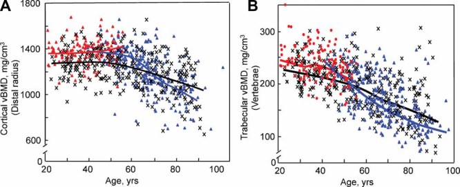
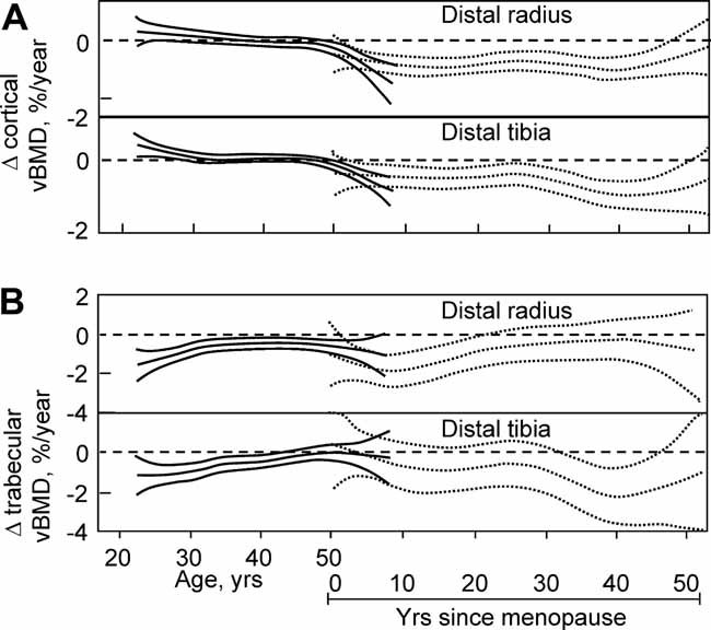
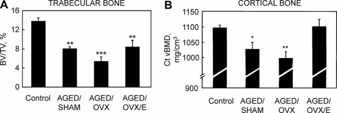
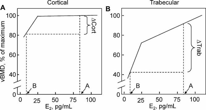
Verification
Claims the paper makes
The claims and statistics of this paper haven’t been checked yet.
Run the check — free, no account needed. It reads the open-access text and checks the sources it cites.
Claims other papers make about this one
Each is checked against this paper’s own text: does it say what the citing paper says it does?
In the citing paper
“One of the most common bone disorders, postmenopausal osteoporosis, arises from an increase in the formation and activity of osteoclasts due to the decrease in estrogen levels [2,3].”
From Anti-Osteoporotic Potential of Water Extract of Anethum graveolens L. Seeds (2023) · cites this paper as [3]
Self-check
Percentages, sums and reported differences re-computed from the paper’s own numbers.
The self-check needs the paper’s full text.
Citations
Cites
Cellular senescence: when bad things happen to good cells
Campisi, J., & d'Adda di Fagagna, F. (2007) · Nature Reviews Molecular Cell Biology · doi:10.1038/nrm2233 · Cited by 4,441
Resveratrol improves health and survival of mice on a high-calorie diet
Baur, J. A., Pearson, K. J., Price, N. L., Jamieson, H. A., Lerin, C., Kalra, A., Prabhu, V. V., Allard, J. S., Lopez-Lluch, G., Lewis, K., et al. (2006) · Nature · doi:10.1038/nature05354 · Cited by 4,290
Sex Steroids and the Construction and Conservation of the Adult Skeleton
Riggs, B. L., Khosla, S., & Melton, L. J. (2002) · Endocrine Reviews · doi:10.1210/edrv.23.3.0465 · Cited by 1,846
Self-citationRole of RANK ligand in mediating increased bone resorption in early postmenopausal women
Eghbali-Fatourechi, G., Khosla, S., Sanyal, A., Boyle, W. J., Lacey, D. L., & Riggs, B. L. (2003) · Journal of Clinical Investigation · doi:10.1172/jci17215 · Cited by 825 · cited 2×
“Thus, by increasing osteoprotegerin (OPG)((45)) and decreasing receptor activator of nuclear kB ligand (RANKL)((46)) production by osteoblastic cells, as well as by suppressive effects on the production of tumor necrosis factor a (TNF-a)((47)) and other proresorptive cytokines,((48)) estrogen reduces osteoclastogenesi…”
BackgroundSelf-citationFalahati-Nini, A., Riggs, B. L., Atkinson, E. J., O’Fallon, W. M., Eastell, R., & Khosla, S. (2000) · Journal of Clinical Investigation · doi:10.1172/jci10942 · Cited by 757 · cited 2×
“Moreover, short-term interventional studies in which men are made hypogonadal and are selectively replaced either with estrogen or testosterone have demonstrated the importance of estrogen in regulating both bone formation and bone resorption in adult men.((30,31))”
BackgroundSelf-citationFSH Directly Regulates Bone Mass
Sun, L., Peng, Y., Sharrow, A. C., Iqbal, J., Zhang, Z., Papachristou, D. J., Zaidi, S., Zhu, L.-L., Yaroslavskiy, B. B., Zhou, H., et al. (2006) · Cell · doi:10.1016/j.cell.2006.01.051 · Cited by 723 · cited 3×
“These investigators also found that osteoclasts and their precursors possessed FSH receptors and that FSH [but not luteinizing hormone (LH)] increased osteoclast formation and function in vitro, and they concluded that high circulating FSH levels caused hypogonadal bone loss.((89)) By contrast, Gao and colleagues((90)…”
BackgroundResultInfluentialHofbauer, L. C., Khosla, S., Dunstan, C. R., Lacey, D. L., Spelsberg, T. C., & Riggs, B. L. (1999) · Endocrinology · doi:10.1210/endo.140.9.7131 · Cited by 692 · cited 2×
“Thus, by increasing osteoprotegerin (OPG)((45)) and decreasing receptor activator of nuclear kB ligand (RANKL)((46)) production by osteoblastic cells, as well as by suppressive effects on the production of tumor necrosis factor a (TNF-a)((47)) and other proresorptive cytokines,((48)) estrogen reduces osteoclastogenesi…”
BackgroundSelf-citationRoggia, C., Gao, Y., Cenci, S., Weitzmann, M. N., Toraldo, G., Isaia, G., & Pacifici, R. (2001) · Proceedings of the National Academy of Sciences · doi:10.1073/pnas.251534698 · Cited by 513
Slemenda, C. W., Longcope, C., Zhou, L., Hui, S. L., Peacock, M., & Johnston, C. C. (1997) · Journal of Clinical Investigation · doi:10.1172/jci119701 · Cited by 455
Androgen therapy in women: An Endocrine Society clinical practice guideline
Wierman, M. E., Basson, R., Davis, S. R., Khosla, S., Miller, K. K., Rosner, W., & Santoro, N. (2006) · The Journal of Clinical Endocrinology & Metabolism · doi:10.1210/jc.2006-1121 · Cited by 301 · cited 1×
“In addition to progesterone, androgen levels also decrease during the menopausal transition((85)) and could contribute to bone loss.”
BackgroundSelf-citation
Research works printed but not linked: 75
- Riggs, 1983, Evidence for two distinct syndromes of involutional osteoporosis, Am J Med., 75, 899, 10.1016/0002-9343(83)90860-4 · DOI
- Riggs, 1998, A unitary model for involutional osteoporosis: estrogen deficiency causes both type I and type II osteoporosis in postmenopausal women and contributes to bone loss in aging men, J Bone Miner Res., 13, 763, 10.1359/jbmr.1998.13.5.763 · DOI
- Manolagas, 2010, From estrogen-centric to aging and oxidative stress: a revised perspective of the pathogenesis of osteoporosis, Endocr Rev., 31, 266, 10.1210/er.2009-0024 · DOI
- Riggs, 2004, Population-based study of age and sex differences in bone volumetric density, size, geometry, and structure at different skeletal sites, J Bone Miner Res., 19, 1945, 10.1359/jbmr.040916 · DOI · Paper page
- Riggs, 2008, A population-based assessment of rates of bone loss at multiple skeletal sites: evidence for substantial trabecular bone loss in young adult women and men, J Bone Miner Res., 23, 205, 10.1359/jbmr.071020 · DOI
- Bonnick, 1998, Bone Densitometry in Clinical Practice, 35, 10.1007/978-1-4757-2780-7 · DOI
- Rockoff, 1969, The relative contribution of trabecular and cortical bone to the strength of human lumbar vertebrae, Calcif Tissue Res., 3, 163, 10.1007/BF02058659 · DOI
- Boutroy, 2008, Finite element analysis based on in vivo HR-pQCT images of the distal radius is associated with wrist fracture in postmenopausal women, J Bone Miner Res., 23, 392, 10.1359/jbmr.071108 · DOI
- Cooper, 1992, Epidemiology of osteoporosis, Trends Endocrinol Metab., 3, 224, 10.1016/1043-2760(92)90032-V · DOI · Paper page
- van Staa, 2001, Epidemiology of fractures in England and Wales, Bone., 29, 517, 10.1016/S8756-3282(01)00614-7 · DOI
- Riggs, 2002, Bone turnover matters: the raloxifene treatment paradox of dramatic decreases in vertebral fractures without commensurate increases in bone density, J Bone Miner Res., 17, 11, 10.1359/jbmr.2002.17.1.11 · DOI
- Richelson, 1984, Relative contributions of aging and estrogen deficiency to postmenopausal bone loss, N Engl J Med., 311, 1273, 10.1056/NEJM198411153112002 · DOI
- Syed, 2010, Effects of chronic estrogen treatment on modulating age-related bone loss in female mice, J Bone Miner Res., 25, 2438, 10.1002/jbmr.129 · DOI
- Nelson, 1982, A longitudinal study of estrous cyclicity in C57BL/6J mice: I. cycle frequency, length and vaginal cytology, Biol Reprod., 27, 327, 10.1095/biolreprod27.2.327 · DOI
- Felicio, 1984, Longitudinal studies of estrous cyclicity in aging C57BL/6J mice: II. cessation of cyclicity and the duration of persistent vaginal cornification, Biol Reprod., 31, 446, 10.1095/biolreprod31.3.446 · DOI
- Glatt, 2007, Age-related changes in trabecular architecture differ in female and male C57BL/6J mice, J Bone Miner Res., 22, 1197, 10.1359/jbmr.070507 · DOI
- Khosla, 1998, Relationship of serum sex steroid levels and bone turnover markers with bone mineral density in men and women: A key role for bioavailable estrogen, J Clin Endocrinol Metab., 83, 2266
- Greendale, 1997, Endogenous sex steroids and bone mineral density in older women and men: the Rancho Bernardo study, J Bone Miner Res., 12, 1833, 10.1359/jbmr.1997.12.11.1833 · DOI
- Center, 1999, Hormonal and biochemica parameters in the determination of osteoporosis in elderly men, J Clin Endocrinol Metab., 84, 3626
- Ongphiphadhanakul, 1998, Serum oestradiol and oestrogen-receptor gene polymorphism are associated with bone mineral density independently of serum testosterone in normal males, Clin Endocrinol., 49, 803, 10.1046/j.1365-2265.1998.00631.x · DOI
- van den Beld, 2000, Measures of bioavailable serum testosterone and estradiol and their relationships with muscle strength, bone density, and body composition in elderly men, J Clin Endocrinol Metab., 85, 3276
- Amin, 2000, Association of hypogonadism and estradiol levels with bone mineral density in elderly men from the Framingham study, Ann Intern Med., 133, 951, 10.7326/0003-4819-133-12-200012190-00010 · DOI
- Khosla, 2001, Relationship of serum sex steroid levels to longitudinal changes in bone density in young versus elderly men, J Clin Endocrinol Metab., 86, 3555, 10.1210/jcem.86.8.7736 · DOI · Paper page
- Gennari, 2003, Longitudinal association between sex hormone levels, bone loss, and bone turnover in elderly men, J Clin Endocrinol Metab., 88, 5327, 10.1210/jc.2003-030736 · DOI · Paper page
- Van Pottelbergh, 2003, Bioavailable estradiol and an aromatase gene polymorphism are determinants of bone mineral density changes in men over 70 years of age, J Clin Endocrinol Metab., 88, 3075, 10.1210/jc.2002-021691 · DOI
- Doran, 2001, Effects of raloxifene, a selective estrogen receptor modulator, on bone turnover markers and serum sex steroid and lipid levels in elderly men, J Bone Miner Res., 16, 2118, 10.1359/jbmr.2001.16.11.2118 · DOI
- Uebelhart, 2004, Raloxifene treatment is associated with increased serum estradiol and decreased bone remodeling in healthy middle-aged men with low sex hormone levels, J Bone Miner Res., 19, 1518, 10.1359/JBMR.040503 · DOI
- Amin, 2006, Estradiol, testosterone, and the risk for hip fractures in elderly men from the Framingham Study, Am J Med., 119, 426, 10.1016/j.amjmed.2005.10.048 · DOI
- Mellstrom, 2008, Older men with low serum estradiol and high serum SHBG have an increased risk of fractures, J Bone Miner Res., 23, 1552, 10.1359/jbmr.080518 · DOI · Paper page
- Khosla, 2005, Relationship of volumetric BMD and structural parameters at different skeletal sites to sex steroid levels in men, J Bone Miner Res., 20, 730, 10.1359/JBMR.041228 · DOI
- Khosla, 2008, Osteoporosis in men, Endocr Rev., 29, 441, 10.1210/er.2008-0002 · DOI
- Lufkin, 1992, Treatment of postmenopausal osteoporosis with transdermal estrogen, Ann Intern Med., 117, 1, 10.7326/0003-4819-117-1-1 · DOI
- Bonewald, 2007, Osteocyte messages from a bony tomb, Cell Metab., 5, 410, 10.1016/j.cmet.2007.05.008 · DOI
- Almeida, 2007, Skeletal involution by age-associated oxidative stress and its acceleration by loss of sex steroids, J Biol Chem., 282, 27285, 10.1074/jbc.M702810200 · DOI
- Tomkinson, 1997, The death of osteocytes via apoptosis accompanies estrogen withdrawal in human bone, J Clin Endocrinol Metab., 82, 3128
- Mirza, 2010, Serum sclerostin levels negatively correlate with parathyroid hormone levels and free estrogen index in postmenopausal women, J Clin Endocrinol Metab., 95, 1991, 10.1210/jc.2009-2283 · DOI
- Modder, 2011, Regulation of circulating sclerostin levels by sex steroids in women and men, J Bone Miner Res., 26, 27, 10.1002/jbmr.128 · DOI
- Armstrong, 2007, WNT/B-Catenin signaling is a component of osteoblastic bone cells' early responses to load-bearing, and requires estrogen receptor a, J Biol Chem., 282, 20715, 10.1074/jbc.M703224200 · DOI
- Pfeilschifter, 2002, Changes in proinflammatory cytokine activity after menopause, Endocr Rev., 23, 90, 10.1210/edrv.23.1.0456 · DOI
- Weitzmann, 2005, The role of T lymphocytes in bone metabolism, Immunol Rev., 208, 154, 10.1111/j.0105-2896.2005.00324.x · DOI
- Srivastava, 1999, Estrogen decreases TNF gene expression by blocking JNK activity and the resulting production of c-Jun and JunD, J Clin Invest., 104, 503, 10.1172/JCI7094 · DOI
- Shevde, 2000, Estrogens suppress RANK ligand-induced osteoclast differentiation via a stromal cell independent mechanism involving c-Jun repression, Proc Natl Acad Sci USA., 97, 7829, 10.1073/pnas.130200197 · DOI · Paper page
- Nakamura, 2007, Estrogen prevents bone loss via estrogen receptor alpha and induction of fas ligand in osteoclasts, Cell., 130, 811, 10.1016/j.cell.2007.07.025 · DOI · Paper page
- Charatcharoenwitthaya, 2007, Effect of blockade of TNF-a and interleukin-1 action on bone resorption in early postmenopausal women, J Bone Miner Res., 22, 724, 10.1359/jbmr.070207 · DOI
- Garnero, 1996, Increased bone turnover in late postmenopausal women is a major determinant of osteoporosis, J Bone Miner Res., 11, 337, 10.1002/jbmr.5650110307 · DOI
- Gregorio, 2001, Attenuation of the self-renewal of transit-amplifying osteoblast progenitors in the murine bone marrow by 17beta-estradiol, J Clin Invest., 107, 803, 10.1172/JCI11653 · DOI
- Okazaki, 2002, Estrogen promotes early osteoblast differentiation and inhibits adipocyte differentiation in mouse bone marrow stromal cell lines that express estrogen receptor (ER) alpha or beta, Endocrinology., 143, 2349, 10.1210/endo.143.6.8854 · DOI
- Dang, 2002, Exposure of KS483 cells to estrogen enhances osteogenesis and inhibits adipogenesis, J Bone Miner Res., 17, 394, 10.1359/jbmr.2002.17.3.394 · DOI
- Bord, 2001, Estrogen receptors alpha and beta are differentially expressed in developing human bone, J Clin Endocrinol Metab., 86, 2309
- Sims, 2002, Deletion of estrogen receptors reveals a regulatory role for estrogen receptors beta in bone remodeling in females but not in males, Bone., 30, 18, 10.1016/S8756-3282(01)00643-3 · DOI
- Hall, 1999, The estrogen receptor beta-isoform (ER beta) of the human estrogen receptor modulates ER alpha transcriptional activity and is a key regulator of the cellular response to estrogens and antiestrogens, Endocrinology., 140, 5566, 10.1210/endo.140.12.7179 · DOI
- Windahl, 2001, Female estrogen receptor B-/- mice are partially protected against age-related trabecular bone loss, J Bone Miner Res., 16, 1388, 10.1359/jbmr.2001.16.8.1388 · DOI
- Genant, 1982, Quantitative computed tomography of vertebral spongiosa: A sensitive method for detecting early bone loss after oophorectomy, Ann Intern Med., 97, 699, 10.7326/0003-4819-97-5-699 · DOI
- Ritchie, 1998, A longitudinal study of calcium homeostasis during human pregnancy and lactation and after resumption of menses, Am J Clin Nutr., 67, 693, 10.1093/ajcn/67.4.693 · DOI
- Sowers, 1993, Changes in bone density with lactation, JAMA., 269, 3130, 10.1001/jama.1993.03500240074029 · DOI
- Naylor, 2000, The effect of pregnancy on bone density and bone turnover, J Bone Miner Res., 15, 129, 10.1359/jbmr.2000.15.1.129 · DOI
- Budy, 1956, Osteogenetic properties of estrogenic hormones, Ann NY Acad Sci., 64, 428, 10.1111/j.1749-6632.1956.tb52464.x · DOI
- Khosla, 2008, Building bone to reverse osteoporosis and repair fractures, J Clin Invest., 118, 421, 10.1172/JCI33612 · DOI
- Finkel, 2009, Recent progress in the biology and physiology of sirtuins, Nature., 460, 587, 10.1038/nature08197 · DOI
- Edwards, 2007, The aging associated gene SIRT-1 regulates osteoclast formation and bone mass in vivo, J Bone Miner Res., 22, S29
- Edwards, 2008, The longevity genen SIRT-1 independently controls both osteoblast and osteoclast function, J Bone Miner Res., 23, S28
- Su, 2007, Forkhead proteins are critical for bone morphogenetic protein-2 regulation and anti-tumor activity of resveratrol, J Biol Chem., 282, 19385, 10.1074/jbc.M702452200 · DOI
- Cummings, 1998, Endogenous hormones and the risk of hip and vertebral fractures among older women, N Engl J Med., 339, 733, 10.1056/NEJM199809103391104 · DOI
- McKane, 1997, Role of estrogen deficiency in pathogenesis of secondary hyperparathyroidism and increased bone resorption in elderly women, Proc Assoc Am Physicians., 109, 174
- Ledger, 1995, Role of parathyroid hormone in mediating nocturnal and age-related increases in bone resorption, J Clin Endocrinol Metab., 80, 3304
- Gennari, 1990, Estrogen preserves a normal intestinal responsiveness to 1,25-dihydroxyvitamin D3 in oophorectomized women, J Clin Endocrinol Metab., 71, 1288, 10.1210/jcem-71-5-1288 · DOI
- McKane, 1995, Mechanism of renal calcium conservation with estrogen replacement therapy in women in early postmenopause - a clinical research center study, J Clin Endocrinol Metab., 80, 3458
- Heshmati, 2002, Role of low levels of endogenous estrogen in regulation of bone resorption in late postmenopausal women, J Bone Miner Res., 17, 172, 10.1359/jbmr.2002.17.1.172 · DOI
- Khosla, 1997, Effects of age and estrogen status on serum parathyroid hormone levels and biochemical markers of bone turnover in women: a population-based study, J Clin Endocrinol Metab., 82, 1522
- Prior, 1990, Progesterone as a bone-trophic hormone, Endocr Rev., 11, 386, 10.1210/edrv-11-2-386 · DOI
- Perrien, 2006, Bone turnover across the menopause transition: correlations with inhibins and follicle-stimulating hormone, J Clin Endocrinol Metab., 91, 1848, 10.1210/jc.2005-2423 · DOI
- Ebeling, 1996, Bone turnover markers and bone density across the menopausal transition, J Clin Endocrinol Metab., 81, 3366
- Sowers, 2006, Hormone predictors of bone mineral density changes during the menopausal transition, J Clin Endocrinal Metab., 91, 1261, 10.1210/jc.2005-1836 · DOI
- Gao, 2007, Altered ovarian function affects skeletal homeostasis independent of the action of follicle-stimulating hormone (FSH), Endocrinology., 148, 2613, 10.1210/en.2006-1404 · DOI
- Drake, 2010, Effects of suppression of follicle stimulating hormone secretion on bone resorption markers in postmenopausal women, J Clin Endocrinol Metab., 95, 5063, 10.1210/jc.2010-1103 · DOI
Cited by
Khosla, S., Oursler, M. J., & Monroe, D. G. (2012) · Trends in Endocrinology & Metabolism · doi:10.1016/j.tem.2012.03.008 · Cited by 943
Self-citationEstrogens and androgens in skeletal physiology and pathophysiology
Almeida, M., Laurent, M. R., Dubois, V., Claessens, F., O'Brien, C. A., Bouillon, R., Vanderschueren, D., & Manolagas, S. C. (2017) · Physiological Reviews · doi:10.1152/physrev.00033.2015 · Cited by 860
Sex Steroid Actions in Male Bone
Vanderschueren, D., Laurent, M. R., Claessens, F., Gielen, E., Lagerquist, M. K., Vandenput, L., Börjesson, A. E., & Ohlsson, C. (2014) · Endocrine Reviews · doi:10.1210/er.2014-1024 · Cited by 327
Van Caenegem, E., Wierckx, K., Taes, Y., Schreiner, T., Vandewalle, S., Toye, K., Lapauw, B., Kaufman, J.-M., & T'Sjoen, G. (2015) · European Journal of Endocrinology · doi:10.1530/eje-14-0586 · Cited by 160
Estrogen receptor-α in osteocytes is important for trabecular bone formation in male mice
Windahl, S. H., Börjesson, A. E., Farman, H. H., Engdahl, C., Movérare-Skrtic, S., Sjögren, K., Lagerquist, M. K., Kindblom, J. M., Koskela, A., Tuukkanen, J., et al. (2013) · Proceedings of the National Academy of Sciences · doi:10.1073/pnas.1220811110 · Cited by 136
Connexin 43 hemichannels protect bone loss during estrogen deficiency
Ma, L., Hua, R., Tian, Y., Cheng, H., Fajardo, R. J., Pearson, J. J., Guda, T., Shropshire, D. B., Gu, S., & Jiang, J. X. (2019) · Bone Research · doi:10.1038/s41413-019-0050-2 · Cited by 48
Vrtačnik, P., Zupan, J., Mlakar, V., Kranjc, T., Marc, J., Kern, B., & Ostanek, B. (2018) · Scientific Reports · doi:10.1038/s41598-018-34255-4 · Cited by 39
Evaluating osteoporosis in chronic kidney disease: Both bone quantity and quality matter
Lloret, M. J., Fusaro, M., Jørgensen, H. S., Haarhaus, M., Gifre, L., Alfieri, C. M., Massó, E., D’Marco, L., Evenepoel, P., & Bover, J. (2024) · Journal of Clinical Medicine · doi:10.3390/jcm13041010 · Cited by 32
Estrogen Deficiency, Postmenopausal Osteoporosis, and Age-Related Bone Loss
Khosla, S., & Pacifici, R. (2013) · Osteoporosis · doi:10.1016/b978-0-12-415853-5.00046-7 · Cited by 31
Panach, L., Serna, E., Tarín, J. J., Cano, A., & García-Pérez, M. Á. (2017) · Osteoporosis International · doi:10.1007/s00198-017-4061-9 · Cited by 13
Osteocyte Wnt/β-catenin pathway activation upon mechanical loading is altered in ovariectomized mice
Jackson, E., Lara-Castillo, N., Akhter, M. P., Dallas, M., Scott, J. M., Ganesh, T., & Johnson, M. L. (2021) · Bone Reports · doi:10.1016/j.bonr.2021.101129 · Cited by 13
Jin, Y., Lee, M., & Park, Y. (2017) · British Journal Of Nutrition · doi:10.1017/s0007114517000344 · Cited by 11
Anti-Osteoporotic Potential of Water Extract of Anethum graveolens L. Seeds
Jang, S.-A., Lee, S.-J., Hwang, Y.-H., & Ha, H. (2023) · Nutrients · doi:10.3390/nu15194302 · Cited by 2
Metadata
- Authors
- Khosla, S., Melton, L. J., & Riggs, B. L.
- Affiliations
- Endocrine Research Unit, College of Medicine, Mayo Clinic, Rochester, MN, USA
- Venue
- Journal of Bone and Mineral Research
- Publisher
- Oxford University Press (OUP)
- Published
- 2010-10-06
- Volume
- 26
- Issue
- 3
- Pages
- 441-451
- ISSN
- 0884-0431
- 1523-4681
- PMC
- PMC3179298
- Cited by
- 305
- References
- 16
- Language
- en
About these checks
Registry, retraction, Cites and statistics results are computed, not judged, with one exception: a printed reference that no index lists is read once by a model before it counts as a citation error. Claims are extracted by a model and kept only when their quote appears verbatim in the text; each cited source is then checked, with a model’s help, for whether it says what the claim relies on. Those verdicts can be wrong — read the claim’s quote and the cited source before relying on one.
Statistics and arithmetic are recomputed, not judged.
Confidence bands are shown only for fields calibrated on our annotated test set. None is calibrated yet, so no band is shown.
What counts as a claim ›How we measure confidence ›How we check this ›
Are you an author, or do you see a problem with this page? Request a review
Data sources
Bibliographic data: Semantic Scholar, OpenAlex, Crossref.
Contains information from the Semantic Scholar Academic Graph Datasets, made available under the ODC Attribution License (ODC-BY 1.0).
Semantic Scholar’s data is described in: Kinney, R., et al. (2023). The Semantic Scholar Open Data Platform. arXiv:2301.10140.
