Cited by 47

The DOI matches its Crossref record; 6 of the 6 references it cites were found in a registry or an index.

Modulation of basal cell fate during productive and transforming HPV-16 infection is mediated by progressive E6-driven depletion of Notch

Kranjec, Christian, Holleywood, Christina, Libert, Diane, Griffin, Heather, Mahmood, Radma, Isaacson, Erin, Doorbar, John·2017·The Journal of Pathology

Abstract

In stratified epithelia such as the epidermis, homeostasis is maintained by the proliferation of cells in the lower epithelial layers and the concomitant loss of differentiated cells from the epithelial surface. These differentiating keratinocytes progressively stratify and form a self‐regenerating multi‐layered barrier that protects the underlying dermis. In such tissue, the continual loss and replacement of differentiated cells also limits the accumulation of oncogenic mutations within the tissue. Inactivating mutations in key driver genes, such as TP53 and NOTCH1, reduce the proportion of differentiating cells allowing for the long‐term persistence of expanding mutant clones in the tissue. Here we show that through the expression of E6, HPV‐16 prevents the early fate commitment of human keratinocytes towards differentiation and confers a strong growth advantage to human keratinocytes. When E6 is expressed either alone or with E7, it promotes keratinocyte proliferation at high cell densities, through the combined inactivation of p53 and Notch1. In organotypic raft culture, the activity of E6 is restricted to the basal layer of the epithelium and is enhanced during the progression from productive to abortive or transforming HPV‐16 infection. Consistent with this, the expression of p53 and cleaved Notch1 becomes progressively more disrupted, and is associated with increased basal cell density and reduced commitment to differentiation. The expression of cleaved Notch1 is similarly disrupted also in HPV‐16‐positive cervical lesions, depending on neoplastic grade. When taken together, these data depict an important role of high‐risk E6 in promoting the persistence of infected keratinocytes in the basal and parabasal layers through the inactivation of gene products that are commonly mutated in non‐HPV‐associated neoplastic squamous epithelia. © 2017 The Authors. The Journal of Pathology published by John Wiley & Sons Ltd on behalf of Pathological Society of Great Britain and Ireland.

Overview

The claims and statistics of this paper haven’t been checked yet.

Run the check — free, no account needed. It reads the open-access text and checks the sources it cites.

A few free checks a day · papers already verified open instantly · results are public

Integrity signals

Registry

Crossref · 10.1002/path.4917

Cites

6 of 6 references verified0 citation errors · 0 not checkable

Statistics

Figures

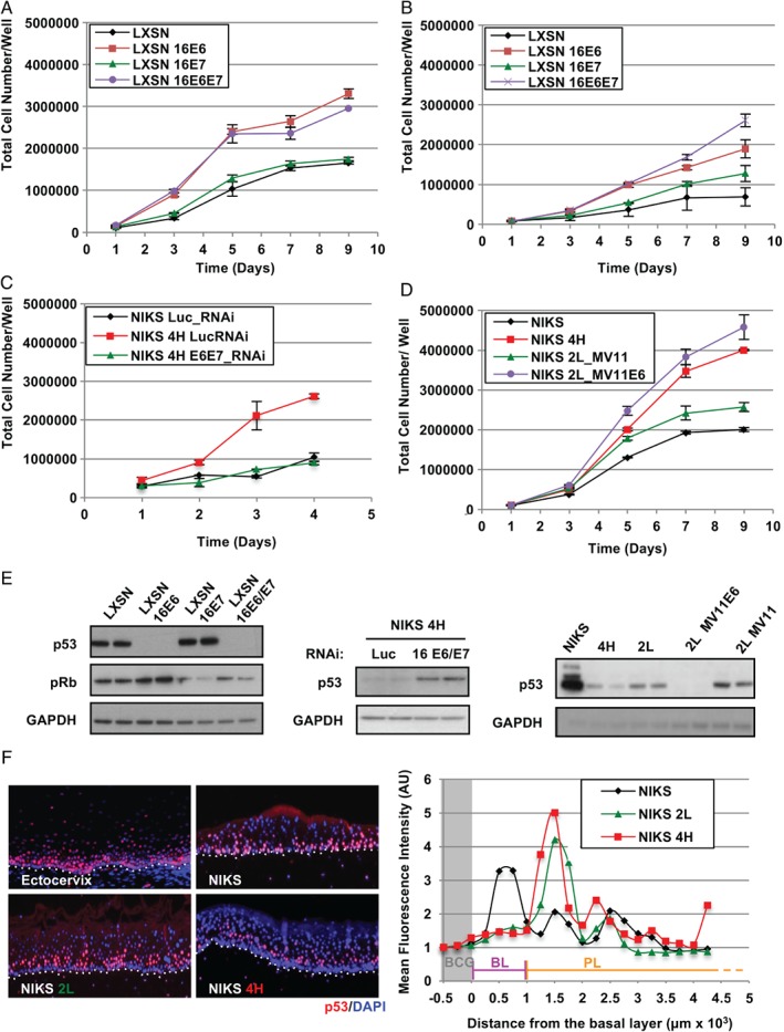
Figure 1 Dominant role of HPV‐16 E6 in driving the growth of NIKS at high cell densities. (A, B) Effects of E6 and E7 expression on the growth pattern of NIKS cells with (A) or without (B) supplemented growth factors. Each point on the plot represents the average from three independent experiments. Error bars represent mean ± SD. (C) NIKS HPV‐16 4H cells were transfected with E6 and E7 RNAi and grown for the indicated time before harvesting and counting. Error bars represent mean ± SD (n = 3). (D) HPV‐16 E6 was ectopically overexpressed in NIKS 2 L (NIKS 2 L MV11_E6) and the effects on cell growth were monitored by growth assay. Error bars represent mean ± SD (n = 3). (E) Representative western blots validating the expression of HPV‐16 E6 and E7 in the experiments in A, C, and D. Levels of p53 and pRb were monitored as surrogate markers for the expression of E6 and E7, respectively. (F) The pattern of expression of p53 was used as a surrogate marker to locate the expression of E6 in raft culture of NIKS 2 L and 4H episomal cell lines. The p53 fluorescence signal was enhanced using tetramethylrhodamine (TMR) tyramide amplification. All sections were counterstained with 4',6‐diamidino‐2‐phenylindole (DAPI). The fluorescent intensity was quantified in the first 0.17 inches of the raft epithelium and normalized with the background signal (BCG) detected immediately underneath the basal layer. BL = basal layer; PL = parabasal layers.
Figure 1 Dominant role of HPV‐16 E6 in driving the growth of NIKS at high cell densities. (A, B) Effects of E6 and E7 expression on the growth pattern of NIKS cells with (A) or without (B) supplemented growth factors. Each point on the plot represents the average from three independent experiments. Error bars represent mean ± SD. (C) NIKS HPV‐16 4H cells were transfected with E6 and E7 RNAi and grown for the indicated time before harvesting and counting. Error bars represent mean ± SD (n = 3). (D) HPV‐16 E6 was ectopically overexpressed in NIKS 2 L (NIKS 2 L MV11_E6) and the effects on cell growth were monitored by growth assay. Error bars represent mean ± SD (n = 3). (E) Representative western blots validating the expression of HPV‐16 E6 and E7 in the experiments in A, C, and D. Levels of p53 and pRb were monitored as surrogate markers for the expression of E6 and E7, respectively. (F) The pattern of expression of p53 was used as a surrogate marker to locate the expression of E6 in raft culture of NIKS 2 L and 4H episomal cell lines. The p53 fluorescence signal was enhanced using tetramethylrhodamine (TMR) tyramide amplification. All sections were counterstained with 4',6‐diamidino‐2‐phenylindole (DAPI). The fluorescent intensity was quantified in the first 0.17 inches of the raft epithelium and normalized with the background signal (BCG) detected immediately underneath the basal layer. BL = basal layer; PL = parabasal layers.CC BY · PubMed Central
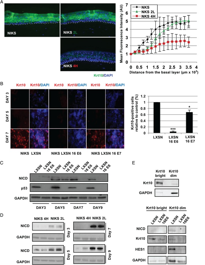
Figure 2 The expression of HPV‐16 E6 prevents the commitment to differentiation of NIKS keratinocytes. (A) Comparison of the timing of expression of the keratin‐10 (Krt10) differentiation marker during epithelial differentiation in organotypic raft cultures of NIKS or the NIKS 2 L (LSIL‐like) and 4H (HSIL‐like) episomal cell lines. Quantification of the Krt10 fluorescence signal during differentiation is shown in the far‐right panel to highlight differences between the different rafts. The fluorescence intensity was normalized against the background signal and plotted against distance from the basal cell layer. Error bars represent mean ± SD (n = 3). (B) Krt10 expression was monitored by immunofluorescence in monolayers of the indicated NIKS cell lines grown to sub‐confluence (day 3), confluence (day 5), and post‐confluence (day 7). The right‐hand panel shows the quantification of Krt10‐positive cells expressed as a percentage of the total cell population at the 7‐day time point relative to the control (LXSN). Ten random fields were acquired for each sample and cells were counted using ImageJ software. (C, D) Western blot analyses of the modulation of NICD by HPV‐16 E6 in NIKS (C) or in NIKS 2 L and 4H (D) across a 9‐day growth assay. (E) Western blot analysis of components of Notch in Krt10‐high and ‐dim FACS‐sorted NIKS cell populations (see also supplementary material, Figure S4).
Figure 2 The expression of HPV‐16 E6 prevents the commitment to differentiation of NIKS keratinocytes. (A) Comparison of the timing of expression of the keratin‐10 (Krt10) differentiation marker during epithelial differentiation in organotypic raft cultures of NIKS or the NIKS 2 L (LSIL‐like) and 4H (HSIL‐like) episomal cell lines. Quantification of the Krt10 fluorescence signal during differentiation is shown in the far‐right panel to highlight differences between the different rafts. The fluorescence intensity was normalized against the background signal and plotted against distance from the basal cell layer. Error bars represent mean ± SD (n = 3). (B) Krt10 expression was monitored by immunofluorescence in monolayers of the indicated NIKS cell lines grown to sub‐confluence (day 3), confluence (day 5), and post‐confluence (day 7). The right‐hand panel shows the quantification of Krt10‐positive cells expressed as a percentage of the total cell population at the 7‐day time point relative to the control (LXSN). Ten random fields were acquired for each sample and cells were counted using ImageJ software. (C, D) Western blot analyses of the modulation of NICD by HPV‐16 E6 in NIKS (C) or in NIKS 2 L and 4H (D) across a 9‐day growth assay. (E) Western blot analysis of components of Notch in Krt10‐high and ‐dim FACS‐sorted NIKS cell populations (see also supplementary material, Figure S4).CC BY · PubMed Central
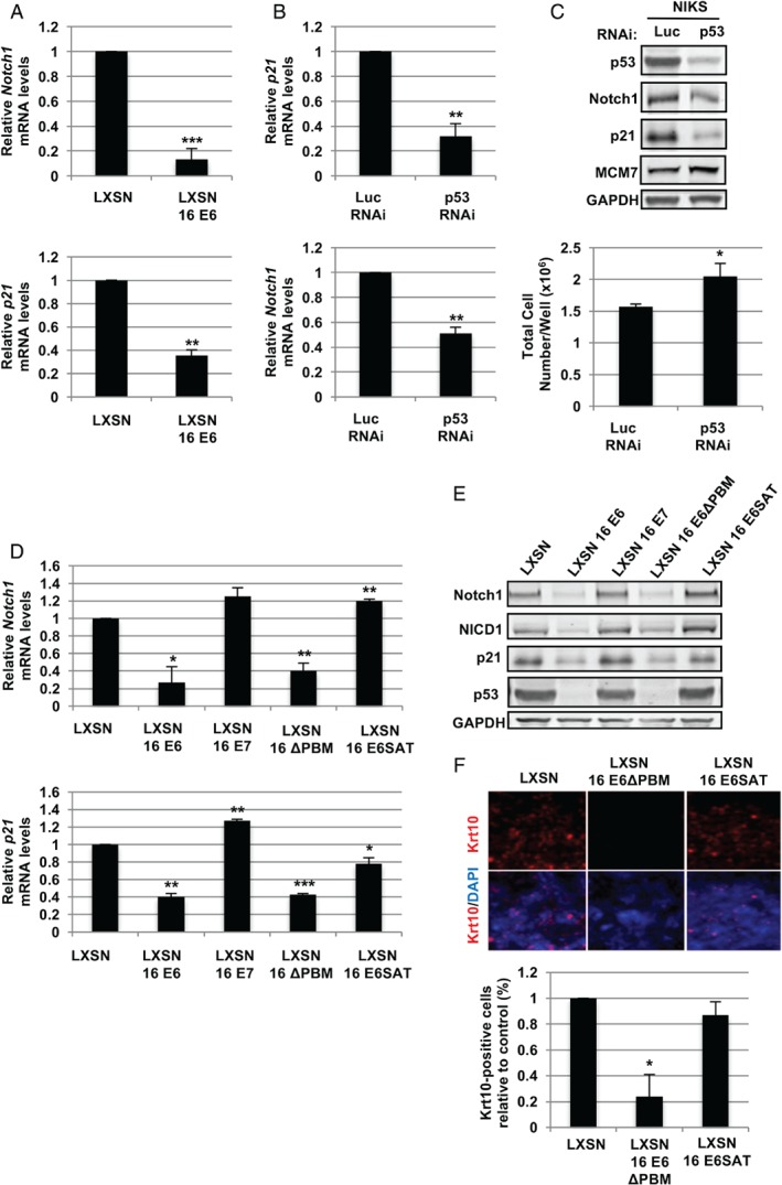
Figure 3 HPV‐16 E6 affects the levels of full‐length and cleaved Notch1 through the degradation of p53. (A, B) RT‐qPCR analysis of the expression of Notch1 and p21 mRNA in control and E6‐expressing cells at post‐confluence (day 7) (A) or in NIKS cells transfected with RNAi to luciferase (control) or p53 (B). Each bar chart represents the average values from three independent experiments. Error bars represent mean ± SD. (C) NIKS cells transfected with RNAi as in panel B were subjected to western blot analysis for the indicated proteins (upper panel). The total cell number of NIKS transfected with control or p53 RNAi was estimated 72 h post‐transfection (see also supplementary material, Figure S5). Error bars represent mean ± SD (n = 3). RT‐qPCR analysis of NOTCH1 and P21 mRNA expression in control NIKS (LXSN) and NIKS expressing E6, E7 or the indicated E6 mutants at post‐confluence (day 7). Error bars represent mean ± SD (n = 3). (E) NIKS cell lines as in panel D were grown to post‐confluence and subjected to western blot analysis for the indicated proteins. (F) Control NIKS or NIKS cells expressing either the wt HPV‐16 E6 or the 16E6 SAT and ΔPBM mutants were grown to post‐confluence prior to fixation. The pattern of Krt10 expression was then analysed by immunofluorescence using Alexa Fluor 594‐conjugated secondary antibodies. The lower panel shows the quantification of Krt10‐positive cells expressed as a percentage of the total cell population at the 7‐day time point relative to the control (LXSN). The quantitative analysis was carried out as in Figure 2B using ImageJ software. Where shown, statistical significance was evaluated using the Student's t‐test.
Figure 3 HPV‐16 E6 affects the levels of full‐length and cleaved Notch1 through the degradation of p53. (A, B) RT‐qPCR analysis of the expression of Notch1 and p21 mRNA in control and E6‐expressing cells at post‐confluence (day 7) (A) or in NIKS cells transfected with RNAi to luciferase (control) or p53 (B). Each bar chart represents the average values from three independent experiments. Error bars represent mean ± SD. (C) NIKS cells transfected with RNAi as in panel B were subjected to western blot analysis for the indicated proteins (upper panel). The total cell number of NIKS transfected with control or p53 RNAi was estimated 72 h post‐transfection (see also supplementary material, Figure S5). Error bars represent mean ± SD (n = 3). RT‐qPCR analysis of NOTCH1 and P21 mRNA expression in control NIKS (LXSN) and NIKS expressing E6, E7 or the indicated E6 mutants at post‐confluence (day 7). Error bars represent mean ± SD (n = 3). (E) NIKS cell lines as in panel D were grown to post‐confluence and subjected to western blot analysis for the indicated proteins. (F) Control NIKS or NIKS cells expressing either the wt HPV‐16 E6 or the 16E6 SAT and ΔPBM mutants were grown to post‐confluence prior to fixation. The pattern of Krt10 expression was then analysed by immunofluorescence using Alexa Fluor 594‐conjugated secondary antibodies. The lower panel shows the quantification of Krt10‐positive cells expressed as a percentage of the total cell population at the 7‐day time point relative to the control (LXSN). The quantitative analysis was carried out as in Figure 2B using ImageJ software. Where shown, statistical significance was evaluated using the Student's t‐test.CC BY · PubMed Central
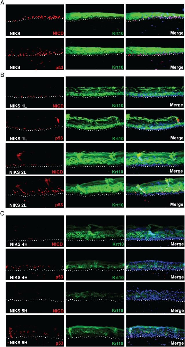
Figure 4 Loss of p53, NICD, and Krt10 expression correlates with abortive infection phenotypes in raft culture. (A–C) Representative images showing the pattern of p53, NICD, and Krt10 expression in organotypic raft culture sections of parental NIKS (A) and LSIL‐like (B) and HSIL‐like (C) NIKS HPV‐16 episomal lines. The fluorescence signal for p53 and NICD was amplified with TMR. Krt10 fluorescence was visualized using Alexa Fluor 488‐conjugated secondary antibodies (see also supplementary material, Figure S9). All sections were counterstained with DAPI.
Figure 4 Loss of p53, NICD, and Krt10 expression correlates with abortive infection phenotypes in raft culture. (A–C) Representative images showing the pattern of p53, NICD, and Krt10 expression in organotypic raft culture sections of parental NIKS (A) and LSIL‐like (B) and HSIL‐like (C) NIKS HPV‐16 episomal lines. The fluorescence signal for p53 and NICD was amplified with TMR. Krt10 fluorescence was visualized using Alexa Fluor 488‐conjugated secondary antibodies (see also supplementary material, Figure S9). All sections were counterstained with DAPI.CC BY · PubMed Central
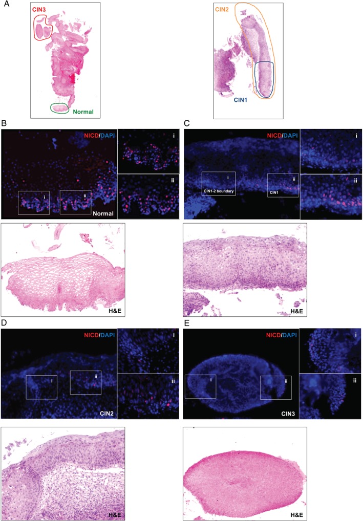
Figure 5 Loss of NICD expression correlates with abortive infections in the human cervix. (A) Low‐power images of H&E‐stained HPV‐16‐positive cervical lesions. Cervical tissue sections were stained with NICD antibodies followed by TMR tyramide fluorescence signal amplification. Tissue sections were counterstained with DAPI. Digital images of stained sections were acquired with a Pannoramic Slide Scanner prior to counterstaining with H&E. Coloured circles with relative CIN grading (according to pathologist's diagnosis) mark the areas shown in detail in panels B–E. (B–E) Magnified images showing the pattern of NICD staining and relative H&E counterstains in normal cervix (B), CIN1 (C), CIN2 (D), and CIN3 (E) (see also supplementary material, Figure S10).
Figure 5 Loss of NICD expression correlates with abortive infections in the human cervix. (A) Low‐power images of H&E‐stained HPV‐16‐positive cervical lesions. Cervical tissue sections were stained with NICD antibodies followed by TMR tyramide fluorescence signal amplification. Tissue sections were counterstained with DAPI. Digital images of stained sections were acquired with a Pannoramic Slide Scanner prior to counterstaining with H&E. Coloured circles with relative CIN grading (according to pathologist's diagnosis) mark the areas shown in detail in panels B–E. (B–E) Magnified images showing the pattern of NICD staining and relative H&E counterstains in normal cervix (B), CIN1 (C), CIN2 (D), and CIN3 (E) (see also supplementary material, Figure S10).CC BY · PubMed Central
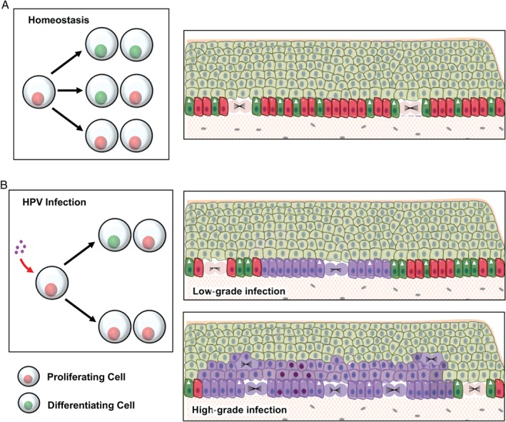
Figure 6 Proposed model for the modulation of keratinocyte cell fate based on the inactivation of p53 and Notch by E6 expression. (A) Homeostasis in the squamous epithelium is determined by the balanced probability of the outcome of each cell division: two differentiating cells, one differentiating and one proliferating progenitor, and two proliferating progenitors (left panel). According to this model, the fate of each division is stochastic; however, across the total population, the odds of having one of the three possible outcomes is balanced. As a result, in the basal layer the proliferation of progenitor cells (red cells with circles) compensates for the loss of cells by terminal differentiation (green cells with arrowhead). (B) Upon infection with HPV‐16, the inactivation of p53 and Notch leads to an unbalanced fate of cell divisions, with a skew towards proliferation (left panel). In low‐grade lesions (upper‐right panel), reduced levels of p53 and Notch in HPV‐infected (purple) cells allow for the maintenance and expansion of the HPV‐infected pool in the basal layer. However, low levels of E6 and E7 expression allow for an adequate level of keratinocyte differentiation able to sustain the viral life cycle. Its deregulation (lower‐right panel) is thought to be associated with an elevation of E6 and E7 expression. Increased E6 levels further reduce the proportion of differentiating cells within the infected basal cell population, allowing for their clonal expansion and persistence with the colonization of large areas of the epithelium. Increased levels of proliferation and long‐term persistence in the epithelium may eventually lead to the accumulation of oncogenic mutations (cells with dark purple nuclei) predisposing to the development of malignancy.
Figure 6 Proposed model for the modulation of keratinocyte cell fate based on the inactivation of p53 and Notch by E6 expression. (A) Homeostasis in the squamous epithelium is determined by the balanced probability of the outcome of each cell division: two differentiating cells, one differentiating and one proliferating progenitor, and two proliferating progenitors (left panel). According to this model, the fate of each division is stochastic; however, across the total population, the odds of having one of the three possible outcomes is balanced. As a result, in the basal layer the proliferation of progenitor cells (red cells with circles) compensates for the loss of cells by terminal differentiation (green cells with arrowhead). (B) Upon infection with HPV‐16, the inactivation of p53 and Notch leads to an unbalanced fate of cell divisions, with a skew towards proliferation (left panel). In low‐grade lesions (upper‐right panel), reduced levels of p53 and Notch in HPV‐infected (purple) cells allow for the maintenance and expansion of the HPV‐infected pool in the basal layer. However, low levels of E6 and E7 expression allow for an adequate level of keratinocyte differentiation able to sustain the viral life cycle. Its deregulation (lower‐right panel) is thought to be associated with an elevation of E6 and E7 expression. Increased E6 levels further reduce the proportion of differentiating cells within the infected basal cell population, allowing for their clonal expansion and persistence with the colonization of large areas of the epithelium. Increased levels of proliferation and long‐term persistence in the epithelium may eventually lead to the accumulation of oncogenic mutations (cells with dark purple nuclei) predisposing to the development of malignancy.CC BY · PubMed Central

Verification

Claims the paper makes

The claims and statistics of this paper haven’t been checked yet.

Run the check — free, no account needed. It reads the open-access text and checks the sources it cites.

A few free checks a day · papers already verified open instantly · results are public

Claims other papers make about this one

Each is checked against this paper’s own text: does it say what the citing paper says it does?

Self-check

Percentages, sums and reported differences re-computed from the paper’s own numbers.

The self-check needs the paper’s full text.

Citations

Cites

Show all 6 references

Research works printed but not linked: 72

  1. 10.1016/j.virol.2010.02.002 · DOI
  2. 10.1016/j.virol.2004.03.033 · DOI
  3. 10.1038/nrc798 · DOI
  4. 10.1016/j.vaccine.2012.06.083 · DOI
  5. 10.1083/jcb.200708185 · DOI
  6. Boyer SN, 1996, E7 protein of human papilloma virus‐16 induces degradation of retinoblastoma protein through the ubiquitin‐proteasome pathway, Cancer Res, 56, 4620
  7. 10.1016/0092-8674(90)90409-8 · DOI · Paper page
  8. Freeman A, 1999, Minichromosome maintenance proteins as biological markers of dysplasia and malignancy, Clin Cancer Res, 5, 2121
  9. 10.1038/modpathol.2015.52 · DOI
  10. 10.1111/j.1752-4571.2008.00063.x · DOI
  11. 10.1016/j.coviro.2016.08.011 · DOI
  12. 10.1038/ncb2963 · DOI
  13. 10.1016/j.ceb.2008.01.010 · DOI
  14. 10.1101/gad.1477606 · DOI
  15. 10.1093/emboj/20.13.3427 · DOI
  16. 10.1242/dev.063610 · DOI · Paper page
  17. 10.1016/S0070-2153(10)92003-6 · DOI
  18. Naganuma S, 2012, Notch receptor inhibition reveals the importance of cyclin D1 and Wnt signaling in invasive esophageal squamous cell carcinoma, Am J Cancer Res, 2, 459
  19. 10.1038/ng1099 · DOI
  20. 10.4161/15384101.2014.988027 · DOI
  21. 10.1038/labinvest.2012.9 · DOI
  22. 10.1002/cyto.a.10072 · DOI
  23. 10.1046/j.1523-1747.2000.00869.x · DOI
  24. 10.1002/ijc.2910430422 · DOI
  25. 10.1002/path.4656 · DOI
  26. 10.1128/JVI.07069-11 · DOI
  27. Lambert PF, 2005, Using an immortalized cell line to study the HPV life cycle in organotypic ‘raft’ cultures, Methods Mol Med, 119, 141
  28. 10.1073/pnas.1002620107 · DOI
  29. 10.1371/journal.pone.0077507 · DOI
  30. 10.1111/j.0022-202X.2005.23655.x · DOI
  31. 10.1046/j.1523-1747.1998.00365.x · DOI
  32. 10.1111/1523-1747.ep12612810 · DOI
  33. 10.1016/S0021-9258(17)44111-1 · DOI
  34. 10.1083/jcb.102.5.1767 · DOI
  35. 10.1101/gad.11.16.2101 · DOI · Paper page
  36. 10.1128/MCB.02119-06 · DOI
  37. 10.1101/gad.1463206 · DOI
  38. 10.1242/jcs.084723 · DOI
  39. 10.1046/j.1523-1747.1999.00780.x · DOI
  40. 10.1101/gad.1484707 · DOI
  41. 10.1038/nature11393 · DOI
  42. 10.1038/sj.onc.1210791 · DOI
  43. 10.1158/1078-0432.CCR-03-0565 · DOI
  44. 10.1101/gad.14.8.913 · DOI
  45. 10.1073/pnas.061032098 · DOI
  46. 10.1016/S0002-9440(10)64814-6 · DOI
  47. 10.1038/sj.onc.1209435 · DOI
  48. 10.1038/sj.onc.1203656 · DOI
  49. Liefer KM, 2000, Down‐regulation of p63 is required for epidermal UV‐B‐induced apoptosis, Cancer Res, 60, 4016
  50. 10.1038/sj.onc.1206536 · DOI
  51. 10.1128/JVI.77.19.10186-10201.2003 · DOI
  52. 10.1371/journal.pone.0067306 · DOI
  53. 10.3390/ijms161125948 · DOI
  54. 10.1016/S1534-5807(04)00098-X · DOI
  55. 10.1111/1523-1747.ep12540786 · DOI
  56. 10.1128/JVI.02527-12 · DOI
  57. 10.1111/1523-1747.ep12876141 · DOI
  58. 10.1016/j.semcancer.2004.04.017 · DOI
  59. 10.4161/cc.6.3.3753 · DOI
  60. 10.1101/gad.1406006 · DOI
  61. 10.18632/oncotarget.3892 · DOI · Paper page
  62. Di Como CJ, 2002, p63 expression profiles in human normal and tumor tissues, Clin Cancer Res, 8, 494
  63. 10.1053/hupa.2001.24324 · DOI
  64. 10.1006/gyno.2000.5953 · DOI
  65. 10.1034/j.1600-0560.2002.290902.x · DOI
  66. 10.1016/S1534-5807(02)00166-1 · DOI
  67. 10.1128/JVI.74.21.10081-10095.2000 · DOI
  68. 10.1128/JVI.66.4.2125-2134.1992 · DOI · Paper page
  69. 10.1128/JVI.02063-08 · DOI
  70. 10.1128/JVI.78.2.821-833.2004 · DOI
  71. 10.1006/viro.1997.8768 · DOI
  72. 10.1016/j.ab.2009.03.017 · DOI

Metadata

Authors
Kranjec, C., Holleywood, C., Libert, D., Griffin, H., Mahmood, R., Isaacson, E., & Doorbar, J.
Affiliations
  • Department of Pathology University of Cambridge, Tennis Court Road Cambridge UK
  • The Francis Crick Institute Mill Hill Laboratory The Ridgeway, Mill Hill London UK
Venue
The Journal of Pathology
Publisher
Wiley
Published
2017-07-24
Volume
242
Issue
4
Pages
448-462
ISSN
  • 0022-3417
  • 1096-9896
Cited by
47
References
6
Language
en
Funders
Medical Research Council (MC_U117584278, MC_PC_13050)

About these checks

Registry, retraction, Cites and statistics results are computed, not judged, with one exception: a printed reference that no index lists is read once by a model before it counts as a citation error. Claims are extracted by a model and kept only when their quote appears verbatim in the text; each cited source is then checked, with a model’s help, for whether it says what the claim relies on. Those verdicts can be wrong — read the claim’s quote and the cited source before relying on one.

Statistics and arithmetic are recomputed, not judged.

Confidence bands are shown only for fields calibrated on our annotated test set. None is calibrated yet, so no band is shown.

What counts as a claim ›How we measure confidence ›How we check this ›

Are you an author, or do you see a problem with this page? Request a review

How to cite

Cite CoChat for the checks on this page, and the paper for its own findings.

This page
CoChat. (2026). Citation check: Modulation of basal cell fate during productive and transforming HPV-16 infection is mediated by progressive E6-driven depletion of Notch. https://app.cochat.ai/papers/10.1002/path.4917
The paper
Kranjec, C., Holleywood, C., Libert, D., Griffin, H., Mahmood, R., Isaacson, E., & Doorbar, J. (2017). Modulation of basal cell fate during productive and transforming HPV-16 infection is mediated by progressive E6-driven depletion of Notch. The Journal of Pathology. https://doi.org/10.1002/path.4917