

Complement as a regulator of adaptive immunity
Open accessThe DOI matches its Crossref record; 101 of the 101 references it cites were found in a registry or an index.
Abstract
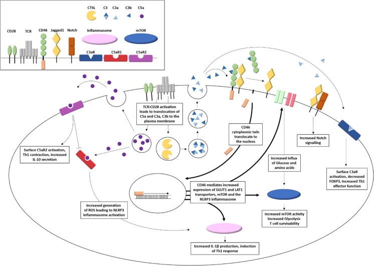
The complement system is an ancient and evolutionarily conserved effector system comprising in mammals over 50 circulating and membrane bound proteins. Complement has long been described as belonging to the innate immune system; however, a number of recent studies have demonstrated its key role in the modulation of the adaptive immune response. This review does not set out to be an exhaustive list of the numerous interactions of the many complement components with adaptive immunity; rather, we will focus more precisely on the role of some complement molecules in the regulation of antigen presenting cells, as well as on their direct effect on the activation of the core adaptive immune cells, B and T lymphocytes. Recent reports on the local production and activation of complement proteins also suggest a major role in the control of effector responses. The crucial role of complement in adaptive immunity is further highlighted by several examples of dysregulation of these pathways in human diseases.
Overview
The claims and statistics of this paper haven’t been checked yet.
Free: it reads the open-access text and checks the first 5 claims that cite a source we can check. Create a free account to check every claim.
Integrity signals
Registry
Cites
Statistics
Figures

Verification
Claims the paper makes
The claims and statistics of this paper haven’t been checked yet.
Free: it reads the open-access text and checks the first 5 claims that cite a source we can check. Create a free account to check every claim.
Claims other papers make about this one
Each is checked against this paper’s own text: does it say what the citing paper says it does?
In the citing paper
“In particular, complement may reduce Treg function (20, 21) and promote T and B cell activation (22, 23).”
From Pre-existing humoral immunity and complement pathway contribute to immunogenicity of adeno-associated virus (AAV) vector in human blood (2022) · cites this paper as [23]
Self-check
Percentages, sums and reported differences re-computed from the paper’s own numbers.
The self-check needs the paper’s full text.
Citations
Cites
Walport, M. J. (2001) · New England Journal of Medicine · doi:10.1056/nejm200104053441406 · Cited by 2,927
Posttranscriptional Control of T Cell Effector Function by Aerobic Glycolysis
Chang, C.-H., Curtis, J. D., Maggi, L. B., Faubert, B., Villarino, A. V., O’Sullivan, D., Huang, S. C.-C., van der Windt, G. J. W., Blagih, J., Qiu, J., et al. (2013) · Cell · doi:10.1016/j.cell.2013.05.016 · Cited by 2,204 · cited 1×
“the cells rise to support the increased energy demands of cellular proliferation and effector functions [81, 82].”
BackgroundThe transcription factor Myc controls metabolic reprogramming upon T lymphocyte activation
Wang, R., Dillon, C. P., Shi, L. Z., Milasta, S., Carter, R., Finkelstein, D., McCormick, L. L., Fitzgerald, P., Chi, H., Munger, J., et al. (2011) · Immunity · doi:10.1016/j.immuni.2011.09.021 · Cited by 2,140 · cited 2×
“This subsequently enhances the expression of the transcription factors myc and HIF1α, resulting in increased expression of the glycolytic machinery required for metabolic reprogramming and effector function [85, 87].”
BackgroundThe CD28 Signaling Pathway Regulates Glucose Metabolism
Frauwirth, K. A., Riley, J. L., Harris, M. H., Parry, R. V., Rathmell, J. C., Plas, D. R., Elstrom, R. L., June, C. H., & Thompson, C. B. (2002) · Immunity · doi:10.1016/s1074-7613(02)00323-0 · Cited by 1,475 · cited 1×
“the cells rise to support the increased energy demands of cellular proliferation and effector functions [81, 82].”
BackgroundRegulatory B Cells: Origin, Phenotype, and Function
Rosser, E. C., & Mauri, C. (2015) · Immunity · doi:10.1016/j.immuni.2015.04.005 · Cited by 1,455 · cited 5×
“Recent studies have highlighted the existence of specific regulatory B cells (Bregs) that also contribute to themaintenance of immune homeostasis in a manner analogous to Tregs [8].”
BackgroundInfluentialC3d of complement as a molecular adjuvant: Bridging innate and acquired immunity
Dempsey, P. W., Allison, M. E. D., Akkaraju, S., Goodnow, C. C., & Fearon, D. T. (1996) · Science · doi:10.1126/science.271.5247.348 · Cited by 1,197
Macintyre, A. N., Gerriets, V. A., Nichols, A. G., Michalek, R. D., Rudolph, M. C., Deoliveira, D., Anderson, S. M., Abel, E. D., Chen, B. J., Hale, L. P., et al. (2014) · Cell Metabolism · doi:10.1016/j.cmet.2014.05.004 · Cited by 1,127
Regulatory T cells exert checks and balances on self tolerance and autoimmunity
Wing, K., & Sakaguchi, S. (2010) · Nature Immunology · doi:10.1038/ni.1818 · Cited by 1,107 · cited 1×
“In counterpoint to these effector Th subsets, a variety of regulatory T cells (Tregs) operate, essential to control immune homeostasis by counteracting overactivation of effector T cells [7].”
BackgroundSinclair, L. V., Rolf, J., Emslie, E., Shi, Y.-B., Taylor, P. M., & Cantrell, D. A. (2013) · Nature Immunology · doi:10.1038/ni.2556 · Cited by 930 · cited 5×
“While a limitation of glucose availability severely impacts T cell activation and effector function [84, 87, 88], T cells display improved effector function with increased glucose [88].”
BackgroundInfluentialJacobs, S. R., Herman, C. E., MacIver, N. J., Wofford, J. A., Wieman, H. L., Hammen, J. J., & Rathmell, J. C. (2008) · The Journal of Immunology · doi:10.4049/jimmunol.180.7.4476 · Cited by 850 · cited 1×
“While a limitation of glucose availability severely impacts T cell activation and effector function [84, 87, 88], T cells display improved effector function with increased glucose [88].”
Background
Cited by
Complosome — the intracellular complement system
West, E. E., & Kemper, C. (2023) · Nature Reviews Nephrology · doi:10.1038/s41581-023-00704-1 · Cited by 253
Justification of specific genetic modifications in pigs for clinical organ xenotransplantation
Cooper, D. K. C., Hara, H., Iwase, H., Yamamoto, T., Li, Q., Ezzelarab, M., Federzoni, E., Dandro, A., & Ayares, D. (2019) · Xenotransplantation · doi:10.1111/xen.12516 · Cited by 178
Role of immune cells in hypertension
Caillon, A., Paradis, P., & Schiffrin, E. L. (2019) · British Journal of Pharmacology · doi:10.1111/bph.14427 · Cited by 163
“The complement system is an important component of the innate immune system that also plays an important role in the induction of adaptive immune responses (Cravedi et al., 2013; Killick et al., 2018).”
BackgroundChronic biofilm-based infections: skewing of the immune response
González, J. F., Hahn, M. M., & Gunn, J. S. (2018) · Pathogens and Disease · doi:10.1093/femspd/fty023 · Cited by 159
Myasthenia gravis: the changing treatment landscape in the era of molecular therapies
Iorio, R. (2024) · Nature Reviews Neurology · doi:10.1038/s41582-023-00916-w · Cited by 153
“One study provided insights into the involvement of complement in adaptive immunity 106 , supporting the hypothesis that inhibiting complement activation can also have an impact on T and B cell subsets in people with MG 107 .”
Metadata
- Authors
- Killick, J., Morisse, G., Sieger, D., & Astier, A. L.
- Affiliations
- Centre for Inflammation Research (GB)
- University of Edinburgh (GB)
- Centre National de la Recherche Scientifique (FR)
- Corresponding author
- Astier, Anne L.
- Venue
- Seminars in Immunopathology
- Published
- 2017-08-25
- Volume
- 40
- Issue
- 1
- Pages
- 37-48
- ISSN
- 1863-2297 (ISSN-L)
- 1863-2300
- PMC
- PMC5794818
- OpenAlex
- W2748098899
- Cited by
- 129
- References
- 101
- Citations by year
- 2026: 9 · 2025: 14 · 2024: 12 · 2023: 19 · 2022: 16 · 2021: 14 · 2020: 14 · 2019: 18 · 2018: 11 · 2017: 2
- FWCI
- 1.26
- Type
- journal article
- Language
- en
- Open access
- hybrid
- doi.org
- Locations
- Seminars in Immunopathology (publishedVersion, cc-by)
- PubMed Central (publishedVersion, cc-by)
- PubMed (publishedVersion)
- HAL (submittedVersion, other-oa)
- Edinburgh Research Explorer (University of Edinburgh) (submittedVersion, cc-by)
- Funders
- Multiple Sclerosis Society (41, 34)
- Topics
- Life Sciences › Immunology and Microbiology › Immunology › Complement system in diseases
- Life Sciences › Immunology and Microbiology › Immunology › Biomarkers in Disease Mechanisms
- Life Sciences › Immunology and Microbiology › Immunology › Immune Response and Inflammation
- Keywords
- complement system, adaptive immunity, antigen-presenting cells, B cells, T cells, complement dysregulation, local complement activation
- MeSH terms
- Animals, Antigen-Presenting Cells, B-Lymphocytes, Carrier Proteins, Cell Communication, Complement System Proteins, Complement Activation, Disease Susceptibility, Humans, Immunologic Factors, Lymphocyte Activation, Protein Binding, T-Lymphocytes, Signal Transduction, Adaptive Immunity, Immunomodulation, Molecular Targeted Therapy
About these checks
Registry, retraction, Cites and statistics results are computed, not judged, with one exception: a printed reference that no index lists is read once by a model before it counts as a citation error. Claims are extracted by a model and kept only when their quote appears verbatim in the text; each cited source is then checked, with a model’s help, for whether it says what the claim relies on. Those verdicts can be wrong — read the claim’s quote and the cited source before relying on one.
Statistics and arithmetic are recomputed, not judged.
Confidence bands are shown only for fields calibrated on our annotated test set. None is calibrated yet, so no band is shown.
What counts as a claim ›How we measure confidence ›How we check this ›
Are you an author, or do you see a problem with this page? Request a review
How to cite
Cite CoChat for the checks on this page, and the paper for its own findings.
- This page
- CoChat. (2026). Citation check: Complement as a regulator of adaptive immunity. https://app.cochat.ai/papers/10.1007/s00281-017-0644-y
- The paper
- Killick, J., Morisse, G., Sieger, D., & Astier, A. L. (2018). Complement as a regulator of adaptive immunity. Seminars in Immunopathology. https://doi.org/10.1007/s00281-017-0644-y
Data sources
Bibliographic data: Semantic Scholar, OpenAlex, Crossref.
Contains information from the Semantic Scholar Academic Graph Datasets, made available under the ODC Attribution License (ODC-BY 1.0).
Semantic Scholar’s data is described in: Kinney, R., et al. (2023). The Semantic Scholar Open Data Platform. arXiv:2301.10140.
