

The DOI matches its Crossref record; 209 of the 209 references it cites were found in a registry or an index.
Development of the serotonergic cells in murine raphe nuclei and their relations with rhombomeric domains
Open accessAbstract
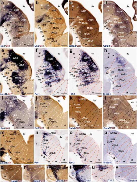
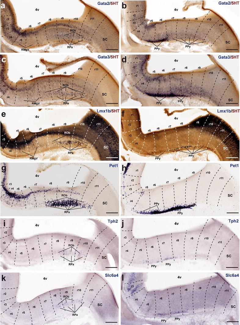
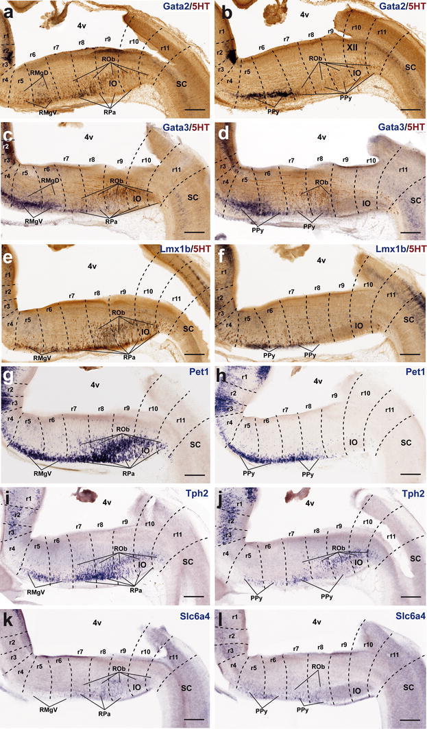
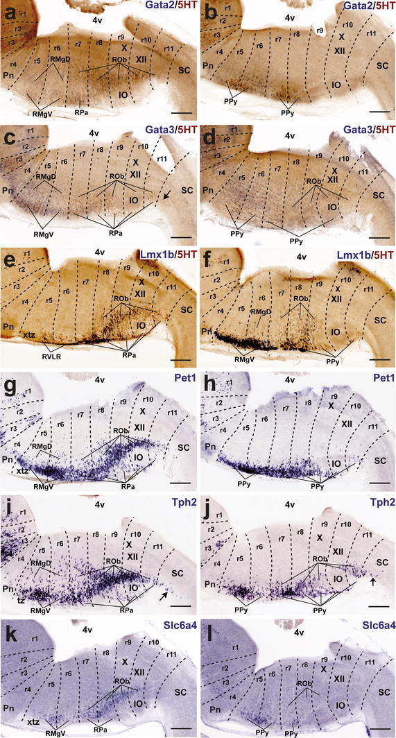
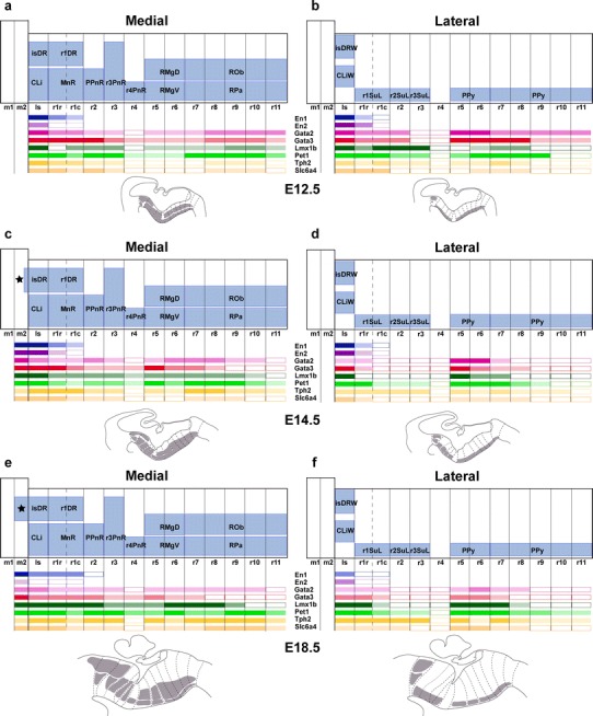
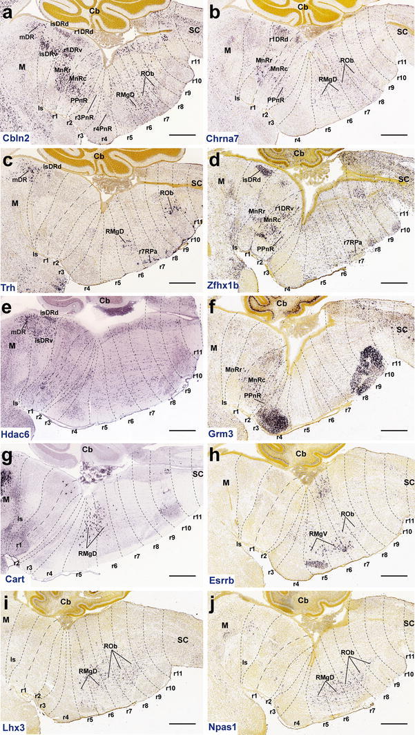
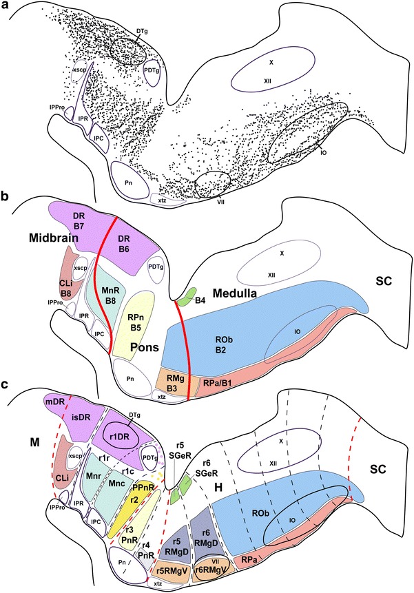
The raphe nuclei represent the origin of central serotonergic projections. The literature distinguishes seven nuclei grouped into rostral and caudal clusters relative to the pons. The boundaries of these nuclei have not been defined precisely enough, particularly with regard to developmental units, notably hindbrain rhombomeres. We hold that a developmental point of view considering rhombomeres may explain observed differences in connectivity and function. There are twelve rhombomeres characterized by particular genetic profiles, and each develops between one and four distinct serotonergic populations. We have studied the distribution of the conventional seven raphe nuclei among these twelve units. To this aim, we correlated 5-HT-immunoreacted neurons with rhombomeric boundary landmarks in sagittal mouse brain sections at different developmental stages. Furthermore, we performed a partial genoarchitectonic analysis of the developing raphe nuclei, mapping all known serotonergic differentiation markers, and compared these results, jointly with others found in the literature, with our map of serotonin-containing populations, in order to examine regional variations in correspondence. Examples of regionally selective gene patterns were identified. As a result, we produced a rhombomeric classification of some 45 serotonergic populations, and suggested a corresponding modified terminology. Only a minor rostral part of the dorsal raphe nucleus lies in the midbrain. Some serotonergic neurons were found in rhombomere 4, contrary to the conventional assumption that it lacks such neurons. We expect that our reclassification of raphe nuclei may be useful for causal analysis of their differential molecular specification, as well as for studies of differential connectivity and function.
Overview
The claims and statistics of this paper haven’t been checked yet.
Run the check — free, no account needed. It reads the open-access text and checks the sources it cites.
Integrity signals
Registry
Cites
Statistics
Figures
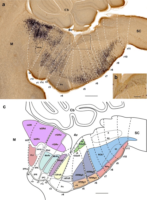
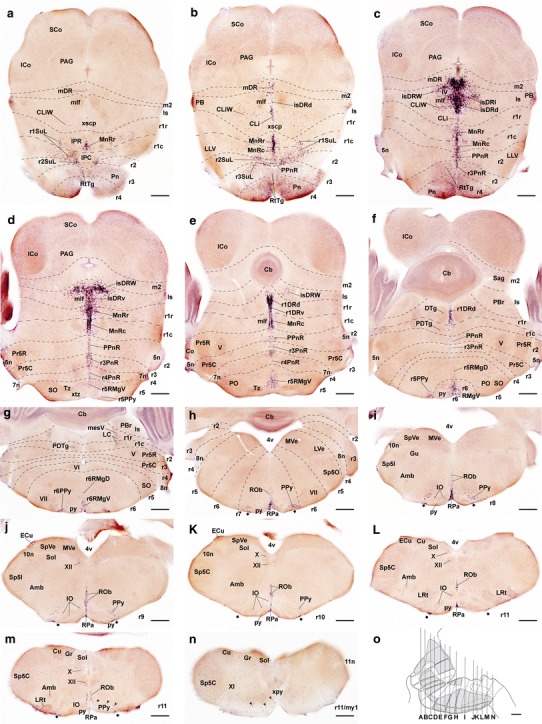
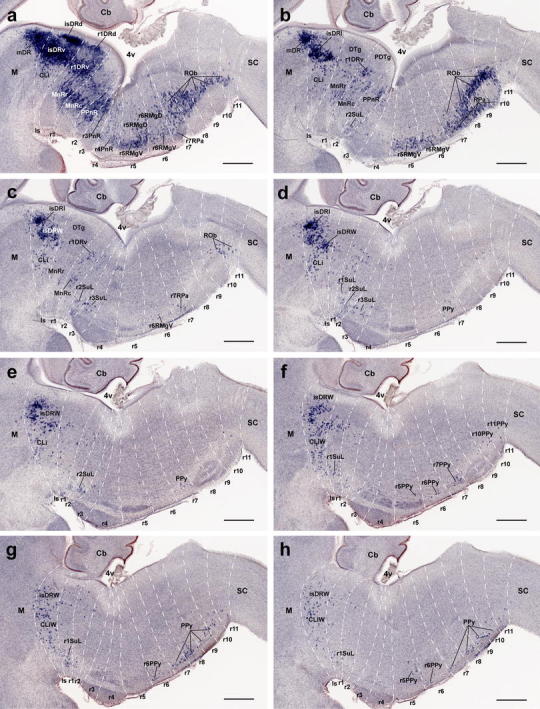
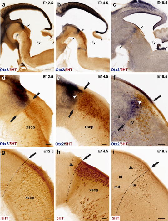
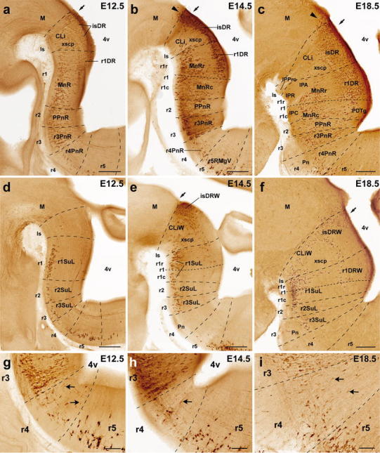
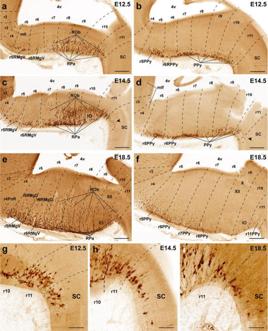
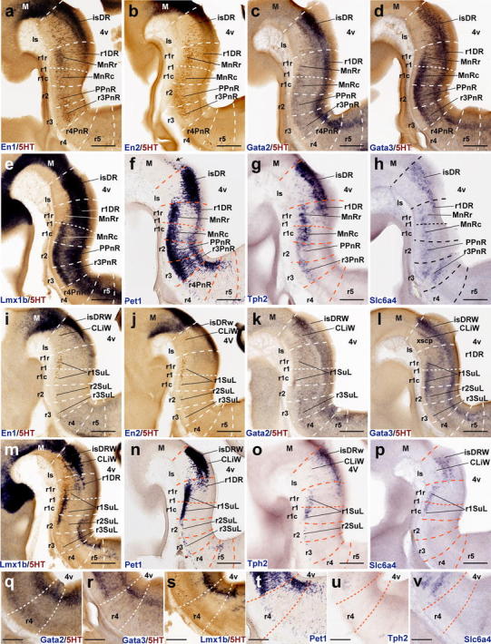
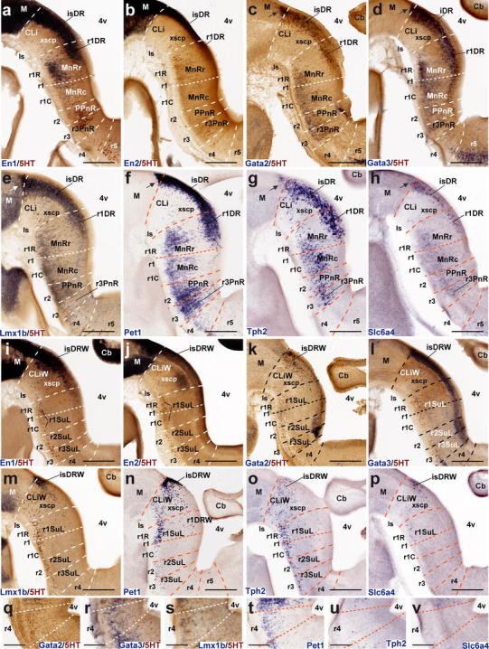
Verification
Claims the paper makes
The claims and statistics of this paper haven’t been checked yet.
Run the check — free, no account needed. It reads the open-access text and checks the sources it cites.
Claims other papers make about this one
Each is checked against this paper’s own text: does it say what the citing paper says it does?
In the citing paper
“DR serotonin neurons derive entirely from rhombomere 1 of the developing mouse brain, whereas MR serotonin neurons derive predominantly from rhombomeres 1, 2, and 3 (Alonso et al., 2013; Bang et al., 2012; Jensen et al., 2008).”
From Single-cell transcriptomes and whole-brain projections of serotonin neurons in the mouse dorsal and median raphe nuclei (2019) · cites this paper as Alonso et al., 2013
In the citing paper
“Evidence has suggested that the DR and MR serotonin systems differ in developmental origin, connectivity, physiology, and behavioral function (Alonso et al., 2013; Calizo et al., 2011; Okaty et al., 2019).”
From Single-cell transcriptomes and whole-brain projections of serotonin neurons in the mouse dorsal and median raphe nuclei (2019) · cites this paper as Alonso et al., 2013
Self-check
Percentages, sums and reported differences re-computed from the paper’s own numbers.
The self-check needs the paper’s full text.
Citations
Cites
The rat brain in stereotaxic coordinates
Palkovits, M. (1983) · Neuropeptides · doi:10.1016/0143-4179(83)90049-5 · Cited by 28,959
The Rat Brain in Stereotaxic Coordinates
Paxinos, G., & Watson, C. (1983) · Elsevier eBooks · doi:10.1016/c2009-0-63235-9 · Cited by 28,585
The mouse brain in stereotaxic coordinates
Konsman, J. (2003) · Psychoneuroendocrinology · doi:10.1016/s0306-4530(03)00088-x · Cited by 16,796
The Mouse Brain in Stereotaxic Coordinates
Paxinos, G. (2001) · Cited by 12,859
Dahlstroem, A., & Fuxé, K. (1964) · PubMed · Cited by 5,133
Handbook of Chemical Neuroanatomy
Stern, C. D. (1986) · Neurochemistry International · doi:10.1016/0197-0186(86)90021-5 · Cited by 4,160
Paxinos, G. (1985) · Cited by 3,715
Structure and function of the brain serotonin system
Jacobs, B. L., & Azmitia, E. C. (1992) · Physiological Reviews · doi:10.1152/physrev.1992.72.1.165 · Cited by 2,574
Steinbusch, H. W. M. (1981) · Neuroscience · doi:10.1016/0306-4522(81)90146-9 · Cited by 2,415
Histologie du système nerveux de l'homme & des vertébrés
y Cajal, S. R. (1909) · Maloine eBooks · doi:10.5962/bhl.title.48637 · Cited by 2,393
Research works printed but not linked: 7
- Azmitia EC, Gannon PJ (1986) Anatomy of the serotonergic system in the primate and sub-primate brain. Adv Neurol 43:407–468
- Cambronero F (1999) Aportaciones a la organización estructural del rombencéfalo de pollo. Dissertation. University of Murcia, Spain
- Castaldi L (1923) Studi sulla struttura e sullo sviluppo del mesencefalo. Richerche in Cavia cobaya. Parte prima. Arch Ital Anat Embriol 21:172–263
- Dahlström A, Fuxe K (1964) Evidence for the existence of monoamine-containing neurons in the central nervous system. Acta Physiol Scand Suppl 232:1–55
- Marburg O (1910) Mikroskopisch-topographischer Atlas des menschlichen Zentralnervensystems. Franz Deuticke, Leipzig
- Swanson LW (1998) Brain maps: structure of the rat brain. 2nd rev edn., Elsevier, Amsterdam
- Swanson LW (2003) Brain architecture. Oxford Univ Press, Oxford/New York
Cited by
Sorted by citations
Embracing diversity in the 5-HT neuronal system
Okaty, B. W., Commons, K. G., & Dymecki, S. M. (2019) · Nature Reviews Neuroscience · doi:10.1038/s41583-019-0151-3 · Cited by 319
Ren, J., Isakova, A., Friedmann, D., Zeng, J., Grutzner, S. M., Pun, A., Zhao, G. Q., Kolluru, S. S., Wang, R., Lin, R., et al. (2019) · eLife · doi:10.7554/elife.49424 · Cited by 304
Muzerelle, A., Scotto-Lomassese, S., Bernard, J. F., Soiza-Reilly, M., & Gaspar, P. (2016) · Brain Structure and Function · doi:10.1007/s00429-014-0924-4 · Cited by 303
A developmental ontology for the mammalian brain based on the prosomeric model
Puelles, L., Harrison, M. L., Paxinos, G., & WATSON, C. R. R. (2013) · Trends in Neurosciences · doi:10.1016/j.tins.2013.06.004 · Cited by 268
Self-citationKumar, N. N., Velić, A., Soliz, J., Shi, Y., Li, K., Wang, S., Weaver, J. L., Sen, J., Abbott, S. B. G., Lazarenko, R. M., et al. (2015) · Science · doi:10.1126/science.aaa0922 · Cited by 231
Puelles, L., & Rubenstein, J. L. R. (2015) · Frontiers in Neuroanatomy · doi:10.3389/fnana.2015.00027 · Cited by 210
Self-citationActivity of Raphé serotonergic neurons controls emotional behaviors
Teissier, A., Chemiakine, A., Inbar, B., Bagchi, S., Ray, R. S., Palmiter, R. D., Dymecki, S. M., Moore, H., & Ansorge, M. S. (2015) · Cell Reports · doi:10.1016/j.celrep.2015.10.061 · Cited by 197
Generation of serotonin neurons from human pluripotent stem cells
Lü, J., Zhong, X., Liu, H., Hao, L., Huang, C. T.-L., Sherafat, M. A., Jones, J. R., Ayala, M., Li, L., & Zhang, S. (2015) · Nature Biotechnology · doi:10.1038/nbt.3435 · Cited by 161
Sego, C., Gonçalves, L., Lima, L. B., Furigo, I. C., Donato, J., & Metzger, M. (2013) · The Journal of Comparative Neurology · doi:10.1002/cne.23533 · Cited by 132
A single-cell transcriptomic and anatomic atlas of mouse dorsal raphe Pet1 neurons
Okaty, B. W., Sturrock, N., Lozoya, Y. E., Chang, Y., Senft, R. A., Lyon, K. A., Alekseyenko, O. V., & Dymecki, S. M. (2020) · eLife · doi:10.7554/elife.55523 · Cited by 122
Identification of serotonergic neuronal modules that affect aggressive behavior
Niederkofler, V., Asher, T. E., Okaty, B. W., Rood, B. D., Narayan, A., Hwa, L. S., Beck, S. G., Miczek, K. A., & Dymecki, S. M. (2016) · Cell Reports · doi:10.1016/j.celrep.2016.10.063 · Cited by 113
Serotonin neuron development: shaping molecular and structural identities
Deneris, E. S., & Gaspar, P. (2017) · Wiley Interdisciplinary Reviews Developmental Biology · doi:10.1002/wdev.301 · Cited by 120
Fernandez, S. P., Cauli, B., Cabezas, C., Muzerelle, A., Poncer, J.-C., & Gaspar, P. (2016) · Brain Structure and Function · doi:10.1007/s00429-015-1142-4 · Cited by 100
Concept of neural genoarchitecture and its genomic fundament
Puelles, L., & Ferrán, J. L. (2012) · Frontiers in Neuroanatomy · doi:10.3389/fnana.2012.00047 · Cited by 102
Self-citationLima, L. B., Bueno, D., Leite, F., Souza, S., Gonçalves, L., Furigo, I. C., Donato, J., & Metzger, M. (2017) · The Journal of Comparative Neurology · doi:10.1002/cne.24217 · Cited by 87
Watson, C., Bartholomaeus, C., & Puelles, L. (2019) · Frontiers in Neuroanatomy · doi:10.3389/fnana.2019.00010 · Cited by 76
Self-citationPuelles, L. (2019) · Frontiers in Neuroanatomy · doi:10.3389/fnana.2019.00020 · Cited by 76
Self-citationCrypto-rhombomeres of the mouse medulla oblongata, defined by molecular and morphological features
Tomás‐Roca, L., Corral-San-Miguel, R., Aroca, P., Puelles, L., & Marı́n, F. (2014) · Brain Structure and Function · doi:10.1007/s00429-014-0938-y · Cited by 76
Self-citationNiederkofler, V., Asher, T. E., & Dymecki, S. M. (2015) · ACS Chemical Neuroscience · doi:10.1021/acschemneuro.5b00021 · Cited by 75
Ascending serotonin neuron diversity under two umbrellas
Commons, K. G. (2016) · Brain Structure and Function · doi:10.1007/s00429-015-1176-7 · Cited by 75
The Serotonin Brainstem Hypothesis for the Sudden Infant Death Syndrome
Kinney, H. C., & Haynes, R. L. (2019) · Journal of Neuropathology & Experimental Neurology · doi:10.1093/jnen/nlz062 · Cited by 72
Molecular anatomy of the thalamic complex and the underlying transcription factors
Nagalski, A., Puelles, L., Dąbrowski, M., Węgierski, T., Kuźnicki, J., & Wisniewska, M. B. (2015) · Brain Structure and Function · doi:10.1007/s00429-015-1052-5 · Cited by 67
Self-citationPlan of the Developing Vertebrate Nervous System
Puelles, L. (2013) · Elsevier eBooks · doi:10.1016/b978-0-12-397265-1.00118-0 · Cited by 64
Self-citationGeneration of caudal-type serotonin neurons and hindbrain-fate organoids from hPSCs
Valiulahi, P., Vidyawan, V., Puspita, L., Oh, Y., Juwono, V. B., Sittipo, P., Friedlander, G., Yahalomi, D., Sohn, J., Lee, Y. K., et al. (2021) · Stem Cell Reports · doi:10.1016/j.stemcr.2021.06.006 · Cited by 63
Rood, B. D., Calizo, L. H., Piel, D., Spangler, Z., Campbell, K., & Beck, S. G. (2014) · Journal of Neuroscience · doi:10.1523/jneurosci.1498-13.2014 · Cited by 64
Mouse Fgf8 ‐Cre‐LacZ lineage analysis defines the territory of the postnatal mammalian isthmus
WATSON, C. R. R., Shimogori, T., & Puelles, L. (2017) · The Journal of Comparative Neurology · doi:10.1002/cne.24242 · Cited by 63
Self-citationXu, P., He, H., Gao, Q., Zhou, Y., Wu, Z., Zhang, X., Sun, L.-Y., Hu, G., Guan, Q., You, Z., et al. (2022) · Journal of Clinical Investigation · doi:10.1172/jci156768 · Cited by 64
Gene Maps and Related Histogenetic Domains in the Forebrain and Midbrain
Puelles, L., Martı́nez, S., Martı́nez-de-la-Torre, M., & Rubenstein, J. L. R. (2014) · Elsevier eBooks · doi:10.1016/b978-0-12-374245-2.00001-2 · Cited by 56
Self-citationDevelopmental studies of avian brain organization
Puelles, L. (2018) · The International Journal of Developmental Biology · doi:10.1387/ijdb.170279lp · Cited by 53
Self-citationRole of Shh in the development of molecularly characterized tegmental nuclei in mouse rhombomere 1
Moreno‐Bravo, J. A., Perez‐Balaguer, A., Martinez-Lopez, J. E., Aroca, P., Puelles, L., Martı́nez, S., & Puelles, E. (2013) · Brain Structure and Function · doi:10.1007/s00429-013-0534-6 · Cited by 46
Self-citationTwo major network domains in the dorsal raphe nucleus
Commons, K. G. (2015) · Journal of Comparative Neurology · doi:10.1002/cne.23748 · Cited by 46
Identification of proliferative progenitors associated with prominent postnatal growth of the pons
Lindquist, R. A., Guinto, C. D., Rodas-Rodriguez, J. L., Fuentealba, L. C., Tate, M. C., Rowitch, D. H., & Álvarez-Buylla, A. (2016) · Nature Communications · doi:10.1038/ncomms11628 · Cited by 39
Acquisition of the Midbrain Dopaminergic Neuronal Identity
Mesman, S., & Smidt, M. P. (2020) · International Journal of Molecular Sciences · doi:10.3390/ijms21134638 · Cited by 36
Haugas, M., Tikker, L., Achim, K., Salminen, M., & Partanen, J. M. (2016) · Development · doi:10.1242/dev.136614 · Cited by 34
Forero, A., Rivero, O., Wäldchen, S., Ku, H.-P., Kiser, D. P., Gärtner, Y., Pennington, L. S., Waider, J., Gaspar, P., Jansch, C., et al. (2017) · Frontiers in Cellular Neuroscience · doi:10.3389/fncel.2017.00307 · Cited by 31
Ferrán, J. L., Ayad, A., Merchán, P., Morales-Delgado, N., Sánchez‐Arrones, L., Alonso, A., Sandoval, J. E., Bardet, S. M., Corral-San-Miguel, R., Sánchez-Guardado, L. Ó., et al. (2015) · Neuromethods · doi:10.1007/978-1-4939-2303-8_5 · Cited by 31
Self-citationNuclear derivatives and axonal projections originating from rhombomere 4 in the mouse hindbrain
Bonito, M. D., Studer, M., & Puelles, L. (2017) · Brain Structure and Function · doi:10.1007/s00429-017-1416-0 · Cited by 31
Self-citationMéndez, S. M. P., Candal, E., Mazan, S., & Rodríguez‐Moldes, I. (2014) · Brain Structure and Function · doi:10.1007/s00429-014-0973-8 · Cited by 28
A New Mammalian Brain Ontology Based on Developmental Gene Expression
WATSON, C. R. R., Mitchelle, A., & Puelles, L. (2016) · Elsevier eBooks · doi:10.1016/b978-0-12-804042-3.00030-0 · Cited by 24
Self-citationSubset specification of central serotonergic neurons
Smidt, M. P., & van Hooft, J. A. (2013) · Frontiers in Cellular Neuroscience · doi:10.3389/fncel.2013.00200 · Cited by 23
Mulvey, B., & Dougherty, J. D. (2021) · Translational Psychiatry · doi:10.1038/s41398-021-01493-6 · Cited by 23
López, J. M., & González, A. (2014) · Brain Behavior and Evolution · doi:10.1159/000358266 · Cited by 23
Embryonic development of selectively vulnerable neurons in Parkinson’s disease
Oliveira, M. A. P., Balling, R., Smidt, M. P., & Fleming, R. M. T. (2017) · npj Parkinson s Disease · doi:10.1038/s41531-017-0022-4 · Cited by 24
Molecular biology of serotonergic systems in avian brains
Fujita, T., Aoki, N., Mori, C., Homma, K. J., & Yamaguchi, S. (2023) · Frontiers in Molecular Neuroscience · doi:10.3389/fnmol.2023.1226645 · Cited by 22
The emerging framework of mammalian auditory hindbrain development
Nothwang, H. G., Ebbers, L., Schlüter, T., & Willaredt, M. A. (2015) · Cell and Tissue Research · doi:10.1007/s00441-014-2110-7 · Cited by 22
Characterization of Induced Pluripotent Stem Cell-derived Human Serotonergic Neurons
Cao, L., Hu, R., Xu, T., Zhang, Z., Li, W., & Lü, J. (2017) · Frontiers in Cellular Neuroscience · doi:10.3389/fncel.2017.00131 · Cited by 21
Liu, Y., Lu, S., & Yao, J. (2017) · Psychiatry and Clinical Neurosciences · doi:10.1111/pcn.12528 · Cited by 22
Kast, R. J., Wu, H.-H., Williams, P., Gaspar, P., & Levitt, P. (2017) · ACS Chemical Neuroscience · doi:10.1021/acschemneuro.7b00020 · Cited by 20
Cav1.2 L-type calcium channels regulate stress coping behavior via serotonin neurons
Ehlinger, D. G., & Commons, K. G. (2018) · Neuropharmacology · doi:10.1016/j.neuropharm.2018.08.033 · Cited by 20
ten Donkelaar, H. J., & Fritzsch, B. (2023) · Springer eBooks · doi:10.1007/978-3-031-26098-8_2 · Cited by 20
Serotonergic Neurons in the Chick Brainstem Express Various Serotonin Receptor Subfamily Genes
Fujita, T., Aoki, N., Mori, C., Fujita, E., Matsushima, T., Homma, K. J., & Yamaguchi, S. (2022) · Frontiers in Physiology · doi:10.3389/fphys.2021.815997 · Cited by 20
Amat, J., Martı́nez-de-la-Torre, M., Trujillo, C. M., Fernández, B., & Puelles, L. (2022) · Brain Behavior and Evolution · doi:10.1159/000524216 · Cited by 19
Self-citationHidalgo‐Sánchez, M., Andreu‐Cervera, A., Villa-Carballar, S., & Echevarrı́a, D. (2022) · Frontiers in Neuroanatomy · doi:10.3389/fnana.2022.826976 · Cited by 19
Lillesaar, C., & Gaspar, P. (2018) · Elsevier eBooks · doi:10.1016/b978-0-12-800050-2.00003-6 · Cited by 18
Shi, Y., Wang, M., Xiao, L., Gui, L., Zheng, W., Bai, L., Su, B., Li, B., Xu, Y., Pan, W. D., et al. (2023) · Frontiers in Cellular Neuroscience · doi:10.3389/fncel.2022.1057887 · Cited by 18
Martı́nez-de-la-Torre, M., Lambertos, A., Peñafiel, R., & Puelles, L. (2017) · Journal of Neuroscience Research · doi:10.1002/jnr.24053 · Cited by 17
Self-citationLópez, J. M., & González, A. (2013) · Brain Structure and Function · doi:10.1007/s00429-013-0661-0 · Cited by 17
Kouwenhoven, W. M., Veenvliet, J. V., van Hooft, J. A., van der Heide, L. P., & Smidt, M. P. (2016) · Biology Open · doi:10.1242/bio.015032 · Cited by 16
An illustrated summary of the prosomeric model
Puelles, L. (2024) · Frontiers in Mammal Science · doi:10.3389/fmamm.2024.1456996 · Cited by 16
Self-citationPuelles, L., Tvrdík, P., & Martı́nez-de-la-Torre, M. (2018) · The Anatomical Record · doi:10.1002/ar.23830 · Cited by 16
Self-citationThe gene regulatory networks underlying formation of the auditory hindbrain
Willaredt, M. A., Schlüter, T., & Nothwang, H. G. (2014) · Cellular and Molecular Life Sciences · doi:10.1007/s00018-014-1759-0 · Cited by 16
Russo, A. M., Lawther, A. J., Prior, B. M., Isbel, L., Somers, W. G., Lesku, J. A., Richdale, A. L., Dissanayake, C., Kent, S., Lowry, C. A., et al. (2018) · Behavioural Brain Research · doi:10.1016/j.bbr.2018.06.019 · Cited by 16
Jansch, C., Ziegler, G. C., Forero, A., Gredy, S., Wäldchen, S., Vitale, M. R., Svirin, E. P., Zöller, J. E. M., Waider, J., Günther, K., et al. (2021) · Journal of Neural Transmission · doi:10.1007/s00702-021-02303-5 · Cited by 15
Zhang, X. L., Spencer, W. C., Tabuchi, N., Kitt, M. M., & Deneris, E. S. (2022) · eLife · doi:10.7554/elife.75970 · Cited by 14
The Midbrain Preisthmus: A Poorly Known Effect of the Isthmic Organizer
Puelles, L., & Hidalgo‐Sánchez, M. (2023) · International Journal of Molecular Sciences · doi:10.3390/ijms24119769 · Cited by 13
Self-citationSoiza‐Reilly, M., & Gaspar, P. (2020) · Handbook of behavioral neuroscience · doi:10.1016/b978-0-444-64125-0.00002-5 · Cited by 13
Chleilat, E., Skatulla, L., Rahhal, B., Hussein, M. T., Feuerstein, M., KRIEGLSTEIN, K. G., & Roussa, E. (2018) · Neuroscience · doi:10.1016/j.neuroscience.2018.04.019 · Cited by 13
Huang, X., Kuang, S., Applegate, T. J., Lin, T.-L., & Cheng, H. W. (2021) · Neuroscience · doi:10.1016/j.neuroscience.2021.08.011 · Cited by 12
Petersen, C. L., Koo, A., Patel, B., & Hurley, L. M. (2020) · Brain Structure and Function · doi:10.1007/s00429-020-02098-3 · Cited by 12
Functional Implications of the Prosomeric Brain Model
Puelles, L. (2024) · Biomolecules · doi:10.3390/biom14030331 · Cited by 11
Self-citationTimothy, M., & Forlano, P. M. (2020) · The Journal of Comparative Neurology · doi:10.1002/cne.24938 · Cited by 11
Developmental Gene Expression Redefines the Mammalian Brain Stem
WATSON, C. R. R., Kirkcaldie, M., & Puelles, L. (2016) · Evolution of Nervous Systems · doi:10.1016/b978-0-12-804042-3.00138-x · Cited by 11
Self-citationSerotonin deficiency induced after brain maturation rescues consequences of early life adversity
Aboagye, B., Weber, T., Merdian, H. L., Bartsch, D., Lesch, K., & Waider, J. (2021) · Scientific Reports · doi:10.1038/s41598-021-83592-4 · Cited by 10
Forebrain Development in Vertebrates
Puelles, L. (2016) · Wiley eBooks · doi:10.1002/9781118316757.ch12 · Cited by 11
Self-citationStrain in the Midbrain: Impact of Traumatic Brain Injury on the Central Serotonin System
O’Connell, C. J., Brown, R. S., Peach, T. M., Traubert, O. D., Schwierling, H. C., Notorgiacomo, G. A., & Robson, M. J. (2024) · Brain Sciences · doi:10.3390/brainsci14010051 · Cited by 10
The development of the serotonergic and dopaminergic systems during chicken mid-late embryogenesis
Huang, X., Kuang, S., Applegate, T. J., Lin, T.-L., & Cheng, H. W. (2019) · Molecular and Cellular Endocrinology · doi:10.1016/j.mce.2019.110472 · Cited by 9
García-Guillén, I. M., Alonso, A., Morales-Delgado, N., Andrés, B., Puelles, L., López‐Bendito, G., Marı́n, F., & Aroca, P. (2020) · Frontiers in Cell and Developmental Biology · doi:10.3389/fcell.2020.588851 · Cited by 10
Self-citationLozano, D. D., González, A., & López, J. M. (2020) · Brain Behavior and Evolution · doi:10.1159/000505473 · Cited by 8
Molecular Segmentation of the Spinal Trigeminal Nucleus in the Adult Mouse Brain
García-Guillén, I. M., Martı́nez-de-la-Torre, M., Puelles, L., Aroca, P., & Marı́n, F. (2021) · Frontiers in Neuroanatomy · doi:10.3389/fnana.2021.785840 · Cited by 8
Self-citationPawlik, B., Schlüter, T., Hartwich, H., Breuel, S., Heepmann, L., & Nothwang, H. G. (2016) · Brain Behavior and Evolution · doi:10.1159/000449447 · Cited by 8
ten Donkelaar, H. J. (2014) · Springer eBooks · doi:10.1007/978-3-642-54687-7_2 · Cited by 8
EZH2 Is Essential for Fate Determination in the Mammalian Isthmic Area
Wever, I., Wagemans, C. M. R. J., & Smidt, M. P. (2019) · Frontiers in Molecular Neuroscience · doi:10.3389/fnmol.2019.00076 · Cited by 8
Nervous System Architecture in Vertebrates
Wullimann, M. F. (2016) · Wiley eBooks · doi:10.1002/9781118316757.ch9 · Cited by 7
Chen, D., Isakova, A., Wan, Z., Wagner, M. J., Wu, Y., & Zhao, B. S. (2026) · Nature Methods · doi:10.1038/s41592-026-03026-9 · Cited by 8
Yu, W., Zhang, R., Zhang, A., & Mei, Y. (2025) · International Journal of Molecular Sciences · doi:10.3390/ijms26031234 · Cited by 7
Puelles, L., Fernández, B., & Martı́nez-de-la-Torre, M. (2014) · Elsevier eBooks · doi:10.1016/b978-0-12-374245-2.00002-4 · Cited by 7
Self-citationJung‐Klawitter, S., & Opladen, T. (2018) · Journal of Inherited Metabolic Disease · doi:10.1007/s10545-018-0225-9 · Cited by 8
The Reticular Formation and the Neuromodulatory Systems
Horn, A. K. E., Němcová, V., ten Donkelaar, H. J., & Overeem, S. (2020) · Springer eBooks · doi:10.1007/978-3-030-41878-6_5 · Cited by 7
Overview of the Development of the Human Brain and Spinal Cord
ten Donkelaar, H. J., Takakuwa, T., Vasung, L., Yamada, S., Shiota, K., & van der Vliet, T. (2023) · Springer eBooks · doi:10.1007/978-3-031-26098-8_1 · Cited by 8
Reelin controls the positioning of brainstem serotonergic raphe neurons
Shehabeldin, R., Lutz, D., Karsak, M., Frotscher, M., KRIEGLSTEIN, K. G., & Sharaf, A. L. (2018) · PLoS ONE · doi:10.1371/journal.pone.0200268 · Cited by 8
Song, Z., Huang, C.-X., Zhang, H., Ye, C., Guan, N. N., & Song, J. (2025) · Science Advances · doi:10.1126/sciadv.adv8128 · Cited by 9
Chleilat, E., Mallmann, R. T., Spanagel, R., Klugbauer, N., KRIEGLSTEIN, K. G., & Roussa, E. (2019) · Frontiers in Cellular Neuroscience · doi:10.3389/fncel.2019.00427 · Cited by 6
Afrin, M., Islam, M. N., Meher, M. M., Jahan, M. R., Nozaki, K., Masumoto, K., Yanai, A., & Shinoda, K. (2025) · Frontiers in Neuroanatomy · doi:10.3389/fnana.2025.1625793 · Cited by 5
The mouse dorsal raphe nucleus as understood by temporal Fgf8 lineage analysis
Guajardo, H. M., Hatini, P. G., & Commons, K. G. (2020) · The Journal of Comparative Neurology · doi:10.1002/cne.25071 · Cited by 5
Perinatal Western-style diet alters serotonergic neurons in the macaque raphe nuclei
Dunn, G. A., Thompson, J. R., Mitchell, A. J., Papadakis, S., Selby, M., Fair, D. A., Gustafsson, H., & Sullivan, E. L. (2023) · Frontiers in Neuroscience · doi:10.3389/fnins.2022.1067479 · Cited by 6
García-Guillén, I. M., Alonso, A., Puelles, L., Marı́n, F., & Aroca, P. (2021) · Frontiers in Neuroanatomy · doi:10.3389/fnana.2021.643320 · Cited by 5
Self-citationLiu, Y., Ng, L., Liu, C., & Forrest, D. (2025) · Endocrinology · doi:10.1210/endocr/bqaf085 · Cited by 4
Malungo, I. B., Mokale, R., Bertelsen, M. F., & Manger, P. R. (2022) · The Anatomical Record · doi:10.1002/ar.25092 · Cited by 2
Overview of the Human Brain and Spinal Cord
ten Donkelaar, H. J. (2011) · Springer eBooks · doi:10.1007/978-3-642-19134-3_1 · Cited by 2
Spinally projecting serotonergic neurons in motor network modulation
Giorgi, A., Heckman, C. J., & Perreault, M. (2025) · Journal of Neurophysiology · doi:10.1152/jn.00139.2025 · Cited by 1
Metadata
- Authors
- Alonso, A., Merchán, P., Sandoval, J. E., Sánchez-Arrones, L., Garcia-Cazorla, A., Artuch, R., Ferrán, J. L., Martínez-de-la-Torre, M., & Puelles, L.
- Affiliations
- Universidad de Murcia (ES)
- Cincinnati Children's Hospital Medical Center (US)
- Institut du Thorax (FR)
- Hospital Sant Joan de Déu Barcelona (ES)
- Corresponding author
- Puelles, Luis
- Venue
- Brain Structure and Function
- Published
- 2012-09-30
- Volume
- 218
- Issue
- 5
- Pages
- 1229-1277
- ISSN
- 1863-2653 (ISSN-L)
- 1863-2661
- PMC
- PMC3748323
- OpenAlex
- W2086520973
- Cited by
- 113
- References
- 208
- Citations by year
- 2026: 4 · 2025: 6 · 2024: 4 · 2023: 6 · 2022: 7 · 2021: 7 · 2020: 10 · 2019: 9 · 2018: 9 · 2017: 11 · 2016: 12 · 2015: 10 · 2014: 10 · 2013: 6 · 2012: 1
- FWCI
- 3.47
- Type
- journal article
- Language
- en
- Open access
- hybrid
- doi.org
- Licence
- cc-by
- Locations
- Brain Structure and Function (publishedVersion, cc-by)
- Europe PMC (PubMed Central) (publishedVersion)
- PubMed Central (publishedVersion, cc-by)
- PubMed (publishedVersion)
- DIGITAL.CSIC (Spanish National Research Council (CSIC)) (submittedVersion)
- Funders
- National Institutes of Health (5r01mh070370-04)
- National Institute of Mental Health (R01 MH070370, 1R01 MH 070370-01A2)
- Topics
- Life Sciences › Biochemistry, Genetics and Molecular Biology › Molecular Biology › Developmental Biology and Gene Regulation
- Life Sciences › Neuroscience › Developmental Neuroscience › Neurogenesis and neuroplasticity mechanisms
- Life Sciences › Neuroscience › Cellular and Molecular Neuroscience › Neurotransmitter Receptor Influence on Behavior
- Keywords
- serotonergic neurons, raphe nuclei, rhombomeres, dorsal raphe nucleus, serotonin immunoreactivity, hindbrain development, rhombomere 4, mouse brain development
- MeSH terms
- Animals, Image Processing, Computer-Assisted, Immunohistochemistry, Terminology as Topic, Raphe Nuclei, Rhombencephalon, Serotonin, In Situ Hybridization, Reverse Transcriptase Polymerase Chain Reaction, Mice, Serotonergic Neurons
About these checks
Registry, retraction, Cites and statistics results are computed, not judged, with one exception: a printed reference that no index lists is read once by a model before it counts as a citation error. Claims are extracted by a model and kept only when their quote appears verbatim in the text; each cited source is then checked, with a model’s help, for whether it says what the claim relies on. Those verdicts can be wrong — read the claim’s quote and the cited source before relying on one.
Statistics and arithmetic are recomputed, not judged.
Confidence bands are shown only for fields calibrated on our annotated test set. None is calibrated yet, so no band is shown.
What counts as a claim ›How we measure confidence ›How we check this ›
Are you an author, or do you see a problem with this page? Request a review
