

The DOI matches its Crossref record; 47 of the 47 references it cites were found in a registry or an index.
Bone marrow mesenchymal stem cell-secreted exosomes carrying microRNA-125b protect against myocardial ischemia reperfusion injury via targeting SIRT7
Open accessAbstract
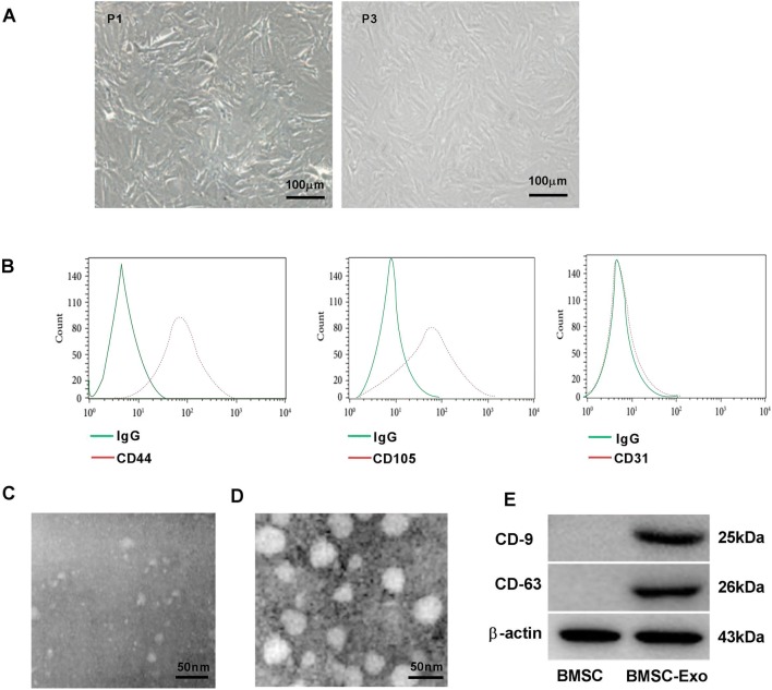
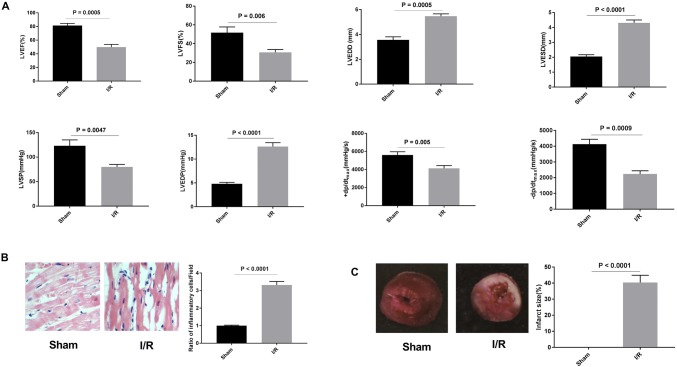
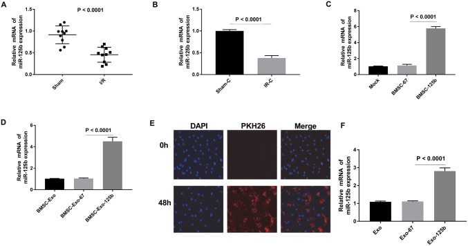
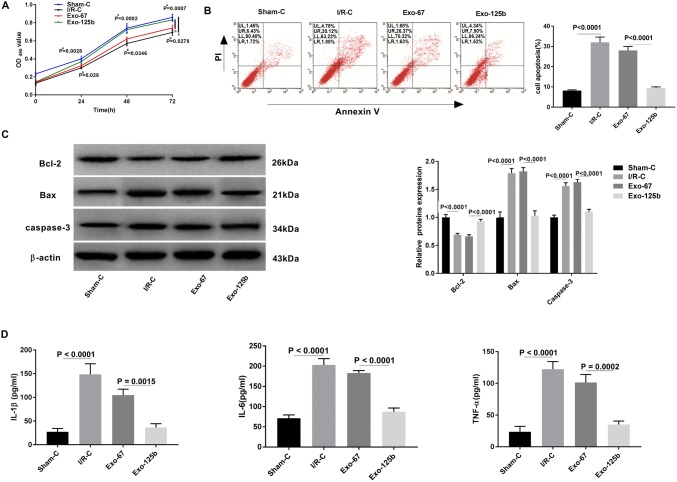
MicroRNA-125b (miR-125b) reduces myocardial infarct area and restrains myocardial ischemia reperfusion injury (I/R). In this study, we aimed to investigate the effect of bone marrow mesenchymal stem cell (BMSC)-derived exosomes carrying miR-125b on I/R rats. The myocardial I/R model in rats was constructed by ligation of the left anterior descending coronary artery (LAD). Rats were randomly divided into I/R and Sham group. Lv-cel-miR-67 (control) or Lv-miR-125b was transfected into BMSCs. Exosomes were extracted from transfected BMSCs, and separately named BMSC-Exo-67, BMSC-Exo-125b, and BMSC-Exo. MTT assay and flow cytometry were used to detect the viability and apoptosis of I/R myocardium cells, respectively. The expression of cell apoptosis proteins and the levels of inflammatory factors were examined by Western blot and ELISA assay, respectively. The target relationship between miR-125b and SIRT7 was predicted by using StarBase3.0, and was confirmed by using dual-luciferase reporter gene assay. qRT-PCR, immunohistochemistry staining, and Western blot were used to evaluate the expression of SIRT7 in myocardium tissues in I/R rats. BMSC-derived exosomes were successfully isolated and identified by TEM and positive expression of CD9 and CD63. The expression of miR-125b was down-regulated in I/R myocardium tissues and cells. BMSC-Exo-125b significantly up-regulated miR-125b in I/R myocardium cells. The intervention of BMSC-Exo-125b significantly increased the cell viability, decreased the apoptotic ratio, down-regulated Bax and caspase-3, up-regulated Bcl-2, and decreased the levels of IL-1β, IL-6, and TNF-α in I/R myocardium cells. SIRT7 was a target of miR-125b, and BMSC-Exo-125b significantly down-regulated SIRT7 in myocardium cells. In addition, the injection of BMSC-Exo-125b alleviated the pathological damages and down-regulated SIRT7 in myocardium tissues of I/R rats. BMSC-derived exosomes carrying miR-125b protected against myocardial I/R by targeting SIRT7.
Overview
The claims and statistics of this paper haven’t been checked yet.
Run the check — free, no account needed. It reads the open-access text and checks the sources it cites.
Integrity signals
Registry
Cites
Statistics
Figures
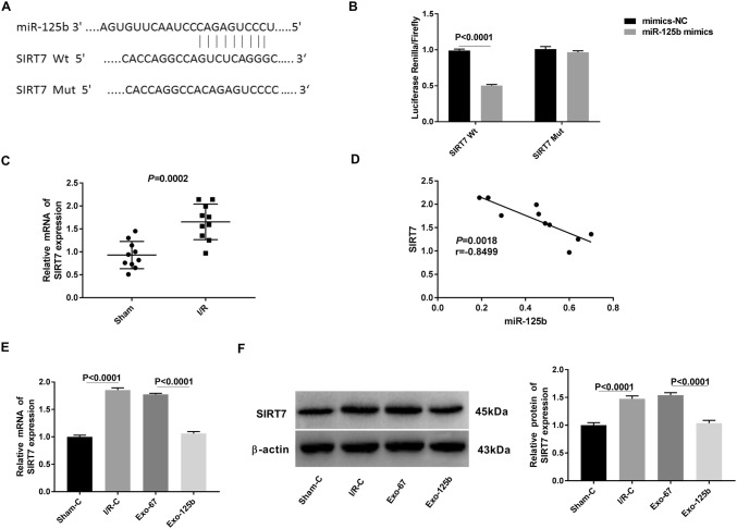
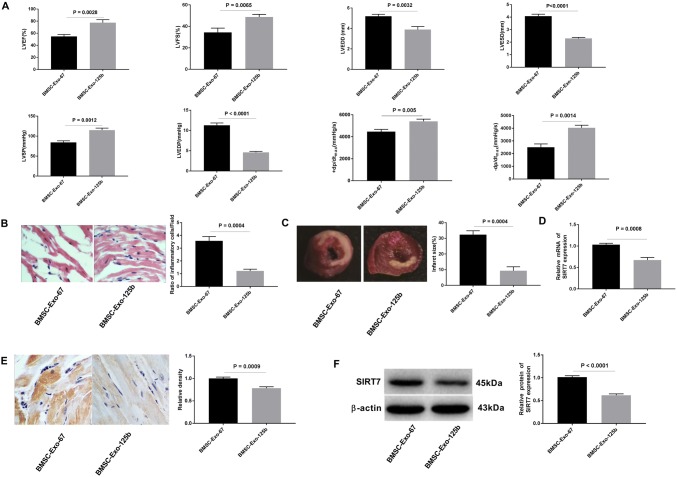
Verification
Claims the paper makes
The claims and statistics of this paper haven’t been checked yet.
Run the check — free, no account needed. It reads the open-access text and checks the sources it cites.
Claims other papers make about this one
Each is checked against this paper’s own text: does it say what the citing paper says it does?
In the citing paper
“Similarly, Chen et al. showed that bone marrow-secreted MSC exosomes carrying miRNA-125b could prevent myocardial ischemia-reperfusion injury by targeting SIRT7 [93].”
From The Therapeutic Potential and Clinical Significance of Exosomes as Carriers of Drug Delivery System (2022) · cites this paper as [93]
Self-check
Percentages, sums and reported differences re-computed from the paper’s own numbers.
The self-check needs the paper’s full text.
Citations
Cites
Valadi, H., Ekström, K., Bossios, A., Sjöstrand, M., Lee, J. J., & Lötvall, J. O. (2007) · Nature Cell Biology · doi:10.1038/ncb1596 · Cited by 12,834
Ischemia and reperfusion—from mechanism to translation
Eltzschig, H. K., & Eckle, T. D. L. G. (2011) · Nature Medicine · doi:10.1038/nm.2507 · Cited by 3,468
Myocardial ischemia-reperfusion injury: a neglected therapeutic target
Hausenloy, D. J., & Yellon, D. M. (2013) · Journal of Clinical Investigation · doi:10.1172/jci62874 · Cited by 2,357
Exosome secreted by MSC reduces myocardial ischemia/reperfusion injury
Lai, R. C., Arslan, F., Lee, M. M., Sze, N. S. K., Choo, A., Chen, T. S., Salto-Tellez, M., Timmers, L., Lee, C. N., El Oakley, R. M., et al. (2010) · Stem Cell Research · doi:10.1016/j.scr.2009.12.003 · Cited by 2,215
Circulating MicroRNAs in Patients With Coronary Artery Disease
Fichtlscherer, S., Rosa, S. D., Fox, H., Schwietz, T., Fischer, A., Liebetrau, C., Weber, M. A., Hamm, C. W., Roexe, T., Müller-Ardogan, M., et al. (2010) · Circulation Research · doi:10.1161/circresaha.109.215566 · Cited by 1,222
Autologous skeletal myoblast transplantation for severe postinfarction left ventricular dysfunction
Menasché, P., Hagège, A. A., Vilquin, J., Desnos, M., Abergel, É., Pouzet, B., Bel, A., Sarateanu, S., Scorsin, M., Schwartz, K., et al. (2003) · Journal of the American College of Cardiology · doi:10.1016/s0735-1097(03)00092-5 · Cited by 1,126
Zhao, J., Li, X., Hu, J., Chen, F., Qiao, S., Sun, X., Gao, L., Xie, J., & Xu, B. (2019) · Cardiovascular Research · doi:10.1093/cvr/cvz040 · Cited by 779
Barile, L., Lionetti, V., Cervio, E., Matteucci, M., Gherghiceanu, M., Popescu, L. M., Torre, T., Siclari, F., Moccetti, T., & Vassalli, G. (2014) · Cardiovascular Research · doi:10.1093/cvr/cvu167 · Cited by 747
Mesenchymal stem cell secretes microparticles enriched in pre-microRNAs
Chen, T. S., Lai, R. C., Lee, M. M., Choo, A. B. H., Lee, C. N., & Lim, S. K. (2009) · Nucleic Acids Research · doi:10.1093/nar/gkp857 · Cited by 672
Vakhrusheva, O., Smolka, C., Gajawada, P., Sawa Kostin, D., Boettger, T., Kubin, T., Braun, T. W., & Bober, E. (2008) · Circulation Research · doi:10.1161/circresaha.107.164558 · Cited by 646
Mammalian Sir2 homolog SIRT7 is an activator of RNA polymerase I transcription
Ford, E., Voit, R., Liszt, G., Magin, C., Grummt, I., & Guarente, L. (2006) · Genes & Development · doi:10.1101/gad.1399706 · Cited by 629
Ren, X.-P., Wu, J., Wang, X., Sartor, M. A., Qian, J., Jones, K., Nicolaou, P. A., Pritchard, T. J., & Fan, G. (2009) · Circulation · doi:10.1161/circulationaha.108.814145 · Cited by 488
Toward MicroRNA–Based Therapeutics for Heart Disease
van Rooij, E., Marshall, W. S., & Olson, E. N. (2008) · Circulation Research · doi:10.1161/circresaha.108.183426 · Cited by 383
Xiao, J., Pan, Y., Li, X. H., Yang, X. Y., Feng, Y. L., Tan, H. H., Jiang, L., Feng, J., & Yu, X. Y. (2016) · Cell Death & Disease · doi:10.1038/cddis.2016.181 · Cited by 373
STRESS signaling pathways that modulate cardiac myocyte apoptosis
Baines, C. P., & Molkentin, J. D. (2004) · Journal of Molecular and Cellular Cardiology · doi:10.1016/j.yjmcc.2004.11.004 · Cited by 334
Wang, X., Zhang, X., Ren, X., Chen, J., Liu, H., Yang, J., Medvedovic, M., Hu, Z., & Fan, G. (2010) · Circulation · doi:10.1161/circulationaha.110.964684 · Cited by 297
Xiao, C., Wang, K., Xu, Y., Hu, H., Zhang, N., Wang, Y., Zhong, Z., Zhao, J., Li, Q., Zhu, D., et al. (2018) · Circulation Research · doi:10.1161/circresaha.118.312758 · Cited by 292
Ma, T., Chen, Y., Chen, Y., Meng, Q., Sun, J., Shao, L., Yu, Y., Huang, H., Hu, Y., Yang, Z., et al. (2018) · Stem Cells International · doi:10.1155/2018/3290372 · Cited by 262
Adding fuel to fire: microRNAs as a new class of mediators of inflammation
Sheedy, F. J., & O'Neill, L. A. J. (2008) · Annals of the Rheumatic Diseases · doi:10.1136/ard.2008.100289 · Cited by 246
The role of microRNA in modulating myocardial ischemia-reperfusion injury
Ye, Y., Perez-Polo, J. R., Qian, J., & Birnbaum, Y. (2010) · Physiological Genomics · doi:10.1152/physiolgenomics.00130.2010 · Cited by 206
Plasma Protein Carbonylation in Haemodialysed Patients: Focus on Diabetes and Gender
Colombo, G., Reggiani, F., Cucchiari, D., Astori, E., Garavaglia, M. L., Portinaro, N. M., Saino, N., Finazzi, S., Milzani, A., Badalamenti, S., et al. (2018) · Oxidative Medicine and Cellular Longevity · doi:10.1155/2018/4149681 · Cited by 174
Wang, X., Ha, T., Zou, J., Ren, D., Liu, L., Zhang, X., Kalbfleisch, J. H., Gao, X., Williams, D. L., & Li, C. (2014) · Cardiovascular Research · doi:10.1093/cvr/cvu044 · Cited by 156
Role of Endothelial Cells in Myocardial Ischemia-Reperfusion Injury
Singhal, A. K., Symons, J. D., Boudina, S., Jaishy, B. P., & Shiu, Y.-T. E. (2010) · Vascular Disease Prevention · doi:10.2174/1874120701007010001 · Cited by 133
Araki, S., Izumiya, Y., Rokutanda, T., Ianni, A., Hanatani, S., Kimura, Y., Onoue, Y., Senokuchi, T., Yoshizawa, T., Yasuda, O., et al. (2015) · Circulation · doi:10.1161/circulationaha.114.014821 · Cited by 121
Diagnostic ability of hand-held echocardiography in ventilated critically ill patients
Vignon, P., Chastagner, C., François, B., Martaillé, J.-F., Normand, S., Bonnivard, M., & Gastinne, H. (2003) · Critical Care · doi:10.1186/cc2360 · Cited by 108
Wang, Y., Zhao, R., Liu, D., Deng, W., Xu, G., Liu, W., Rong, J., Long, X., Ge, J., & Shi, B. (2018) · Oxidative Medicine and Cellular Longevity · doi:10.1155/2018/4971261 · Cited by 108
Wang, C., Zhu, G., He, W., Yin, H., Fan, L., Gou, X., & Li, X. (2019) · The FASEB Journal · doi:10.1096/fj.201801821r · Cited by 102
Yang, M., Chen, J., Zhao, J., & Meng, M. (2014) · PLoS ONE · doi:10.1371/journal.pone.0108024 · Cited by 87
Ischemic postconditioning inhibits apoptosis after renal ischemia/reperfusion injury in rat
Chen, H., Xing, B., Liu, X., Zhan, B., Zhou, J., Zhu, H., & Chen, Z. (2008) · Transplant International · doi:10.1111/j.1432-2277.2007.00606.x · Cited by 82
Zhao, D., Pan, F., Sun, Y., Qin, Z., Zhang, Z., Tan, Y., Gao, E., Lau, W. B., Ma, X., Yang, J. Z., et al. (2018) · Cell Death and Disease · doi:10.1038/s41419-018-0726-3 · Cited by 71
Zhong-yang, S. (2013) · World Journal of Gastroenterology · doi:10.3748/wjg.v19.i23.3583 · Cited by 70
MicroRNA-15a/b are up-regulated in response to myocardial ischemia/reperfusion injury
Li-Feng, L., Liang, Z., Zhen-Rong, L., Liu, X., Bai, J., Jie, C., Chen, C., & Yu, W. (2012) · 老年心脏病学杂志:英文版 · doi:10.3724/sp.j.1263.2012.00028 · Cited by 66
Jin, G., Qiu, G., Wu, D., Hu, Y., Qiao, P., Fan, C., & Gao, F. (2013) · International Journal of Molecular Medicine · doi:10.3892/ijmm.2013.1340 · Cited by 64
Bone Marrow Mesenchymal Stem Cells Reduce Intestinal Ischemia/Reperfusion Injuries in Rats
Jiang, H., Qu, L., Li, Y., Gu, L., Shi, Y., Zhang, J., Zhu, W., & Li, J. (2009) · Journal of Surgical Research · doi:10.1016/j.jss.2009.07.035 · Cited by 60
Zhang, Y., Wang, B., Fu, X., Guan, S., Han, W., Zhang, J., Gan, Q., Fang, W., Ying, W., & Qu, X. (2016) · Europe PMC (PubMed Central) · Cited by 55
Inflammatory response post-myocardial infarction and reperfusion: a new therapeutic target?
Bonvini, R. F., Hendiri, T., & Camenzind, E. (2005) · European Heart Journal Supplements · doi:10.1093/eurheartj/sui077 · Cited by 54
Ma, R., Hu, X., Gangying, H., Yi, C., Changjiang, Z., Xuefei, L., Li, Y., & Jiang, H. (2015) · PLoS ONE · doi:10.1371/journal.pone.0143064 · Cited by 44
Penna, C. R. R., Brancaccio, M., Tullio, F., Rubinetto, C., Perrelli, M.-G., Angotti, C., Pagliaro, P., & Tarone, G. (2014) · Basic Research in Cardiology · doi:10.1007/s00395-014-0418-9 · Cited by 43
miR-125b suppresses the proliferation of hepatocellular carcinoma cells by targeting Sirtuin7.
Zhao, L., & Wang, W. (2015) · PubMed · Cited by 43
Sirtuin7 has an oncogenic potential via promoting the growth of cholangiocarcinoma cells
Li, W., Sun, Z., Chen, C., Wang, L., Geng, Z., & Tao, J. (2018) · Biomedicine & Pharmacotherapy · doi:10.1016/j.biopha.2018.02.007 · Cited by 25
Anti-inflammatory effect of exendin-4 postconditioning during myocardial ischemia and reperfusion
Du, X., Hu, X., & Wei, J. (2014) · Molecular Biology Reports · doi:10.1007/s11033-014-3252-0 · Cited by 21
Lv, J., Tian, J., Zheng, G., & Zhao, J. (2017) · Journal of Biochemical and Molecular Toxicology · doi:10.1002/jbt.21955 · Cited by 20
Ganguly, R., Lytwyn, M., & Pierce, G. N. (2013) · Current Pharmaceutical Design · doi:10.2174/138161281939131127111018 · Cited by 12
Zhang, W., Yang, C., Guo, W., Guo, X., Bian, J., Zhou, Q.-Z., Chen, M., Zhou, J., Chen, Z., Wang, P., et al. (2018) · Europe PMC (PubMed Central) · doi:10.3969/j.issn.1673-4254.2018.08.02 · Cited by 12
Chen, C., Yang, J., James, I., Zhang, H.-Y., & Besner, G. E. (2014) · Surgery · doi:10.1016/j.surg.2014.01.013 · Cited by 11
Zhang, Z., Zhang, H., Li, H., Chen, X., Liu, M., Liu, D., Zhao, Y., & Kong, X. (2014) · Molecular and Cellular Biochemistry · doi:10.1007/s11010-014-2025-3 · Cited by 5
Transendocardial, autologous bone-marrow cell transplant in severe, chronic ischemic heart failure
Dohman, H., Perin, E. C., Sousa, A., Silva, S., Tinoco, C., Esporcatte, R., Rangel, F. O. D., Campos, L., Fernandes, M., & Dohmann, H. J. F. (2003) · Critical Care · doi:10.1186/cc2208 · Cited by 2
Cited by
Sorted by citations
Exosome engineering: Current progress in cargo loading and targeted delivery
Fu, S., Wang, Y., Xia, X., & Zheng, J. C. (2020) · NanoImpact · doi:10.1016/j.impact.2020.100261 · Cited by 444
Exosomal microRNAs derived from mesenchymal stem cells: cell-to-cell messages
Asgarpour, K., Shojaei, Z., Amiri, F., Ai, J., Mahjoubin‐Tehran, M., Ghasemi, F., Arefnezhad, R., Hamblin, M. R., & Mirzaei, H. (2020) · Cell Communication and Signaling · doi:10.1186/s12964-020-00650-6 · Cited by 224
Extracellular vesicles as a drug delivery system: A systematic review of preclinical studies
de Castilla, P. E. M., Tong, L., Huang, C., Sofias, A. M., Pastorin, G., Chen, X. S., Storm, G., Schiffelers, R. M., & Wang, J. (2021) · Advanced Drug Delivery Reviews · doi:10.1016/j.addr.2021.05.011 · Cited by 219
Extracellular vesicles for tissue repair and regeneration: Evidence, challenges and opportunities
Nagelkerke, A., Ojansivu, M., van der Koog, L., Whittaker, T. E., Cunnane, E. M., da Silva, A. M., Dekker, N., & Stevens, M. M. (2021) · Advanced Drug Delivery Reviews · doi:10.1016/j.addr.2021.04.013 · Cited by 211
Ma, Z.-J., Yang, J.-J., Lu, Y.-B., Liu, Z.-Y., & Wang, X.-X. (2020) · World Journal of Stem Cells · doi:10.4252/wjsc.v12.i8.814 · Cited by 155
Metadata
- Authors
- Chen, Q., Liu, Y., Ding, X., Li, Q., Qiu, F., Wang, M., Shen, Z., Zheng, H., & Fu, G.
- Affiliations
- Sir Run Run Shaw Hospital (CN)
- Nanjing Drum Tower Hospital (CN)
- Zhejiang Provincial People's Hospital (CN)
- Corresponding author
- Zheng, Hao
- Fu, Guosheng
- Venue
- Molecular and Cellular Biochemistry
- Published
- 2019-12-19
- Volume
- 465
- Issue
- 1-2
- Pages
- 103-114
- ISSN
- 0300-8177 (ISSN-L)
- 1573-4919
- PMC
- PMC6955239
- OpenAlex
- W2996170603
- Cited by
- 146
- References
- 47
- Citations by year
- 2026: 12 · 2025: 19 · 2024: 24 · 2023: 26 · 2022: 30 · 2021: 26 · 2020: 9
- FWCI
- 5.37
- Type
- journal article
- Language
- en
- Open access
- hybrid
- doi.org
- Locations
- Molecular and Cellular Biochemistry (publishedVersion, cc-by)
- Europe PMC (PubMed Central) (publishedVersion, cc-by)
- PubMed Central (publishedVersion, cc-by)
- PubMed (publishedVersion)
- Funders
- Effects and mechanisms of microRNA and O-GlcNAcylation on paracrine function of diabetic endothelial progenitor cell (Y18H020004)
- The mechanism research identifing Oct4 upregulating Cripto-1 mediated osteoblast differentiation of valve interstitial cells during the pathogenesis of aortic valve calcification (81800342)
- Topics
- Life Sciences › Biochemistry, Genetics and Molecular Biology › Molecular Biology › Extracellular vesicles in disease
- Health Sciences › Medicine › Cardiology and Cardiovascular Medicine › Cardiac Ischemia and Reperfusion
- Life Sciences › Biochemistry, Genetics and Molecular Biology › Cancer Research › MicroRNA in disease regulation
- Keywords
- bone marrow mesenchymal stem cells, exosomes, miR-125b, SIRT7, myocardial ischemia-reperfusion injury, cardioprotection, apoptosis, inflammation, stem cell-derived exosomes, myocardial infarction, Bax, caspase-3, Bcl-2, inflammatory cytokines, microRNA target regulation, exosome delivery
- MeSH terms
- Animals, Bone Marrow Cells, Male, Myocardial Reperfusion Injury, Cytokines, Rats, Sprague-Dawley, Apoptosis, MicroRNAs, Sirtuins, Rats, Exosomes, Mesenchymal Stem Cells
About these checks
Registry, retraction, Cites and statistics results are computed, not judged, with one exception: a printed reference that no index lists is read once by a model before it counts as a citation error. Claims are extracted by a model and kept only when their quote appears verbatim in the text; each cited source is then checked, with a model’s help, for whether it says what the claim relies on. Those verdicts can be wrong — read the claim’s quote and the cited source before relying on one.
Statistics and arithmetic are recomputed, not judged.
Confidence bands are shown only for fields calibrated on our annotated test set. None is calibrated yet, so no band is shown.
What counts as a claim ›How we measure confidence ›How we check this ›
Are you an author, or do you see a problem with this page? Request a review
