Student or educator? Sign up with your school email and get 6 months of Pro free.
CoChat

The DOI matches its Crossref record; 128 of the 132 references it cites were found in a registry or an index.

The Basics of Brain Development

Open access
Stiles, Joan, Jernigan, Terry L. · 2010 · Neuropsychology Review

Abstract

Over the past several decades, significant advances have been made in our understanding of the basic stages and mechanisms of mammalian brain development. Studies elucidating the neurobiology of brain development span the levels of neural organization from the macroanatomic, to the cellular, to the molecular. Together this large body of work provides a picture of brain development as the product of a complex series of dynamic and adaptive processes operating within a highly constrained, genetically organized but constantly changing context. The view of brain development that has emerged from the developmental neurobiology literature presents both challenges and opportunities to psychologists seeking to understand the fundamental processes that underlie social and cognitive development, and the neural systems that mediate them. This chapter is intended to provide an overview of some very basic principles of brain development, drawn from contemporary developmental neurobiology, that may be of use to investigators from a wide range of disciplines.

Overview

The claims and statistics of this paper haven’t been checked yet.

Free: it reads the open-access text and checks the first 5 claims that cite a source we can check. Create a free account to check every claim.

A few free checks a day · papers already verified open instantly · results are public

Integrity signals

Registry

Crossref · 10.1007/s11065-010-9148-4

Cites

128 of 128 references verified0 citation errors · 0 not checkable

Statistics

Figures

Fig. 1 Human embryo at Carnegie Stage 23, the end of the embryonic period (GW8). It is 30 mm long. Image from the Kyoto Collection reproduced with permission of Prof Kohei Shiota, Graduate School of Medicine, Kyoto University, and obtained with permission of Dr. Mark Hill, University of New South Wales, http://embryology.med.unsw.edu.au/embryo.htm
Fig. 1 Human embryo at Carnegie Stage 23, the end of the embryonic period (GW8). It is 30 mm long. Image from the Kyoto Collection reproduced with permission of Prof Kohei Shiota, Graduate School of Medicine, Kyoto University, and obtained with permission of Dr. Mark Hill, University of New South Wales, http://embryology.med.unsw.edu.au/embryo.htmCC BY-NC · PubMed Central
Fig. 2 Schematic drawing of a neuron. Each neuron a single large axon. At the distal tip of the axon is a growth cone that serves to guide the axon to targeted brain regions. Once the axon reaches the target site, synapses, or points of connection, form between the axon and the target neuron. The synapse allows electrochemical signals to be transmitted to the target neuron. Each neuron also has a complex arbor of dendrites that receive information from other neurons. Image in the public domain uploaded from: http://upload.wikimedia.org/wikipedia/commons/7/72/Neuron-figure-notext.svg. Original image from Nicolas Rougier
Fig. 2 Schematic drawing of a neuron. Each neuron a single large axon. At the distal tip of the axon is a growth cone that serves to guide the axon to targeted brain regions. Once the axon reaches the target site, synapses, or points of connection, form between the axon and the target neuron. The synapse allows electrochemical signals to be transmitted to the target neuron. Each neuron also has a complex arbor of dendrites that receive information from other neurons. Image in the public domain uploaded from: http://upload.wikimedia.org/wikipedia/commons/7/72/Neuron-figure-notext.svg. Original image from Nicolas RougierCC BY-NC · PubMed Central
Fig. 3 Two views of the human brain. a. Lateral view (rostral end is left, caudal is right) shows an apparently uniform surface marked by gyri and sulcal folds (Right hemisphere of J. Piłsudski’s brain, lateral view, image in the public domain). b. Coronal cross-section (cut at approximately the level of the dotted line in A) stained for cell bodies that mark neurons. The neocortex is the thin mantel layer (dark purple) on the surface of the brain. The white areas are connecting fiber pathways. Image reproduced with permission from http://www.brains.rad.msu.edu which is supported by the U.S. National Science Foundation. Images obtained with permission from Wiki Commons, http://commons.wikimedia.org/wiki
Fig. 3 Two views of the human brain. a. Lateral view (rostral end is left, caudal is right) shows an apparently uniform surface marked by gyri and sulcal folds (Right hemisphere of J. Piłsudski’s brain, lateral view, image in the public domain). b. Coronal cross-section (cut at approximately the level of the dotted line in A) stained for cell bodies that mark neurons. The neocortex is the thin mantel layer (dark purple) on the surface of the brain. The white areas are connecting fiber pathways. Image reproduced with permission from http://www.brains.rad.msu.edu which is supported by the U.S. National Science Foundation. Images obtained with permission from Wiki Commons, http://commons.wikimedia.org/wikiCC BY-NC · PubMed Central
Fig. 4 The major spatial dimensions of the E13 embryo. a. The dorsal surface view of the embryo on E13 is shown in the first panel. The wall of the amniotic sac has been cut away to reveal the dorsal surface (epiblast layer) of the embryo. The rostral (“head”) end of the embryo is on the top of this figure, and the caudal (“tail”) end is at the bottom. b. A lateral cross-section of the embryo and placenta at E13. On E13, the two-layered embryo is located centrally between two major placental sacs. The amniotic sac (which later in development will surround the embryo) is located above the embryo, and the yolk sac is located below. The rostral end of the embryo is to the right in this figure. To place the embryo shown in the first panel of A within the context of the lateral view of the embryo and placenta shown in B, it is necessary to first rotate the embryo so that the rostral end faces right (second panel of A), and then rotate the embryo in depth so that the dorsal surface faces up (last panel of A). C. The comparable rostral-caudal and dorsal-ventral spatial axes of an infant. The spatial axes of a crawling infant are comparable to the position of the embryo in B. Illustrations by Matthew Stiles Davis reprinted by permission of the publisher from THE FUNDAMENTALS OF BRAIN DEVELOPMENT: INTEGRATING NATURE AND NURTURE by Joan Stiles, Cambridge, Mass.: Harvard University Press, Copyright © 2008 by the President and Fellows of Harvard College
Fig. 4 The major spatial dimensions of the E13 embryo. a. The dorsal surface view of the embryo on E13 is shown in the first panel. The wall of the amniotic sac has been cut away to reveal the dorsal surface (epiblast layer) of the embryo. The rostral (“head”) end of the embryo is on the top of this figure, and the caudal (“tail”) end is at the bottom. b. A lateral cross-section of the embryo and placenta at E13. On E13, the two-layered embryo is located centrally between two major placental sacs. The amniotic sac (which later in development will surround the embryo) is located above the embryo, and the yolk sac is located below. The rostral end of the embryo is to the right in this figure. To place the embryo shown in the first panel of A within the context of the lateral view of the embryo and placenta shown in B, it is necessary to first rotate the embryo so that the rostral end faces right (second panel of A), and then rotate the embryo in depth so that the dorsal surface faces up (last panel of A). C. The comparable rostral-caudal and dorsal-ventral spatial axes of an infant. The spatial axes of a crawling infant are comparable to the position of the embryo in B. Illustrations by Matthew Stiles Davis reprinted by permission of the publisher from THE FUNDAMENTALS OF BRAIN DEVELOPMENT: INTEGRATING NATURE AND NURTURE by Joan Stiles, Cambridge, Mass.: Harvard University Press, Copyright © 2008 by the President and Fellows of Harvard CollegeCC BY-NC · PubMed Central
Fig. 5 The major events of gastulation occur between E13 and E20. a. The onset of gastrulation is marked by the formation of the primitive streak and the primitive node. The primitive streak provides an opening to deeper embryonic layers. The primitive node is a critical molecular signaling center. On E13, cells from the epiblast layer begin to migrate toward the primitive node and streak (blue arrows). The dotted line indicates the cross-sectional view shown in panel B. b. The migrating cells first move to the primitive streak and then change direction and move down and under the upper layer (blue arrows). As the cells pass the node they receive molecular signals that induce gene expression in the migrating cells. By the end of gastrulation, the hypoblast layer is replaced by the newly formed endodermal layer and the epiblast layer by the ectodermal layer. Between these layers the mesodermal layer forms. c. Once under the upper layer, the cells change direction and begin migrating rostrally under the upper layer (blue arrows). The first cells to migrate form the most rostral regions of the newly forming endodermal and mesodermal layers. Later migrating cells form progressively more caudal regions of the layers. d. Cells that migrate along the axial midline send molecular signals that induce cells in the overlying epiblast layer to differentiate into neuroectodermal cells (red band) which are the neural progenitor cells. Migrating cells also receive a second set of signals from the node that induce anterior or posterior fate in different subpopulations of the neurectodermal cells. Early migrating cells signal anterior fate in the progenitor cells, while late migrating cells signal posterior fate. Illustrations by Matthew Stiles Davis reprinted by permission of the publisher from THE FUNDAMENTALS OF BRAIN DEVELOPMENT: INTEGRATING NATURE AND NURTURE by Joan Stiles, Cambridge, Mass.: Harvard University Press, Copyright © 2008 by the President and Fellows of Harvard College
Fig. 5 The major events of gastulation occur between E13 and E20. a. The onset of gastrulation is marked by the formation of the primitive streak and the primitive node. The primitive streak provides an opening to deeper embryonic layers. The primitive node is a critical molecular signaling center. On E13, cells from the epiblast layer begin to migrate toward the primitive node and streak (blue arrows). The dotted line indicates the cross-sectional view shown in panel B. b. The migrating cells first move to the primitive streak and then change direction and move down and under the upper layer (blue arrows). As the cells pass the node they receive molecular signals that induce gene expression in the migrating cells. By the end of gastrulation, the hypoblast layer is replaced by the newly formed endodermal layer and the epiblast layer by the ectodermal layer. Between these layers the mesodermal layer forms. c. Once under the upper layer, the cells change direction and begin migrating rostrally under the upper layer (blue arrows). The first cells to migrate form the most rostral regions of the newly forming endodermal and mesodermal layers. Later migrating cells form progressively more caudal regions of the layers. d. Cells that migrate along the axial midline send molecular signals that induce cells in the overlying epiblast layer to differentiate into neuroectodermal cells (red band) which are the neural progenitor cells. Migrating cells also receive a second set of signals from the node that induce anterior or posterior fate in different subpopulations of the neurectodermal cells. Early migrating cells signal anterior fate in the progenitor cells, while late migrating cells signal posterior fate. Illustrations by Matthew Stiles Davis reprinted by permission of the publisher from THE FUNDAMENTALS OF BRAIN DEVELOPMENT: INTEGRATING NATURE AND NURTURE by Joan Stiles, Cambridge, Mass.: Harvard University Press, Copyright © 2008 by the President and Fellows of Harvard CollegeCC BY-NC · PubMed Central
Fig. 6 Changes in the morphology of the embryo in the embryonic period. The formation of the neural tube occurs between E19 and E29. a. The emergence of the neural ridges is observed on E19. b. The ridges fold over to begin the process of neural tube formation. c. Closure of the neural tube begins on E22 in central regions of the newly forming neural tube. d. Closure continues in rostral and caudal direction. The anterior neuropore closes on E25, and the posterior on E27. e. Following the closure of the neural tube, the embryo begins to expand particularly in anterior regions. The primary vesicles are evident by E28. These include the Prosencephalon, Mesencephalon, and Rhombencephalon. f. By E49 the secondary vesicles emerge. The Prosencephalon differentiates into the Telencephalon and Diencephalon, and the Rhombencephalon into the Metencephalon and Myelencephalon. Illustrations by Matthew Stiles Davis reprinted by permission of the publisher from THE FUNDAMENTALS OF BRAIN DEVELOPMENT: INTEGRATING NATURE AND NURTURE by Joan Stiles, Cambridge, Mass.: Harvard University Press, Copyright © 2008 by the President and Fellows of Harvard College
Fig. 6 Changes in the morphology of the embryo in the embryonic period. The formation of the neural tube occurs between E19 and E29. a. The emergence of the neural ridges is observed on E19. b. The ridges fold over to begin the process of neural tube formation. c. Closure of the neural tube begins on E22 in central regions of the newly forming neural tube. d. Closure continues in rostral and caudal direction. The anterior neuropore closes on E25, and the posterior on E27. e. Following the closure of the neural tube, the embryo begins to expand particularly in anterior regions. The primary vesicles are evident by E28. These include the Prosencephalon, Mesencephalon, and Rhombencephalon. f. By E49 the secondary vesicles emerge. The Prosencephalon differentiates into the Telencephalon and Diencephalon, and the Rhombencephalon into the Metencephalon and Myelencephalon. Illustrations by Matthew Stiles Davis reprinted by permission of the publisher from THE FUNDAMENTALS OF BRAIN DEVELOPMENT: INTEGRATING NATURE AND NURTURE by Joan Stiles, Cambridge, Mass.: Harvard University Press, Copyright © 2008 by the President and Fellows of Harvard CollegeCC BY-NC · PubMed Central
Fig. 7 The effects of different concentrations of Emx2 and Pax6 on the development of sensorimotor cortical areas. It is the combination of the specific concentration of each molecule that determines the identity of the cortical region. Mutations that affect the quantities of either molecule alter cortical patterning. Adapted with permission from Bishop et al. (2002). "Distinct actions of Emx1, Emx2, and Pax6 in regulating the specification of areas in the developing neocortex." J Neurosci 22(17): 7627–7638, Fig. 1
Fig. 7 The effects of different concentrations of Emx2 and Pax6 on the development of sensorimotor cortical areas. It is the combination of the specific concentration of each molecule that determines the identity of the cortical region. Mutations that affect the quantities of either molecule alter cortical patterning. Adapted with permission from Bishop et al. (2002). "Distinct actions of Emx1, Emx2, and Pax6 in regulating the specification of areas in the developing neocortex." J Neurosci 22(17): 7627–7638, Fig. 1CC BY-NC · PubMed Central
Fig. 8 Different modes of neuronal migration to the neocortex. a. Neuron migration by somal translocation where cell extends a cytoplasmic process and attaches to the outside of the brain compartment (pial surface), and then the nucleus moves up into the brain area. b. Neuron migraton radial glial guide. Radial glial provides scaffold for neuron to migrate along. c. Neuron migration from second proliferative zone in ganglionic eminences by tangential migration (arrows indicate direction of migration for different neuron populations). Figures A and B adapted with permission from Nadarajah et al. (2003). Neuronal Migration in the Developing Cerebral Cortex: Observations Based on Real- time Imaging. Cerebral Cortex, 13, 607–611. Figure 5. Figure C adapted from illustrations by Matthew Stiles Davis reprinted by permission of the publisher from THE FUNDAMENTALS OF BRAIN DEVELOPMENT: INTEGRATING NATURE AND NURTURE by Joan Stiles, Cambridge, Mass.: Harvard University Press, Copyright © 2008 by the President and Fellows of Harvard College
Fig. 8 Different modes of neuronal migration to the neocortex. a. Neuron migration by somal translocation where cell extends a cytoplasmic process and attaches to the outside of the brain compartment (pial surface), and then the nucleus moves up into the brain area. b. Neuron migraton radial glial guide. Radial glial provides scaffold for neuron to migrate along. c. Neuron migration from second proliferative zone in ganglionic eminences by tangential migration (arrows indicate direction of migration for different neuron populations). Figures A and B adapted with permission from Nadarajah et al. (2003). Neuronal Migration in the Developing Cerebral Cortex: Observations Based on Real- time Imaging. Cerebral Cortex, 13, 607–611. Figure 5. Figure C adapted from illustrations by Matthew Stiles Davis reprinted by permission of the publisher from THE FUNDAMENTALS OF BRAIN DEVELOPMENT: INTEGRATING NATURE AND NURTURE by Joan Stiles, Cambridge, Mass.: Harvard University Press, Copyright © 2008 by the President and Fellows of Harvard CollegeCC BY-NC · PubMed Central
Fig. 9 a. The earliest produced neurons migrate to the deepest cortical layers (dark blue). Subsequently migrating neurons migrate to successively more superficial layers (lighter blues) creating an inside out order of migration. Adapted with permission from Cooper (2008). Trends in Neuroscience, 31(3), 113-19. b. As shown in the first panel, the first neurons migrate from the ventricular zone (VZ) to form the preplate (PP). As shown in the second panel, the next neurons split the PP into the marginal zone (MZ) an the subplate (SP), both transient brain structures. The mature brain, shown in the third panel, has six well developed cortical layers (I-VI), but none of the embryonic structures (MZ, SP, VZ). The intermediate zone (IZ) has become a mature white matter layer (WM). Illustrations by Matthew Stiles Davis reprinted by permission of the publisher from THE FUNDAMENTALS OF BRAIN DEVELOPMENT: INTEGRATING NATURE AND NURTURE by Joan Stiles, Cambridge, Mass.: Harvard University Press, Copyright © 2008 by the President and Fellows of Harvard College
Fig. 9 a. The earliest produced neurons migrate to the deepest cortical layers (dark blue). Subsequently migrating neurons migrate to successively more superficial layers (lighter blues) creating an inside out order of migration. Adapted with permission from Cooper (2008). Trends in Neuroscience, 31(3), 113-19. b. As shown in the first panel, the first neurons migrate from the ventricular zone (VZ) to form the preplate (PP). As shown in the second panel, the next neurons split the PP into the marginal zone (MZ) an the subplate (SP), both transient brain structures. The mature brain, shown in the third panel, has six well developed cortical layers (I-VI), but none of the embryonic structures (MZ, SP, VZ). The intermediate zone (IZ) has become a mature white matter layer (WM). Illustrations by Matthew Stiles Davis reprinted by permission of the publisher from THE FUNDAMENTALS OF BRAIN DEVELOPMENT: INTEGRATING NATURE AND NURTURE by Joan Stiles, Cambridge, Mass.: Harvard University Press, Copyright © 2008 by the President and Fellows of Harvard CollegeCC BY-NC · PubMed Central
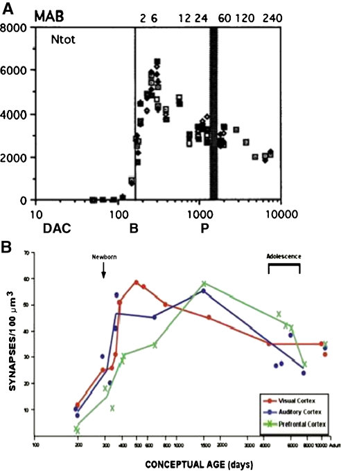
Fig. 10 Synaptic connectivity in the primate brain exhibits initial exuberant production followed by gradual pruning. a. In primate brain, the number of synaptic contacts per probe was plotted along a logarithmic scale as a function of days after conception (DAC). Months after birth (MAB) are indicated along the top of the graph, birth (B) at 166 days post conception is indicated by the thin vertical line and puberty (P) at 3–4 years by the thick vertical line. Reprinted with permission from Bourgeois and Rakic (1993). “Changes of synaptic density in the primary visual cortex of the macaque monkey from fetal to adult stage.” Journal of Neuroscience 13(7): 2801–2820, Fig. 3. b. In human brains, counts of the number of synapses per constant volume of tissue were measured as a function of pre- and postnatal age. Adapted with permission from Huttenlocher and Dabholkar (1997). Regional differences in synaptogenesis in human cerebral cortex. Journal of Comparative Neurology, 387, 167–178, Fig. 2
Fig. 10 Synaptic connectivity in the primate brain exhibits initial exuberant production followed by gradual pruning. a. In primate brain, the number of synaptic contacts per probe was plotted along a logarithmic scale as a function of days after conception (DAC). Months after birth (MAB) are indicated along the top of the graph, birth (B) at 166 days post conception is indicated by the thin vertical line and puberty (P) at 3–4 years by the thick vertical line. Reprinted with permission from Bourgeois and Rakic (1993). “Changes of synaptic density in the primary visual cortex of the macaque monkey from fetal to adult stage.” Journal of Neuroscience 13(7): 2801–2820, Fig. 3. b. In human brains, counts of the number of synapses per constant volume of tissue were measured as a function of pre- and postnatal age. Adapted with permission from Huttenlocher and Dabholkar (1997). Regional differences in synaptogenesis in human cerebral cortex. Journal of Comparative Neurology, 387, 167–178, Fig. 2CC BY-NC · PubMed Central
Fig. 11 Estimated volumes of brain structures in normal volunteers are plotted against age. The volumes in the figures are presented as standardized residuals (removing variability associated with volume of the supratentorial cranial vault). They are, from left, volumes of frontal cortex, thalamus, nucleus accumbens, and cerebral white matter. Note the rapid age-related change (and striking individual differences) in the childhood and adolescent age-range. (Figures modified from Jernigan & Gamst, Neurobiology of Aging, 26 (9), 1271–1274, 2005)
Fig. 11 Estimated volumes of brain structures in normal volunteers are plotted against age. The volumes in the figures are presented as standardized residuals (removing variability associated with volume of the supratentorial cranial vault). They are, from left, volumes of frontal cortex, thalamus, nucleus accumbens, and cerebral white matter. Note the rapid age-related change (and striking individual differences) in the childhood and adolescent age-range. (Figures modified from Jernigan & Gamst, Neurobiology of Aging, 26 (9), 1271–1274, 2005)CC BY-NC · PubMed Central
Fig. 12 Cross sectional data from Lebel et al. (2008, Lebel and Beaulieu 2009) showing robust FA increases in 4 major fiber tracts; note rapid change in FA in young school-aged children and variability in the pace at which FA in the different tracts approaches asymptote. Reprinted with permission from Lebel et al. (2008). “Microstructural maturation of the human brain from childhood to adulthood.” Neuroimage 40(3): 1044–1055
Fig. 12 Cross sectional data from Lebel et al. (2008, Lebel and Beaulieu 2009) showing robust FA increases in 4 major fiber tracts; note rapid change in FA in young school-aged children and variability in the pace at which FA in the different tracts approaches asymptote. Reprinted with permission from Lebel et al. (2008). “Microstructural maturation of the human brain from childhood to adulthood.” Neuroimage 40(3): 1044–1055CC BY-NC · PubMed Central
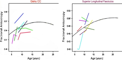
Fig. 13 Individual trajectories for sequential measurements of FA in the genu of the corpus callosum (left) and the superior longitudinal fasciculus (SLF) (right), redrawn from Lebel et al. (2008, Lebel and Beaulieu 2009), illustrating individual differences
Fig. 13 Individual trajectories for sequential measurements of FA in the genu of the corpus callosum (left) and the superior longitudinal fasciculus (SLF) (right), redrawn from Lebel et al. (2008, Lebel and Beaulieu 2009), illustrating individual differencesCC BY-NC · PubMed Central
Fig. 14 Autoradiographs of the ocular dominance columns (ODC) in two young monkeys. A radioactive transneuronal dye was injected into one eye and taken up by neurons in the input layer of primary visual cortex (PVC). a. The normal patterning of the ODC in a 6-week old monkey. ODCs from each eye are equal and adultlike. b. ODC patterning from an animal that was monocularly deprived at 2 weeks of age. The nondeprived eye was injected with the tracer at 18 months of age. ODC for the nondeprived eye (light bands) expand and while those of the deprived eye (dark bands) shrink showing clear dominance of the nondeprived eye in PVC. Adapted from LeVay et al. (1980). Journal of Comparative Neurology, 191, 1–51, Figs. 5 and 6
Fig. 14 Autoradiographs of the ocular dominance columns (ODC) in two young monkeys. A radioactive transneuronal dye was injected into one eye and taken up by neurons in the input layer of primary visual cortex (PVC). a. The normal patterning of the ODC in a 6-week old monkey. ODCs from each eye are equal and adultlike. b. ODC patterning from an animal that was monocularly deprived at 2 weeks of age. The nondeprived eye was injected with the tracer at 18 months of age. ODC for the nondeprived eye (light bands) expand and while those of the deprived eye (dark bands) shrink showing clear dominance of the nondeprived eye in PVC. Adapted from LeVay et al. (1980). Journal of Comparative Neurology, 191, 1–51, Figs. 5 and 6CC BY-NC · PubMed Central

Verification

Claims the paper makes

The claims and statistics of this paper haven’t been checked yet.

Free: it reads the open-access text and checks the first 5 claims that cite a source we can check. Create a free account to check every claim.

A few free checks a day · papers already verified open instantly · results are public

Claims other papers make about this one

Each is checked against this paper’s own text: does it say what the citing paper says it does?

Self-check

Percentages, sums and reported differences re-computed from the paper’s own numbers.

The self-check needs the paper’s full text.

Citations

Cites

Show all 132 references

Research works printed but not linked: 1

  1. Lebel, C., & Beaulieu, C. (2009). Longitudinal diffusion tensor imaging of healthy brain development in children. Honolulu: ISMRM Annual Meeting.

Cited by

See all 2,009

Metadata

Authors
Stiles, J., & Jernigan, T. L.
Affiliations
University of California San Diego (US)
Corresponding author
Jernigan, Terry L.
Venue
Neuropsychology Review
Published
2010-11-03
Volume
20
Issue
4
Pages
327-348
ISSN
  • 1040-7308 (ISSN-L)
  • 1573-6660
OpenAlex
W2115599658
Cited by
2,009
References
132
Citations by year
2026: 142 · 2025: 184 · 2024: 199 · 2023: 209 · 2022: 184 · 2021: 197 · 2020: 195 · 2019: 155 · 2018: 158 · 2017: 122 · 2016: 92 · 2015: 64 · 2014: 49 · 2013: 24 · 2012: 23
FWCI
1.43
Type
journal article
Language
en
Open access
Licence
cc-by-nc
Locations
  • Neuropsychology Review (publishedVersion, cc-by-nc)
  • Europe PMC (PubMed Central) (publishedVersion, cc-by-nc)
  • PubMed Central (publishedVersion, cc-by-nc)
  • PubMed (publishedVersion)
  • eScholarship (California Digital Library) (submittedVersion, public-domain)
  • eScholarship (California Digital Library) (submittedVersion)
Funders
  • Lundbeckfonden (R32‐A3161)
  • H. Lundbeck A/S
  • National Institutes of Health (5r01hd025077-10)
  • U.S. Public Health Service (R32-A3161)
  • University of California, San Diego (RC2DA029475)
  • National Institute on Drug Abuse (RC2DA029475)
  • National Institute of Child Health and Human Development
  • Eunice Kennedy Shriver National Institute of Child Health and Human Development (R01HD060595, R01 HD 25077)
Topics
  • Life Sciences › Neuroscience › Developmental Neuroscience › Neurogenesis and neuroplasticity mechanisms
  • Life Sciences › Biochemistry, Genetics and Molecular Biology › Genetics › Genetics and Neurodevelopmental Disorders
  • Social Sciences › Psychology › Developmental and Educational Psychology › Child and Animal Learning Development
Keywords
brain development, developmental neuroscience, mammalian brain, neuronal organization, social development (developmental psychology), cognitive development, neural systems, genetic constraints, developmental mechanisms
MeSH terms
Animals, Brain, Cell Differentiation, Diagnostic Imaging, Humans, Nerve Net, Neural Pathways, Neurobiology, Neurons, Gene Expression Regulation, Developmental, Body Patterning, Neurogenesis

About these checks

Registry, retraction, Cites and statistics results are computed, not judged, with one exception: a printed reference that no index lists is read once by a model before it counts as a citation error. Claims are extracted by a model and kept only when their quote appears verbatim in the text; each cited source is then checked, with a model’s help, for whether it says what the claim relies on. Those verdicts can be wrong — read the claim’s quote and the cited source before relying on one.

Statistics and arithmetic are recomputed, not judged.

Confidence bands are shown only for fields calibrated on our annotated test set. None is calibrated yet, so no band is shown.

What counts as a claim ›How we measure confidence ›How we check this ›

Are you an author, or do you see a problem with this page? Request a review

How to cite

Cite CoChat for the checks on this page, and the paper for its own findings.

This page
CoChat. (2026). Citation check: The Basics of Brain Development. https://app.cochat.ai/papers/10.1007/s11065-010-9148-4
The paper
Stiles, J., & Jernigan, T. L. (2010). The Basics of Brain Development. Neuropsychology Review. https://doi.org/10.1007/s11065-010-9148-4

Data sources

Bibliographic data: Semantic Scholar, OpenAlex, Crossref.

Contains information from the Semantic Scholar Academic Graph Datasets, made available under the ODC Attribution License (ODC-BY 1.0).

Semantic Scholar’s data is described in: Kinney, R., et al. (2023). The Semantic Scholar Open Data Platform. arXiv:2301.10140.