

The Basics of Brain Development
Open accessThe DOI matches its Crossref record; 128 of the 132 references it cites were found in a registry or an index.
Abstract
Over the past several decades, significant advances have been made in our understanding of the basic stages and mechanisms of mammalian brain development. Studies elucidating the neurobiology of brain development span the levels of neural organization from the macroanatomic, to the cellular, to the molecular. Together this large body of work provides a picture of brain development as the product of a complex series of dynamic and adaptive processes operating within a highly constrained, genetically organized but constantly changing context. The view of brain development that has emerged from the developmental neurobiology literature presents both challenges and opportunities to psychologists seeking to understand the fundamental processes that underlie social and cognitive development, and the neural systems that mediate them. This chapter is intended to provide an overview of some very basic principles of brain development, drawn from contemporary developmental neurobiology, that may be of use to investigators from a wide range of disciplines.
Overview
The claims and statistics of this paper haven’t been checked yet.
Free: it reads the open-access text and checks the first 5 claims that cite a source we can check. Create a free account to check every claim.
Integrity signals
Registry
Cites
Statistics
Figures





Verification
Claims the paper makes
The claims and statistics of this paper haven’t been checked yet.
Free: it reads the open-access text and checks the first 5 claims that cite a source we can check. Create a free account to check every claim.
Claims other papers make about this one
Each is checked against this paper’s own text: does it say what the citing paper says it does?
The source backs part of the claim, not all of it — or backs it for a different population, condition or comparison.
In the citing paper
“We now know from recent studies that it is the birth and migration of neurons derived from oRGCs that play a role in the development of the primary sulci (superior frontal, inferior frontal and precentral) in week 25–26 of gestation [23, 76].”
From Development of prefrontal cortex (2022) · cites this paper as [23]
Experimental — accuracy not yet measuredIn the citing paper
“This intricate structural-functional interplay underscores that the ontogeny of functional networks likely reflects programmed neurodevelopmental events (e.g. neurogenesis, cell death, myelination, pruning, synaptic plasticity, and glial development) (Innocenti and Price, 2005; Stiles and Jernigan, 2010)”
From Development of large-scale functional networks from birth to adulthood: A guide to the neuroimaging literature (2017) · cites this paper as Stiles and Jernigan, 2010
In the citing paper
“The brain’s major fiber pathways become consolidated through myelination, though their presence is largely established prenatally (Stiles and Jernigan, 2010).”
From Development of large-scale functional networks from birth to adulthood: A guide to the neuroimaging literature (2017) · cites this paper as Stiles and Jernigan, 2010
Self-check
Percentages, sums and reported differences re-computed from the paper’s own numbers.
The self-check needs the paper’s full text.
Citations
Cites
Philosophical transactions of the Royal Society of London. B
Press, H. (1886) · Cited by 10,080
MR diffusion tensor spectroscopy and imaging
Basser, P. J., Mattiello, J., & LeBihan, D. (1994) · Biophysical Journal · doi:10.1016/s0006-3495(94)80775-1 · Cited by 5,970
Dynamic mapping of human cortical development during childhood through early adulthood
Gogtay, N., Giedd, J. N., Lusk, L., Hayashi, K. M., Greenstein, D., Vaituzis, A. C., Nugent, T. F., Herman, D. H., Clasen, L. S., Toga, A. W., et al. (2004) · Proceedings of the National Academy of Sciences · doi:10.1073/pnas.0402680101 · Cited by 5,604
(2005) · Choice Reviews Online · doi:10.5860/choice.43-0626 · Cited by 4,674
Neurotrophins: Roles in Neuronal Development and Function
Huang, E. J., & Reichardt, L. F. (2001) · Annual Review of Neuroscience · doi:10.1146/annurev.neuro.24.1.677 · Cited by 4,394
Ferrier lecture. Functional architecture of macaque monkey visual cortex
Hubel, D. H., & Wiesel, T. N. (1977) · Proceedings of the Royal Society of London. Series B. Biological Sciences · doi:10.1098/rspb.1977.0085 · Cited by 2,608 · cited 1×
“The seminal studies of Hubel and Weisel (Hubel and Wiesel 1977; Hubel et al. 1977) showed that monocular visual deprivation in the early postnatal period can substantially alter basic patterns of organization within primary visual cortex (PVC).”
Background(1995) · Choice Reviews Online · doi:10.5860/choice.32-5931 · Cited by 2,311
Mode of cell migration to the superficial layers of fetal monkey neocortex
Rakic, P. (1972) · Journal of Comparative Neurology · doi:10.1002/cne.901450105 · Cited by 2,170 · cited 1×
“Because of the greater distances, neurons require what was originally identified as a special population of cells within the VZ called “radial glial guides” to support their migration (Rakic 1972).”
BackgroundRegional Development of the Brain in Early Life
(1968) · Archives of Disease in Childhood · doi:10.1136/adc.43.229.388 · Cited by 2,090
Neurons derived from radial glial cells establish radial units in neocortex
Noctor, S. C., Flint, A. C., Weissman, T. A., Dammerman, R. S., & Kriegstein, A. R. (2001) · Nature · doi:10.1038/35055553 · Cited by 2,016 · cited 2×
“…radial glial guides were originally thought to be a special, transient population of cells, it has recently been discovered that the cells that provide the scaffolding are actually the neural progenitor cells (Noctor et al. 2001; Noctor et al. 2002; Parnavelas et al. 2002; Weissman et al. 2003).”
BackgroundBrain development in children and adolescents: Insights from anatomical magnetic resonance imaging
Lenroot, R. K., & Giedd, J. N. (2006) · Neuroscience & Biobehavioral Reviews · doi:10.1016/j.neubiorev.2006.06.001 · Cited by 1,863 · cited 1×
“The brain increases in size by four-fold during the preschool period, reaching approximately 90% of adult volume by age 6 (Reiss et al. 1996; Iwasaki et al. 1997; Courchesne et al. 2000; Kennedy and Dehay 2001; Paus et al. 2001; Kennedy et al. 2002; Lenroot and Giedd 2006).”
(1967) · Medical Entomology and Zoology · Cited by 1,713
Neuronal subtype specification in the cerebral cortex
Molyneaux, B. J., Arlotta, P., Menezes, J. R. L., & Macklis, J. D. (2007) · Nature Reviews Neuroscience · doi:10.1038/nrn2151 · Cited by 1,652
Plasticity of ocular dominance columns in monkey striate cortex
Hubel, D. H., Wiesel, T. N., LeVay, S., Barlow, H. B., & Gaze, R. M. (1977) · Philosophical Transactions of the Royal Society of London. B, Biological Sciences · doi:10.1098/rstb.1977.0050 · Cited by 1,642
Longitudinal Mapping of Cortical Thickness and Brain Growth in Normal Children
Sowell, E. R., Thompson, P. M., Leonard, C. M., Welcome, S. E., Kan, E., & Toga, A. W. (2004) · The Journal of Neuroscience · doi:10.1523/jneurosci.1798-04.2004 · Cited by 1,481 · cited 3×
“Using mapping methods, Sowell et al. (2004) reported similarities in the patterns of brain growth and cortical density reductions and interpreted this as evidence that local cortical thinning might bear a direct relationship to myelination of nearby fiber tracts; but the nature of this relationship…”
BackgroundMethodIn vivo evidence for post-adolescent brain maturation in frontal and striatal regions
Sowell, E. R., Thompson, P. M., Holmes, C. J., Jernigan, T. L., & Toga, A. W. (1999) · Nature Neuroscience · doi:10.1038/13154 · Cited by 1,439 · cited 1×
“More recent MR morphometry studies have provided more anatomical detail by employing mapping methods for visualizing the pattern of age-related change (Giedd, Snell and et al. 1996; Giedd, Vaituzis and et al. 1996; Sowell et al. 1999a; Sowell et al. 1999b; Sowell et al. 2002).”
BackgroundSelf-citationExperience and brain development
Greenough, W. T., Black, J. E., & Wallace, C. S. (1987) · Child Development · doi:10.2307/1130197 · Cited by 1,398
Microstructural maturation of the human brain from childhood to adulthood
Lebel, C., Walker, L., Leemans, A., Phillips, L., & Beaulieu, C. (2008) · NeuroImage · doi:10.1016/j.neuroimage.2007.12.053 · Cited by 1,398 · cited 3×
“Lebel et al. (2008, Lebel and Beaulieu 2009 ) reported diffusion imaging results in a large group of typically developing children and young adults.”
BackgroundResultInfluentialPfefferbaum, A., Mathalon, D. H., Sullivan, E. V., Rawles, J. M., Zipursky, R. B., & Lim, K. O. (1994) · Archives of Neurology · doi:10.1001/archneur.1994.00540210046012 · Cited by 1,363 · cited 2×
“…studies comparing brain morphology in children and adults showed that graymatter volumes, both in the cerebral cortex and in subcortical nuclei, were considerably larger in school-aged children than in young adults (Jernigan and Tallal 1990; Jernigan et al. 1991; Pfefferbaum et al. 1994).”
BackgroundTranslating developmental time across mammalian species
Clancy, B., Darlington, R. B., & Finlay, B. L. (2001) · Neuroscience · doi:10.1016/s0306-4522(01)00171-3 · Cited by 1,221
The development of ocular dominance columns in normal and visually deprived monkeys
Le Vay, S., Wiesel, T. N., & Hubel, D. H. (1980) · Journal of Comparative Neurology · doi:10.1002/cne.901910102 · Cited by 1,123 · cited 1×
“ODC for the nondeprived eye (light bands) expand and while those of the deprived eye (dark bands) shrink showing clear dominance of the nondeprived eye in PVC. Adapted from LeVay et al. (1980) .”
BackgroundGyral development of the human brain
Chi, J. G., Dooling, E. C., & Gilles, F. H. (1977) · Annals of Neurology · doi:10.1002/ana.410010109 · Cited by 1,091 · cited 6×
“At the end of neurulation the embryo is 3 to 5 mm long, and by the end of the GW8 it grows to 27 to 31 mm, a tenfold increase.”
BackgroundInfluentialA long, remarkable journey: Tangential migration in the telencephalon
Marín, O., & Rubenstein, J. L. R. (2001) · Nature Reviews Neuroscience · doi:10.1038/35097509 · Cited by 1,059
Courchesne, E., Chisum, H. J., Townsend, J., Cowles, A., Covington, J., Egaas, B., Harwood, M., Hinds, S., & Press, G. A. (2000) · Radiology · doi:10.1148/radiology.216.3.r00au37672 · Cited by 1,049 · cited 1×
“This question was addressed by McConnell and colleagues (McConnell and Kaznowski 1991; Frantz and McConnell 1996; Desai and McConnell 2000) in a series of studies that asked whether different subsets of progenitor cells produce specific kinds of neurons, or if the same progenitor is capable of…”
BackgroundQuantitative magnetic resonance imaging of human brain development: ages 4–18
Giedd, J. N., Snell, J. W., Lange, N., Rajapakse, J. C., Casey, B. J., Kozuch, P. L., Vaituzis, A. C., Vauss, Y. C., Hamburger, S. D., Kaysen, D., et al. (1996) · Cerebral Cortex · doi:10.1093/cercor/6.4.551 · Cited by 1,024
Development of the human cerebral cortex: Boulder Committee revisited
Bystron, I., Blakemore, C., & Rakic, P. (2008) · Nature Reviews Neuroscience · doi:10.1038/nrn2252 · Cited by 977
Bayer, S. A., Altman, J., Russo, R. J., & Zhang, X. (1993) · PubMed · Cited by 912
Asymmetric Inheritance of Radial Glial Fibers by Cortical Neurons
Miyata, T., Kawaguchi, A., Okano, H., & Ogawa, M. (2001) · Neuron · doi:10.1016/s0896-6273(01)00420-2 · Cited by 892
Toga, A. W., Thompson, P. M., & Sowell, E. R. (2006) · Trends in Neurosciences · doi:10.1016/j.tins.2006.01.007 · Cited by 872 · cited 1×
“These findings were confirmed and extended by later studies (see Toga et al. 2006 for a review), but the underlying tissue alterations remain a matter of speculation.”
ResultMaturation of white matter in the human brain: A review of magnetic resonance studies.
Paus, T., Collins, D. L., Evans, A. C., Leonard, G., Pike, B., & Zijdenbos, A. (2001) · Brain Research Bulletin · doi:10.1016/s0361-9230(00)00434-2 · Cited by 863 · cited 1×
“The brain increases in size by four-fold during the preschool period, reaching approximately 90% of adult volume by age 6 (Reiss et al. 1996; Iwasaki et al. 1997; Courchesne et al. 2000; Kennedy and Dehay 2001; Paus et al. 2001; Kennedy et al. 2002; Lenroot and Giedd 2006).”
BackgroundPurinergic signalling in neuron–glia interactions
Fields, R. D., & Burnstock, G. (2006) · Nature Reviews Neuroscience · doi:10.1038/nrn1928 · Cited by 857 · cited 1×
“Neuron-glia signaling mechanisms mediating effects of action potentials on oligodendrocyte differentiation and myelination have been reported (see Fields and Burnstock 2006 for review); therefore it is plausible that increasing activity in neural circuits plays a role both in myelination and in…”
BackgroundBarnea-Goraly, N., Menon, V., Eckert, M., Tamm, L., Bammer, R., Karchemskiy, A., Dant, C. C., & Reiss, A. L. (2005) · Cerebral Cortex · doi:10.1093/cercor/bhi062 · Cited by 848 · cited 1×
“These MRI signal changes reflect alterations in tissue chemistry that are presumed to mark the proliferation of oligodendrocytes and deposition of myelin, and they reveal much about the timing and anatomical distribution of these processes (Barkovich 2000; Barkovich 2005 ).”
BackgroundSegmental patterns of neuronal development in the chick hindbrain
Lumsden, A., & Keynes, R. (1989) · Nature · doi:10.1038/337424a0 · Cited by 846
Brain development, gender and IQ in children: A volumetric imaging study
Reiss, A. L., Abrams, M. T., Singer, H. S., Ross, J. L., & Denckla, M. B. (1996) · Brain · doi:10.1093/brain/119.5.1763 · Cited by 844
Cell Cycle Dependence of Laminar Determination in Developing Neocortex
McConnell, S. K., & Kaznowski, C. E. (1991) · Science · doi:10.1126/science.1925583 · Cited by 709
Sowell, E. R., Trauner, D. A., Gamst, A., & Jernigan, T. L. (2002) · Developmental Medicine and Child Neurology · doi:10.1017/s0012162201001591 · Cited by 701 · cited 3×
“More recent MR morphometry studies have provided more anatomical detail by employing mapping methods for visualizing the pattern of age-related change (Giedd, Snell and et al. 1996; Giedd, Vaituzis and et al. 1996; Sowell et al. 1999a; Sowell et al. 1999b; Sowell et al. 2002).”
BackgroundSelf-citationThe genetic basis of mammalian neurulation
Copp, A. J., Greene, N. D. E., & Murdoch, J. N. (2003) · Nature Reviews Genetics · doi:10.1038/nrg1181 · Cited by 697 · cited 1×
“Over the course of several days, the ridges rise, fold inward and fuse to form a hollow tube (Copp et al. 2003).”
BackgroundModes of neuronal migration in the developing cerebral cortex
Nadarajah, B., & Parnavelas, J. G. (2002) · Nature Reviews Neuroscience · doi:10.1038/nrn845 · Cited by 688 · cited 2×
“The migrating neurons attach themselves to the radial glial guide and move along the cellular scaffold out into the developing cortical plate (Nadarajah and Parnavelas 2002).”
BackgroundSynaptogenesis in the Prefrontal Cortex of Rhesus Monkeys
Bourgeois, J.-P., Goldman-Rakic, P. S., & Rakic, P. (1994) · Cerebral Cortex · doi:10.1093/cercor/4.1.78 · Cited by 687 · cited 2×
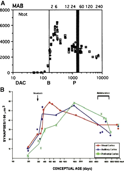
“At the macroscropic level, studies of both monkeys and humans have documented widespread exuberant production of connections throughout all brain regions in the early postnatal period (Zecevic et al. 1989; Bourgeois and Rakic 1993; Bourgeois et al. 1994; Huttenlocher and de Courten 1987; Huttenlocher and Dabholkar 199…”
BackgroundPatterning and plasticity of the cerebral cortex
Sur, M., & Rubenstein, J. L. R. (2005) · Science · doi:10.1126/science.1112070 · Cited by 677
Role of the Reelin Signaling Pathway in Central Nervous System Development
Rice, D. S., & Curran, T. (2001) · Annual Review of Neuroscience · doi:10.1146/annurev.neuro.24.1.1005 · Cited by 667 · cited 1×
“The cortex of animals with defects in Reelin signaling lack laminar structure; the preplate fails to split, and the neurons simply conglomerate under the abnormal preplate (Rice and Curran 2001).”
BackgroundDistinct cortical migrations from the medial and lateral ganglionic eminences
Anderson, S. A., Marín, O., Horn, C., Jennings, K., & Rubenstein, J. L. R. (2001) · Development · doi:10.1242/dev.128.3.353 · Cited by 657
The timing of cortical neurogenesis is encoded within lineages of individual progenitor cells
Shen, Q., Wang, Y., Dimos, J. T., Fasano, C. A., Phoenix, T. N., Lemischka, I. R., Ivanova, N. B., Stifani, S., Morrisey, E. E., & Temple, S. (2006) · Nature Neuroscience · doi:10.1038/nn1694 · Cited by 625
Noctor, S. C., Flint, A. C., Weissman, T. A., Wong, W. S., Clinton, B. K., & Kriegstein, A. R. (2002) · The Journal of Neuroscience · doi:10.1523/jneurosci.22-08-03161.2002 · Cited by 623 · cited 2×
“…radial glial guides were originally thought to be a special, transient population of cells, it has recently been discovered that the cells that provide the scaffolding are actually the neural progenitor cells (Noctor et al. 2001; Noctor et al. 2002; Parnavelas et al. 2002; Weissman et al. 2003).”
BackgroundArea Patterning of the Mammalian Cortex
O'Leary, D. D. M., Chou, S.-J., & Sahara, S. (2007) · Neuron · doi:10.1016/j.neuron.2007.10.010 · Cited by 583
Regulation of Area Identity in the Mammalian Neocortex by Emx2 and Pax6
Bishop, K. M., Goudreau, G., & O'Leary, D. D. M. (2000) · Science · doi:10.1126/science.288.5464.344 · Cited by 575
Bourgeois, J., & Rakic, P. (1993) · The Journal of Neuroscience · doi:10.1523/jneurosci.13-07-02801.1993 · Cited by 567 · cited 2×
“At the macroscropic level, studies of both monkeys and humans have documented widespread exuberant production of connections throughout all brain regions in the early postnatal period (Zecevic et al. 1989; Bourgeois and Rakic 1993; Bourgeois et al. 1994; Huttenlocher and de Courten 1987; Huttenlocher and Dabholkar 199…”
BackgroundThe development of cerebral connections during the first 20–45 weeks’ gestation
Kostović, I., & Jovanov-Milošević, N. (2006) · Seminars in Fetal and Neonatal Medicine · doi:10.1016/j.siny.2006.07.001 · Cited by 567
The caudal ganglionic eminence is a source of distinct cortical and subcortical cell populations
Nery, S., Fishell, G., & Corbin, J. G. (2002) · Nature Neuroscience · doi:10.1038/nn971 · Cited by 558
Sowell, E. R., Thompson, P. M., Holmes, C. J., Batth, R., Jernigan, T. L., & Toga, A. W. (1999) · NeuroImage · doi:10.1006/nimg.1999.0436 · Cited by 554 · cited 1×
“More recent MR morphometry studies have provided more anatomical detail by employing mapping methods for visualizing the pattern of age-related change (Giedd, Snell and et al. 1996; Giedd, Vaituzis and et al. 1996; Sowell et al. 1999a; Sowell et al. 1999b; Sowell et al. 2002).”
BackgroundSelf-citationConcepts of myelin and myelination in neuroradiology.
Barkovich, A. J. (2000) · PubMed · Cited by 539
The development of synapses in striate cortex of man.
Huttenlocher, P. R., & de Courten, C. (1987) · PubMed · Cited by 524
Exuberance in the development of cortical networks
Innocenti, G. M., & Price, D. J. (2005) · Nature Reviews Neuroscience · doi:10.1038/nrn1790 · Cited by 512
Østby, Y., Tamnes, C. K., Fjell, A. M., Westlye, L. T., Due-Tønnessen, P., & Walhovd, K. B. (2009) · The Journal of Neuroscience · doi:10.1523/jneurosci.1242-09.2009 · Cited by 480 · cited 1×
“A recent study [Ostby et al. 2009] confirmed these observations in a large cross-sectional sample and provided concurrent estimates of cortical surface area and cortical thickness.”
ResultLin, S.-C., & Bergles, D. E. (2004) · Nature Neuroscience · doi:10.1038/nn1162 · Cited by 463 · cited 1×
“An intriguing new line of evidence also suggests that a subset of the OPCs dispersed throughout the brain form excitatory and inhibitory connections with neurons, and thus may contribute actively and directly to neural signaling (Lin and Bergles 2004).”
BackgroundExperimentally Induced Visual Projections into Auditory Thalamus and Cortex
Sur, M., Garraghty, P. E., & Roe, A. W. (1988) · Science · doi:10.1126/science.2462279 · Cited by 459 · cited 1×
“For example, Sur and colleagues (Sur et al. 1988; Pallas et al. 1990), surgically eliminated the major input pathway to primary auditory cortex (PAC) in one day old ferrets to determine what would happen to this important sensory area in the absence of input.”
BackgroundMultiple origins of Cajal-Retzius cells at the borders of the developing pallium
Bielle, F., Griveau, A., Narboux-Nême, N., Vigneau, S., Sigrist, M., Arber, S., Wassef, M., & Pierani, A. (2005) · Nature Neuroscience · doi:10.1038/nn1511 · Cited by 459
The life, death, and replacement of oligodendrocytes in the adult CNS
McTigue, D. M., & Tripathi, R. B. (2008) · Journal of Neurochemistry · doi:10.1111/j.1471-4159.2008.05570.x · Cited by 455 · cited 2×
“…many excess oligodendrocytes undergo apoptosis a few days after differentiating, and there is evidence that this process depends on signals from nearby axons, such that the number of surviving oligodendrocytes matches the local axonal surface area (see McTigue and Tripathi 2008, for review).”
BackgroundNeural activity and the dynamics of central nervous system development
Hua, J. Y., & Smith, S. J. (2004) · Nature Neuroscience · doi:10.1038/nn1218 · Cited by 450
MATURATION OF HUMAN CEREBRUM OBSERVED IN VIVO DURING ADOLESCENCE
JERNIGAN, T. L., TRAUNER, D. A., HESSELINK, J. R., & TALLAL, P. A. (1991) · Brain · doi:10.1093/brain/114.5.2037 · Cited by 444
Self-citationExperience-driven brain plasticity: Beyond the synapse
MARKHAM, J. A., & GREENOUGH, W. T. (2004) · Neuron Glia Biology · doi:10.1017/s1740925x05000219 · Cited by 437 · cited 2×
“Greenough has shown that simply rearing animals in either impoverished (standard laboratory cage) or enriched environments (large enclosures with interesting and changing landmarks and multiple littermates) affects the development of a wide range of brain structures and functions (Black et al. 1987; Greenough and Chan…”
BackgroundStanfield, B. B., O'Leary, D. D. M., & Fricks, C. (1982) · Nature · doi:10.1038/298371a0 · Cited by 436
Hermoye, L., Saint-Martin, C., Cosnard, G., Lee, S.-K., Kim, J., Nassogne, M.-C., Menten, R., Clapuyt, P., Donohue, P. K., Hua, K., et al. (2006) · NeuroImage · doi:10.1016/j.neuroimage.2005.08.017 · Cited by 425
Diffusion tensor imaging of brain development
Hüppi, P. S., & Dubois, J. (2006) · Seminars in Fetal and Neonatal Medicine · doi:10.1016/j.siny.2006.07.006 · Cited by 405
The central nervous system; structure and function
(1993) · Comparative Biochemistry and Physiology Part A: Physiology · doi:10.1016/0300-9629(93)90291-b · Cited by 400
The neurotrophic theory and naturally occurring motoneuron death
Oppenheim, R. W. (1989) · Trends in Neurosciences · doi:10.1016/0166-2236(89)90021-0 · Cited by 398 · cited 2×
“One factor that protects against the apoptosis cascade is uptake of neurotrophic substances (Levi-Montalcini 1964; Oppenheim 1989).”
BackgroundADAPTIVE ROLES OF PROGRAMMED CELL DEATH DURING NERVOUS SYSTEM DEVELOPMENT
Buss, R. R., Sun, W., & Oppenheim, R. W. (2006) · Annual Review of Neuroscience · doi:10.1146/annurev.neuro.29.051605.112800 · Cited by 397
Progressive restriction in fate potential by neural progenitors during cerebral cortical development
Desai, A. R., & McConnell, S. K. (2000) · Development · doi:10.1242/dev.127.13.2863 · Cited by 383
The determination of projection neuron identity in the developing cerebral cortex
Leone, D. P., Srinivasan, K., Chen, B., Alcamo, E., & McConnell, S. K. (2008) · Current Opinion in Neurobiology · doi:10.1016/j.conb.2008.05.006 · Cited by 382
Development and plasticity of cortical areas and networks
Sur, M., & Leamey, C. A. (2001) · Nature Reviews Neuroscience · doi:10.1038/35067562 · Cited by 372 · cited 9×
“Within the typical primary visual pathway, inputs from the two eyes remain segregated from the retina to the thalamus to PVC.”
BackgroundInfluentialVisual behaviour mediated by retinal projections directed to the auditory pathway
von Melchner, L., Pallas, S. L., & Sur, M. (2000) · Nature · doi:10.1038/35009102 · Cited by 370
Restriction of Late Cerebral Cortical Progenitors to an Upper-Layer Fate
Frantz, G. D., & McConnell, S. K. (1996) · Neuron · doi:10.1016/s0896-6273(00)80280-9 · Cited by 356
Diffusion tensor imaging of neurodevelopment in children and young adults.
Snook, L., Paulson, L.-A., Roy, D., Phillips, L., & Beaulieu, C. (2005) · NeuroImage · doi:10.1016/j.neuroimage.2005.03.016 · Cited by 350
Levi‐Montalcini, R. (1964) · Annals of the New York Academy of Sciences · doi:10.1111/j.1749-6632.1964.tb33978.x · Cited by 316
Telencephalic cells take a tangent: non-radial migration in the mammalian forebrain
Corbin, J. G., Nery, S., & Fishell, G. (2001) · Nature Neuroscience · doi:10.1038/nn749 · Cited by 298
Diffusion Weighting by the Trace of the Diffusion Tensor within a Single Scan
Mori, S., & Van Zijl, P. C. M. (1995) · Magnetic Resonance in Medicine · doi:10.1002/mrm.1910330107 · Cited by 278 · cited 1×
“Only recently, however, has it been possible to examine the maturation of fiber tracts directly, using diffusion tensor imaging (DTI) (Basser et al. 1994; Mori and van Zijl 1995 ).”
MethodBishop, K. M., Rubenstein, J. L. R., & O'Leary, D. D. M. (2002) · The Journal of Neuroscience · doi:10.1523/jneurosci.22-17-07627.2002 · Cited by 260 · cited 4×
“…spinal column, such that by the end of the embryonic period in GW8 primitive patterning of sensorimotor regions within the neocortex is established (Bishop et al. 2002), major compartments within diencephalic and midbrain regions have differentiated (Nakamura et al. 2005; Kiecker and Lumsden…”
BackgroundInfluentialNeuronal migration mechanisms in development and disease
Valiente, M., & Marín, O. (2010) · Current Opinion in Neurobiology · doi:10.1016/j.conb.2009.12.003 · Cited by 255 · cited 2×
“The CR cells produce a molecular signal, called Reelin, that is part of the pathway that signals neurons when to stop migrating and take up their positions in cortex (Bielle et al. 2005; Huang 2009; Valiente and Marin 2010).”
BackgroundChanges in synaptic density in motor cortex of rhesus monkey during fetal and postnatal life
Zecevic, N., Bourgeois, J.-P., & Rakic, P. (1989) · Developmental Brain Research · doi:10.1016/0165-3806(89)90124-7 · Cited by 253 · cited 2×
“At the macroscropic level, studies of both monkeys and humans have documented widespread exuberant production of connections throughout all brain regions in the early postnatal period (Zecevic et al. 1989; Bourgeois and Rakic 1993; Bourgeois et al. 1994; Huttenlocher and de Courten 1987; Huttenlocher and Dabholkar 199…”
BackgroundA mechanism for inside-out lamination in the neocortex
Cooper, J. A. (2008) · Trends in Neurosciences · doi:10.1016/j.tins.2007.12.003 · Cited by 236 · cited 1×
“The migration of neurons into the developing neocortex results in the formation of an orderly 6-layered structure (Cooper 2008).”
BackgroundHamasaki, T., Leingärtner, A., Ringstedt, T., & O'Leary, D. D. M. (2004) · Neuron · doi:10.1016/j.neuron.2004.07.016 · Cited by 234
Cell migration in the normal and pathological postnatal mammalian brain
Cayre, M., Canoll, P., & Goldman, J. E. (2009) · Progress in Neurobiology · doi:10.1016/j.pneurobio.2009.02.001 · Cited by 234
Neuronal Migration in the Developing Cerebral Cortex: Observations Based on Real-time Imaging
Nadarajah, B. (2003) · Cerebral Cortex · doi:10.1093/cercor/13.6.607 · Cited by 231 · cited 2×
“Figures A and B adapted with permission from Nadarajah et al. (2003).”
MethodGenetic regulation of arealization of the neocortex
O’Leary, D. D., & Sahara, S. (2008) · Current Opinion in Neurobiology · doi:10.1016/j.conb.2008.05.011 · Cited by 224
Cell death in early neural development: beyond the neurotrophic theory
de la Rosa, E. J., & de Pablo, F. (2000) · Trends in Neurosciences · doi:10.1016/s0166-2236(00)01628-3 · Cited by 221 · cited 1×
“…an essential role in eliminating cell populations that serve only a transient function in brain development, such as cells of the MZ or SP. Importantly there is strong evidence of high levels of cell death in the neural progenitor population (de la Rosa and de Pablo 2000; Yeo and Gautier 2004).”
BackgroundDiffusion Tensor Imaging and Tractography of Human Brain Development
Mukherjee, P., & McKinstry, R. C. (2006) · Neuroimaging Clinics of North America · doi:10.1016/j.nic.2005.11.004 · Cited by 221 · cited 2×
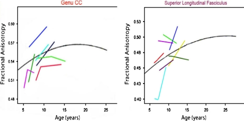
“example, suggests that FA reaches asymptote earliest in long projection, then commissural, and finally association fibers, the latter continuing to exhibit age-related FA increases well into adulthood (see Huppi and Dubois 2006; Mukherjee and McKinstry 2006 for reviews; Cascio et al. 2007).”
BackgroundSchneider, J. F. L., Il’yasov, K. A., Hennig, J., & Martin, E. (2004) · Neuroradiology · doi:10.1007/s00234-003-1154-2 · Cited by 219 · cited 1×
“Nevertheless, there is growing evidence that alterations reflected in and measurable with diffusion imaging continue throughout childhood and adolescence (Schneider et al. 2004; Barnea-Goraly et al. 2005; Snook et al. 2005).”
BackgroundHedgehog signaling from the ZLI regulates diencephalic regional identity
Kiecker, C., & Lumsden, A. (2004) · Nature Neuroscience · doi:10.1038/nn1338 · Cited by 216 · cited 2×
“…is established (Bishop et al. 2002), major compartments within diencephalic and midbrain regions have differentiated (Nakamura et al. 2005; Kiecker and Lumsden 2004), and the segmental organization of the hindbrain and spinal column have been specified (Lumsden and Keynes 1989; Gavalas et…”
BackgroundStanfield, B. B., & O'Leary, D. D. M. (1985) · Journal of Comparative Neurology · doi:10.1002/cne.902380210 · Cited by 206 · cited 1×
“Exuberant connectivity has been documented in pathways as diverse as the corpus callosum, thalamocortical pathways, corticospinal tract and pathways linking the temporal lobe and the limbic system (Stanfield et al. 1982; Stanfield and O’Leary 1985; Innocenti and Price 2005).”
BackgroundLATE CHILDHOOD CHANGES IN BRAIN MORPHOLOGY OBSERVABLE WITH MRI
Jernigan, T. L., & Tallal, P. (1990) · Developmental Medicine & Child Neurology · doi:10.1111/j.1469-8749.1990.tb16956.x · Cited by 202
Self-citationProgrammed cell death in the developing human telencephalon
Rakic, S., & Zecevic, N. (2000) · European Journal of Neuroscience · doi:10.1046/j.1460-9568.2000.00153.x · Cited by 202 · cited 2×
“progenitor cell compartments in the human brain (Rakic and Zecevic 2000).”
BackgroundAsymmetric cell division during neurogenesis in Drosophila and vertebrates
Wodarz, A., & Huttner, W. B. (2003) · Mechanisms of Development · doi:10.1016/j.mod.2003.06.003 · Cited by 202 · cited 1×
“In neural progenitors, asymmetrical cell division produces one neural progenitor and one neuron (Wodarz and Huttner 2003).”
BackgroundAn Introduction to Modern Genetics
Waddington, C. H. (2016) · Routledge eBooks · doi:10.4324/9781315665412 · Cited by 200
Human Cortex Development: Estimates of Neuronal Numbers Indicate Major Loss Late During Gestation
Rabinowicz, T., de Courten-Myers, G. M., Petetot, J. M.-C., XI, G., & de los Reyes, E. (1996) · Journal of Neuropathology and Experimental Neurology · doi:10.1097/00005072-199603000-00007 · Cited by 184 · cited 1×
“Across the cortex, rates of apoptosis within all layers is high, reaching 70% in some regions (Rabinowicz et al. 1996).”
BackgroundEarly neural cell death: dying to become neurons
Yeo, W., & Gautier, J. (2004) · Developmental Biology · doi:10.1016/j.ydbio.2004.07.026 · Cited by 177 · cited 2×
“…an essential role in eliminating cell populations that serve only a transient function in brain development, such as cells of the MZ or SP. Importantly there is strong evidence of high levels of cell death in the neural progenitor population (de la Rosa and de Pablo 2000; Yeo and Gautier 2004).”
BackgroundJones, T. A., & Greenough, W. T. (1996) · Neurobiology of Learning and Memory · doi:10.1006/nlme.1996.0005 · Cited by 174
Human cortex development: estimates of neuronal numbers indicate major loss late during gestation.
Rabinowicz, T., de Courten‐Myers, G. M., Petétot, J. M.-C., Xi, G., & Reyes, E. D. L. (1996) · PubMed · Cited by 169
CASCIO, C. J., GERIG, G., & PIVEN, J. (2007) · Journal of the American Academy of Child & Adolescent Psychiatry · doi:10.1097/01.chi.0000246064.93200.e8 · Cited by 167
Isthmus organizer for midbrain and hindbrain development
Nakamura, H., Katahira, T., Matsunaga, E., & Sato, T. (2005) · Brain Research Reviews · doi:10.1016/j.brainresrev.2004.10.005 · Cited by 159 · cited 2×
“2002), major compartments within diencephalic and midbrain regions have differentiated (Nakamura et al. 2005; Kiecker and Lumsden 2004), and the segmental organization of the hindbrain and spinal column have been specified (Lumsden and Keynes 1989; Gavalas et al.”
BackgroundNeurogenic Radial Glial Cells in Reptile, Rodent and Human: from Mitosis to Migration
Weissman, T. (2003) · Cerebral Cortex · doi:10.1093/cercor/13.6.550 · Cited by 158 · cited 2×
“…radial glial guides were originally thought to be a special, transient population of cells, it has recently been discovered that the cells that provide the scaffolding are actually the neural progenitor cells (Noctor et al. 2001; Noctor et al. 2002; Parnavelas et al. 2002; Weissman et al. 2003).”
BackgroundComplex experience promotes capillary formation in young rat visual cortex
Black, J., Sirevaag, A. M., & Greenough, W. T. (1987) · Neuroscience Letters · doi:10.1016/0304-3940(87)90113-3 · Cited by 155 · cited 2×
“Greenough has shown that simply rearing animals in either impoverished (standard laboratory cage) or enriched environments (large enclosures with interesting and changing landmarks and multiple littermates) affects the development of a wide range of brain structures and functions (Black et al. 1987; Greenough and Chan…”
BackgroundPlasticity of Synapse Structure and Pattern in the Cerebral Cortex
Greenough, W. T., & Chang, F. (1988) · Cerebral cortex · doi:10.1007/978-1-4615-6619-9_11 · Cited by 149 · cited 1×
“…cage) or enriched environments (large enclosures with interesting and changing landmarks and multiple littermates) affects the development of a wide range of brain structures and functions (Black et al. 1987; Greenough and Chang 1988; Jones and Greenough 1996; Markham and Greenough 2004).”
BackgroundGradients in the Brain: The Control of the Development of Form and Function in the Cerebral Cortex
Sansom, S. N., & Livesey, F. J. (2009) · Cold Spring Harbor Perspectives in Biology · doi:10.1101/cshperspect.a002519 · Cited by 148 · cited 1×
“As was the case with Pax6 and Emx2, blocking the expression of these genes results in dramatic alteration in the sensorimotor organization of the neocortex (O’Leary et al. 2007; Zembrzycki et al. 2007; O’Leary and Sahara 2008; Sansom and Livesey 2009).”
ResultAbsolute eigenvalue diffusion tensor analysis for human brain maturation
Suzuki, Y., Matsuzawa, H., Kwee, I. L., & Nakada, T. (2003) · NMR in Biomedicine · doi:10.1002/nbm.848 · Cited by 144
The Fundamentals of Brain Development: Integrating Nature and Nurture
Stiles, J. (2008) · doi:10.2307/j.ctv1pncndb · Cited by 142
Self-citationMagnetic resonance techniques in the assessment of myelin and myelination
Barkovich, A. J. (2005) · Journal of Inherited Metabolic Disease · doi:10.1007/s10545-005-5952-z · Cited by 140 · cited 1×
“These MRI signal changes reflect alterations in tissue chemistry that are presumed to mark the proliferation of oligodendrocytes and deposition of myelin, and they reveal much about the timing and anatomical distribution of these processes (Barkovich 2000; Barkovich 2005).”
BackgroundChanges in volume with age—consistency and interpretation of observed effects
Jernigan, T. L., & Gamst, A. C. (2005) · Neurobiology of Aging · doi:10.1016/j.neurobiolaging.2005.05.016 · Cited by 135 · cited 2×
“The plots illustrate results from an extended age-range for volumes of particular brain structures (modified from Jernigan and Gamst 2005 ).”
BackgroundSelf-citationThe fundamentals of brain development: integrating nature and nurture
(2009) · Choice Reviews Online · doi:10.5860/choice.46-3233 · Cited by 131 · cited 5×
“42 days post conception (Bystron et al. 2008; Stiles 2008) and is largely complete by midgestation.”
BackgroundInfluentialSelf-citationMorange, M., & Cobb, M. A. (2001) · Cited by 130
Gavalas, A., Ruhrberg, C., Livet, J., Henderson, C. E., & Krumlauf, R. (2003) · Development · doi:10.1242/dev.00802 · Cited by 124 · cited 1×
“…established (Bishop et al. 2002), major compartments within diencephalic and midbrain regions have differentiated (Nakamura et al. 2005; Kiecker and Lumsden 2004), and the segmental organization of the hindbrain and spinal column have been specified (Lumsden and Keynes 1989; Gavalas et al. 2003).”
BackgroundThe Central Nervous System –Structure and Function
Jellinger, K. A. (2004) · European Journal of Neurology · doi:10.1111/j.1468-1331.2004.00810.x · Cited by 117
Cell cycle dependence of laminar determination in developing neocortex
McConnell, S. K., & Kaznowski, C. E. (1992) · Trends in Cell Biology · doi:10.1016/0962-8924(92)90150-l · Cited by 115
Genetic interplay between the transcription factors Sp8 and Emx2 in the patterning of the forebrain
Zembrzycki, A., Griesel, G., Stoykova, A., & Mansouri, A. (2007) · Neural Development · doi:10.1186/1749-8104-2-8 · Cited by 109
Molecular regulation of neuronal migration during neocortical development
Huang, Z. (2009) · Molecular and Cellular Neuroscience · doi:10.1016/j.mcn.2009.06.003 · Cited by 102 · cited 4×
“The CR cells produce a molecular signal, called Reelin, that is part of the pathway that signals neurons when to stop migrating and take up their positions in cortex (Bielle et al. 2005; Huang 2009; Valiente and Marin 2010).”
BackgroundInfluentialPallas, S. L., Roe, A. W., & Sur, M. (1990) · Journal of Comparative Neurology · doi:10.1002/cne.902980105 · Cited by 100 · cited 1×
“For example, Sur and colleagues (Sur et al. 1988; Pallas et al. 1990), surgically eliminated the major input pathway to primary auditory cortex (PAC) in one day old ferrets to determine what would happen to this important sensory area in the absence of input.”
BackgroundRole of programmed cell death in normal neuronal development and function
Buss, R. R., & Oppenheim, R. W. (2004) · Anatomical Science International · doi:10.1111/j.1447-073x.2004.00088.x · Cited by 94 · cited 1×
“Although the evidence is somewhat limited, cell death may serve as a mechanism for correcting errors in neuronal production or migration (Buss and Oppenheim 2004).”
BackgroundCorticogenesis in human and nonhuman primates.
RAKIC, P. T. (1995) · Cited by 84
An Introduction to Modern Genetics.
Just, T., & Waddington, C. (1939) · The American Midland Naturalist · doi:10.2307/2420355 · Cited by 82
Volumetric quantification of brain development using MRI
Iwasaki, N., Hamano, K., Okada, Y., Horigome, Y., Nakayama, J., Takeya, T., Takita, H., & Nose, T. (1997) · Neuroradiology · doi:10.1007/s002340050517 · Cited by 65
Brown, M. C., Keynes, R. J., & Lumsden, A. (2002) · Research Portal (King's College London) · Cited by 53
Magnusson, D. (2000) · Oxford University Press eBooks · doi:10.1037/10518-006 · Cited by 49
Chapter 6 The origin and migration of cortical neurons
Parnavelas, J. G., Alifragis, P., & Nadarajah, B. (2002) · Progress in brain research · doi:10.1016/s0079-6123(02)36008-4 · Cited by 39
Basic principles of MRI and morphometry studies of human brain development
Kennedy, D. N., Makris, N., Herbert, M. R., Takahashi, T., & Caviness, V. S. (2002) · Developmental Science · doi:10.1111/1467-7687.00366 · Cited by 33
Gyral development of the human brain.
Chi, J. G., & Dooling, E. C. (1976) · PubMed · Cited by 24
A long remarkable journey : tangential migration in the telencephalon : nature reviews
Marín, Ó. (2001) · Neuroscience · Cited by 5
Giedd (1996) · doi:10.1002/(sici)1096-9861(19960304)366:2<223::aid-cne3>3.0.co;2-7
Neocortical neuron number in humans: Effect of sex and age
(1997) · doi:10.1002/(sici)1096-9861(19970728)384:2<312::aid-cne10>3.0.co;2-k · cited 1×
“The mature brain is composed of more than 100 billion neurons (Pakkenberg and Gundersen 1997).”
BackgroundHuttenlocher (1997) · doi:10.1002/(sici)1096-9861(19971020)387:2<167::aid-cne1>3.0.co;2-z · cited 5×
“…this period (Yakovlev and Lecours 1967), and more limited, but persuasive, evidence for reduction of synaptic density in cortex during childhood (Huttenlocher and Dabholkar 1997), but it remains unclear to what extent these factors, and perhaps others, contribute to the changing morphology…”
BackgroundMethodInfluential
Research works printed but not linked: 1
- Lebel, C., & Beaulieu, C. (2009). Longitudinal diffusion tensor imaging of healthy brain development in children. Honolulu: ISMRM Annual Meeting.
Cited by
The central nervous system and the gut microbiome
Sharon, G., Sampson, T. R., Geschwind, D. H., & Mazmanian, S. K. (2016) · Cell · doi:10.1016/j.cell.2016.10.027 · Cited by 1,513
“In the cerebral cortex, which is most elaborated in humans (Geschwind and Rakic, 2013), neural progenitor cells (NPCs; also known as radial glial cells) proliferate in the ventricular zone.”
BackgroundInfluentialMaternal immune activation and abnormal brain development across CNS disorders
Knuesel, I., Chicha, L., Britschgi, M., Schobel, S. A., Bodmer, M., Hellings, J. A., Toovey, S., & Prinssen, E. P. (2014) · Nature Reviews Neurology · doi:10.1038/nrneurol.2014.187 · Cited by 880
Dubois, J., Dehaene-Lambertz, G., Kulikova, S., Poupon, C., Hüppi, P. S., & Hertz-Pannier, L. (2014) · Neuroscience · doi:10.1016/j.neuroscience.2013.12.044 · Cited by 851
“All these processes of overproduction/elimination are essential to sustain the functional networks plasticity (Stiles and Jernigan 2010).”
BackgroundThe Cellular and Molecular Landscapes of the Developing Human Central Nervous System
Silbereis, J. C., Pochareddy, S., Zhu, Y., Li, M., & Sestan, N. (2016) · Neuron · doi:10.1016/j.neuron.2015.12.008 · Cited by 818
“…progression of myelination, and tractography over the same ages have been noted in certain neurological and psychiatric disorders (Lee et al., 2014; Stiles and Jernigan, 2010; Tau and Peterson, 2010), highlighting the importance of the appropriate regulation of these prolonged human…”
InfluentialFilipello, F., Morini, R., Corradini, I., Zerbi, V., Canzi, A., Michalski, B., Erreni, M., Markicevic, M., Starvaggi-Cucuzza, C., Otero, K., et al. (2018) · Immunity · doi:10.1016/j.immuni.2018.04.016 · Cited by 695
“Patients included in the age window between 5 and 23 years (i.e., the developmental period coinciding with synaptic pruning in humans; Stiles and Jernigan, 2010) displayed a significant reduction of TREM2 protein expression relative to healthy subjects (Figures 7A and 7B).”
Background
Metadata
- Authors
- Stiles, J., & Jernigan, T. L.
- Affiliations
- University of California San Diego (US)
- Corresponding author
- Jernigan, Terry L.
- ORCID
- Jernigan, Terry L.
- Venue
- Neuropsychology Review
- Published
- 2010-11-03
- Volume
- 20
- Issue
- 4
- Pages
- 327-348
- ISSN
- 1040-7308 (ISSN-L)
- 1573-6660
- PMC
- PMC2989000
- OpenAlex
- W2115599658
- Cited by
- 2,009
- References
- 132
- Citations by year
- 2026: 142 · 2025: 184 · 2024: 199 · 2023: 209 · 2022: 184 · 2021: 197 · 2020: 195 · 2019: 155 · 2018: 158 · 2017: 122 · 2016: 92 · 2015: 64 · 2014: 49 · 2013: 24 · 2012: 23
- FWCI
- 1.43
- Type
- journal article
- Language
- en
- Open access
- hybrid
- doi.org
- Licence
- cc-by-nc
- Locations
- Neuropsychology Review (publishedVersion, cc-by-nc)
- Europe PMC (PubMed Central) (publishedVersion, cc-by-nc)
- PubMed Central (publishedVersion, cc-by-nc)
- PubMed (publishedVersion)
- eScholarship (California Digital Library) (submittedVersion, public-domain)
- eScholarship (California Digital Library) (submittedVersion)
- Funders
- Lundbeckfonden (R32‐A3161)
- H. Lundbeck A/S
- National Institutes of Health (5r01hd025077-10)
- U.S. Public Health Service (R32-A3161)
- University of California, San Diego (RC2DA029475)
- National Institute on Drug Abuse (RC2DA029475)
- National Institute of Child Health and Human Development
- Eunice Kennedy Shriver National Institute of Child Health and Human Development (R01HD060595, R01 HD 25077)
- Topics
- Life Sciences › Neuroscience › Developmental Neuroscience › Neurogenesis and neuroplasticity mechanisms
- Life Sciences › Biochemistry, Genetics and Molecular Biology › Genetics › Genetics and Neurodevelopmental Disorders
- Social Sciences › Psychology › Developmental and Educational Psychology › Child and Animal Learning Development
- Keywords
- brain development, developmental neuroscience, mammalian brain, neuronal organization, social development (developmental psychology), cognitive development, neural systems, genetic constraints, developmental mechanisms
- MeSH terms
- Animals, Brain, Cell Differentiation, Diagnostic Imaging, Humans, Nerve Net, Neural Pathways, Neurobiology, Neurons, Gene Expression Regulation, Developmental, Body Patterning, Neurogenesis
About these checks
Registry, retraction, Cites and statistics results are computed, not judged, with one exception: a printed reference that no index lists is read once by a model before it counts as a citation error. Claims are extracted by a model and kept only when their quote appears verbatim in the text; each cited source is then checked, with a model’s help, for whether it says what the claim relies on. Those verdicts can be wrong — read the claim’s quote and the cited source before relying on one.
Statistics and arithmetic are recomputed, not judged.
Confidence bands are shown only for fields calibrated on our annotated test set. None is calibrated yet, so no band is shown.
What counts as a claim ›How we measure confidence ›How we check this ›
Are you an author, or do you see a problem with this page? Request a review
Data sources
Bibliographic data: Semantic Scholar, OpenAlex, Crossref.
Contains information from the Semantic Scholar Academic Graph Datasets, made available under the ODC Attribution License (ODC-BY 1.0).
Semantic Scholar’s data is described in: Kinney, R., et al. (2023). The Semantic Scholar Open Data Platform. arXiv:2301.10140.
