

The DOI matches its Crossref record; 404 of the 406 references it cites were found in a registry or an index; 196 of the 345 sources its claims cite were checked for support.
Development of prefrontal cortex
Open accessAbstract
During evolution, the cerebral cortex advances by increasing in surface and the introduction of new cytoarchitectonic areas among which the prefrontal cortex (PFC) is considered to be the substrate of highest cognitive functions. Although neurons of the PFC are generated before birth, the differentiation of its neurons and development of synaptic connections in humans extend to the 3rd decade of life. During this period, synapses as well as neurotransmitter systems including their receptors and transporters, are initially overproduced followed by selective elimination. Advanced methods applied to human and animal models, enable investigation of the cellular mechanisms and role of specific genes, non-coding regulatory elements and signaling molecules in control of prefrontal neuronal production and phenotypic fate, as well as neuronal migration to establish layering of the PFC. Likewise, various genetic approaches in combination with functional assays and immunohistochemical and imaging methods reveal roles of neurotransmitter systems during maturation of the PFC. Disruption, or even a slight slowing of the rate of neuronal production, migration and synaptogenesis by genetic or environmental factors, can induce gross as well as subtle changes that eventually can lead to cognitive impairment. An understanding of the development and evolution of the PFC provide insight into the pathogenesis and treatment of congenital neuropsychiatric diseases as well as idiopathic developmental disorders that cause intellectual disabilities.
Overview
CoChat Summary
## TL;DR
This is a narrative review of how the human prefrontal cortex (PFC) develops, from early patterning and neuronal migration to synaptogenesis, neurotransmitter system maturation, and evolutionary expansion. It also links disrupted PFC development to neurodevelopmental and psychiatric disorders, arguing that slowing or disrupting these processes can produce cognitive and behavioral impairment.
## Key findings
- In humans, PFC neuron differentiation and synaptic development extend into the 3rd decade of life, and neurotransmitter systems are initially overproduced followed by selective elimination/pruning.
- The PFC is described as exceptionally expanded in humans, occupying about 30% of the cortical surface, and the human frontal lobe is said to have evolved three times larger than that of great apes.
- Human PFC synaptic density is reported to spike around 3.5 years of age and be almost double the net density of the adult PFC; in early childhood the PFC has a 2–3 fold higher density of dendritic spines than the adult PFC.
- The paper states that disruption or slowing of neuronal production, migration, and synaptogenesis by genetic or environmental factors can lead to gross and subtle changes that eventually cause cognitive impairment.
## Methods
- This is a narrative review, not an original experimental study.
- It synthesizes evidence from human postmortem tissue, animal models (especially rodents and nonhuman primates), and advanced methods such as immunohistology, functional assays, imaging, genome-wide transcriptome/epigenome profiling, and stem cell/organoid approaches.
- The main approach is integrative literature review across developmental biology, neurochemistry, evolution, and neurodevelopmental disorders.
## Limitations
- The paper itself notes that most neurotransmitter development studies come from rodents, while primate and human prefrontal connectivity is more regionally specific; this limits direct translation.
- It explicitly says that later stages of development in brain organoid approaches still need to be optimized.
- Evident from the text: this is a broad synthesis without new primary data, so its conclusions depend on the quality and comparability of the cited studies.
Model-written summary. Check it against the paper before relying on it.
Integrity signals
Registry
Cites
Statistics
Figures



Verification
Claims the paper makes
97 Valid · 5 Not valid · 24 Not checked · 10 Own results and interpretations
Some cited sources support this claim and some don’t — read each source’s verdict below.
In the paper
“The pyramidal excitatory neurons born in the VZ and SVZ of the prefrontal subdomains, similar to other cortical areas, start to migrate radially toward the proper position in the CP under the influence of Fgfs [50, 59, 60].”
§ Development of prefrontal cortex > The early stages of PFC development > Prefrontal expansion and lamination
What the sources say
- Not checked — no full text of the sourcecites [50] Noctor SC, Flint AC, Weissman TA, Dammerman RS, Kriegstein AR. Neurons derived from radial glial cells establish radial units in neocortex. Nature. 2001;409:714–20. source
- Partly supportedcites [59] Nobrega-Pereira S, Marin O. Transcriptional control of neuronal migration in the developing mouse brain. Cereb Cortex. 2009;19:i107–13. sourceExperimental — accuracy not yet measured
The sections “Role of Transcription Factors in the Migration of Cortical Projection Neurons” and “Transcription Factors as Multitasking Regulators during Brain Development” describe cortical projection neurons migrating radially and reaching the cortical plate, with progenitors in the VZ/SVZ. They do not specify prefrontal subdomains or say that Fgfs influence this migration; the factors discussed instead include Ngn1/2. Page numbers were unavailable in our copy. (LLM, conf=0.87)
- Disputedcites [60] Paredes MF, James D, Gil-Perotin S, Kim H, Cotter JA, Ng C, et al. Extensive migration of young neurons into the infant human frontal lobe. Science. 2016;354:6308. sourceExperimental — accuracy not yet measured
The Abstract and Discussion describe young neurons migrating tangentially into the infant frontal lobe and contributing to inhibitory circuits, rather than pyramidal excitatory neurons migrating radially. The section “Postnatally migrating neurons differentiate into interneurons” reports that these cells lack markers of excitatory neurons. Our copy does not identify Fgfs as an influence on their migration. (LLM, conf=0.90)
Checked by a reasoning model — may be wrong.
plainstates a cause- Asserted: passes — The authors put it forward as true.
- Checkable: passes — It can be checked against evidence.
- Load-bearing: unclear — It is unclear whether the paper’s conclusions depend on it.
- Based: passes — The paper gives a reason why it holds.
Some cited sources support this claim and some don’t — read each source’s verdict below.
In the paper
“Other monogenic syndromes like Kleefstra, KBG, WitKoS, Angelman, Coffin-Sirris, Rubinstein-Taybi, Phelan-McDermid, Smith–Magenis Syndrome and Kabuki syndrome also have a clear prefrontal component in their behavioral and cognitive phenotype [272–280].”
§ Development of prefrontal cortex > PFC development and mental illness > From a genetic point of view
What the sources say
- Partly supportedcites [272] Alarcon JM, Malleret G, Touzani K, Vronskaya S, Ishii S, Kandel ER, et al. Chromatin acetylation, memory, and LTP are impaired in CBP+/- mice: a model for the cognitive deficit in Rubinstein-Taybi syndrome and its amelioration. Neuron. 2004;42:947–59. sourceExperimental — accuracy not yet measured
The section on CBP+/− mice as a model for Rubinstein–Taybi syndrome (p. 8) describes short attention spans and difficulty planning and executing motor acts, supporting the behavioral aspect of the claim for that syndrome. It does not establish a prefrontal component; the same passage says future studies are needed to identify the brain regions involved. This citation is assessed for Rubinstein–Taybi syndrome, not the other syndromes covered by the citation range. (LLM, conf=0.84)
- Partly supportedcites [273] van Dongen LCM, Wingbermuhle E, van der Veld WM, Vermeulen K, Bos-Roubos AG, Ockeloen CW, et al. Exploring the behavioral and cognitive phenotype of KBG syndrome. Genes Brain Behav. 2019;18:e12553. sourceExperimental — accuracy not yet measured
For KBG syndrome, the Discussion reports difficulties with sustained attention, inhibition and cognitive flexibility, alongside impulsive behavior. These findings support a behavioral and cognitive component consistent with the citing sentence, but the paper does not establish that it is specifically prefrontal; it also does not study the other listed syndromes. (LLM, conf=0.82)
- Disputedcites [274] van Dongen LCM, Wingbermuhle PAM, van der Veld WM, Stumpel C, Kleefstra T, Egger JIM. Exploring the cognitive phenotype of Kabuki (Niikawa-Kuroki) syndrome. J Intellect Disabil Res. 2019;63:498–506. sourceExperimental — accuracy not yet measured
For Kabuki syndrome—the syndrome this source studies—the Abstract Results report no weaknesses in planning or cognitive flexibility relative to a genetic control group. The Discussion describes executive functioning as relatively unaffected, rather than a clear prefrontal weakness. The paper does not assess prefrontal anatomy or the other syndromes listed. (LLM, conf=0.84)
Checked by a reasoning model — may be wrong.
- Not checked — no full text of the sourcecites [275] Schut EHS, Alonso A, Smits S, Khamassi M, Samanta A, Negwer M, et al. The Object Space Task reveals increased expression of cumulative memory in a mouse model of Kleefstra syndrome. Neurobiol Learn Mem. 2020;173:107265. source
- Partly supportedcites [276] Egger JI, Zwanenburg RJ, van Ravenswaaij-Arts CM, Kleefstra T, Verhoeven WM. Neuropsychological phenotype and psychopathology in seven adult patients with Phelan-McDermid syndrome: implications for treatment strategy. Genes Brain Behav. 2016;15:395–404. sourceExperimental — accuracy not yet measured
The Discussion (page unavailable) identifies executive and social-cognitive deficits in Phelan–McDermid syndrome, supporting a cognitive and behavioral component of the claim. The Methods mention the (orbito)prefrontal cortex in a general explanation of alexithymia, but the paper does not establish a clear prefrontal component specifically for Phelan–McDermid syndrome. This source does not address the other syndromes in the list. (LLM, conf=0.82)
- Partly supportedcites [277] Sidorov MS, Judson MC, Kim H, Rougie M, Ferrer AI, Nikolova VD, et al. Enhanced operant extinction and prefrontal excitability in a mouse model of angelman syndrome. J Neurosci. 2018;38:2671–82. sourceExperimental — accuracy not yet measured
The Abstract and Discussion report exaggerated operant extinction and hyperexcitable prefrontal neurons in Angelman syndrome model mice, supporting a prefrontal component for the Angelman part of the cited list. The paper does not establish that component as clear in people with Angelman syndrome; its Introduction says prefrontal dysfunction likely contributes to cognitive impairment, and the Abstract notes that two other prefrontally dependent tasks were largely normal. (LLM, conf=0.84)
- Partly supportedcites [278] Laje G, Bernert R, Morse R, Pao M, Smith AC. Pharmacological treatment of disruptive behavior in Smith-Magenis syndrome. Am J Med Genet Part C Semin Med Genet. 2010;154C:463–8. sourceExperimental — accuracy not yet measured
The Introduction describes Smith–Magenis syndrome as a genetic syndrome with intellectual disability and distinctive neurobehavioral problems; the Discussion also mentions inattention and impulsivity. But neither passage identifies a prefrontal component of its behavioral or cognitive phenotype. This source supports the Smith–Magenis phenotype portion of the multi-syndrome sentence, not its prefrontal characterization. (LLM, conf=0.84)
- Partly supportedcites [279] Laje G, Morse R, Richter W, Ball J, Pao M, Smith AC. Autism spectrum features in Smith-Magenis syndrome. Am J Med Genet Part C Semin Med Genet. 2010;154C:456–62. sourceExperimental — accuracy not yet measured
The Introduction describes cognitive impairment, distractibility, and a characteristic behavioral phenotype in Smith–Magenis syndrome. The Discussion reports autistic-spectrum symptoms, but neither passage establishes a clear prefrontal component. This verdict concerns Smith–Magenis syndrome only, the syndrome addressed by this source. (LLM, conf=0.85)
- Could not verifycites [280] Proulx E, Young EJ, Osborne LR, Lambe EK. Enhanced prefrontal serotonin 5-HT(1A) currents in a mouse model of Williams-Beuren syndrome with low innate anxiety. J Neurodev Disord. 2010;2:99–108. sourceExperimental — accuracy not yet measured
The Introduction (page unavailable) discusses Williams-Beuren syndrome and its mouse models, not the named syndromes. Searches for the syndrome names and for a prefrontal component of their behavioral and cognitive phenotypes found no passage addressing the claim. (LLM, conf=0.76)
boostedstates an association- Asserted: passes — The authors put it forward as true.
- Checkable: passes — It can be checked against evidence.
- Load-bearing: unclear — It is unclear whether the paper’s conclusions depend on it.
- Based: passes — The paper gives a reason why it holds.
- Partly supported
Some cited sources support this claim and some don’t — read each source’s verdict below.
In the paper
“For some of these syndromes it has recently been shown that deficits in the structural development of the PFC underlie these problems [281–287].”
§ Development of prefrontal cortex > PFC development and mental illness > From a genetic point of view
What the sources say
- Partly supportedcites [281] Witteveen JS, Willemsen MH, Dombroski TC, van Bakel NH, Nillesen WM, van Hulten JA, et al. Haploinsufficiency of MeCP2-interacting transcriptional co-repressor SIN3A causes mild intellectual disability by affecting the development of cortical integrity. Nat Genet. 2016;48:877–87. sourceExperimental — accuracy not yet measured
The Abstract (page not provided) links SIN3A haploinsufficiency to mild syndromic intellectual disability and reports altered cortical development in mice. The Figure 1 caption (page not provided) describes seemingly underdeveloped frontal lobes in one person, but cautions that the MRI quality was suboptimal. These findings do not establish that structural deficits specifically in the PFC underlie the syndrome’s problems. (LLM, conf=0.84)
- Disputedcites [282] Frega M, Linda K, Keller JM, Gumus-Akay G, Mossink B, van Rhijn JR, et al. Neuronal network dysfunction in a model for Kleefstra syndrome mediated by enhanced NMDAR signaling. Nat Commun. 2019;10:4928. sourceExperimental — accuracy not yet measured
In Results, under “Single-cell level characterization of KS neurons” (page unavailable), the paper reports no significant differences in somatodendritic morphology between Kleefstra-syndrome and control cortical neurons. It studies neuronal network dysfunction, not structural development of the PFC, and therefore does not show that PFC structural deficits underlie the problems described. (LLM, conf=0.88)
Checked by a reasoning model — may be wrong.
- Partly supportedcites [283] Boisgontier J, Tacchella JM, Lemaitre H, Lehman N, Saitovitch A, Gatinois V, et al. Anatomical and functional abnormalities on MRI in kabuki syndrome. NeuroImage Clin. 2019;21:101610. sourceExperimental — accuracy not yet measured
The Discussion (page unavailable) reports reduced grey matter in a prefrontal region in patients with Kabuki syndrome and says frontal abnormalities may explain some motor impairments. The Conclusion (page unavailable) likewise describes a possible brain substrate for fine-motor deficits. The study does not establish that disrupted PFC development underlies those problems; it presents the link as a possibility. (LLM, conf=0.86)
- Partly supportedcites [284] Jacot-Descombes S, Keshav NU, Dickstein DL, Wicinski B, Janssen WGM, Hiester LL, et al. Altered synaptic ultrastructure in the prefrontal cortex of Shank3-deficient rats. Mol Autism. 2020;11:89. sourceExperimental — accuracy not yet measured
The Conclusions report altered synaptic ultrastructure in the medial PFC of Shank3-deficient rats and say the changes may have implications for attentional deficits. The paper does not show that disrupted PFC structural development causes those deficits; its conclusion is tentative and based on a rat model, not a demonstration across monogenic syndromes. (LLM, conf=0.86)
- Partly supportedcites [285] Celen C, Chuang JC, Luo X, Nijem N, Walker AK, Chen F, et al. Arid1b haploinsufficient mice reveal neuropsychiatric phenotypes and reversible causes of growth impairment. eLife. 2017;6:e25730:1–22. sourceExperimental — accuracy not yet measured
The Results section (‘Arid1b haploinsufficiency resulted in neuroanatomical and gene expression abnormalities’; page unavailable) reports reduced cortical thickness and other brain abnormalities in Arid1b-mutant mice, alongside behavioral phenotypes. It calls these findings correlates, however; it does not identify a PFC-specific developmental deficit or show that such a deficit underlies the behavioral problems. (LLM, conf=0.84)
- Partly supportedcites [286] Rotaru DC, van Woerden GM, Wallaard I, Elgersma Y. Adult Ube3a gene reinstatement restores the electrophysiological deficits of prefrontal cortex layer 5 neurons in a mouse model of angelman syndrome. J Neurosci. 2018;38:8011–30. sourceExperimental — accuracy not yet measured
The Introduction reports altered orbitofrontal gray-matter volume in people with Angelman syndrome and suggests that some behavioral problems may depend on PFC function. But the discussion describes impaired development of PFC projections as a possibility not tested in this study; it does not establish that structural PFC deficits underlie the behavioral problems. (LLM, conf=0.86)
- Partly supportedcites [287] Huang WH, Wang DC, Allen WE, Klope M, Hu H, Shamloo M, et al. Early adolescent Rai1 reactivation reverses transcriptional and social interaction deficits in a mouse model of Smith-Magenis syndrome. Proc Natl Acad Sci USA. 2018;115:10744–9. sourceExperimental — accuracy not yet measured
The Results section ('Reduced Spine Density in Prefrontal Cortex Neurons'; page unavailable) reports reduced dendritic spine density in the medial PFC of one-month-old Rai1-deficient mice. The Discussion (page unavailable) says this structural change *may* contribute to a social-interaction deficit; it does not establish that deficient PFC structural development underlies the problems, or extend that conclusion beyond this mouse model. (LLM, conf=0.87)
plainstates a cause- Asserted: passes — The authors put it forward as true.
- Checkable: passes — It can be checked against evidence.
- Load-bearing: unclear — It is unclear whether the paper’s conclusions depend on it.
- Based: passes — The paper gives a reason why it holds.
- Partly supported
The cited sources support only part of this claim — read each source’s verdict below.
In the paper
“The most ventral subdomain of the mPFC is the infralimbic cortex (IL) and is involved in coping with chronic stress eventually leading to structural changes and prefrontal dysfunction [5–11].”
§ Development of prefrontal cortex > PFC definition
What the sources say
- Partly supportedcites [5] Flak JN, Solomon MB, Jankord R, Krause EG, Herman JP. Identification of chronic stress-activated regions reveals a potential recruited circuit in rat brain. Eur J Neurosci. 2012;36:2547–55. sourceExperimental — accuracy not yet measured
The Results (Upstream Limbic Structures) report increased FosB/ΔFosB expression in the infralimbic cortex after chronic variable stress. The Discussion suggests that prefrontal activation may produce lasting changes in gene transcription and contribute to dysfunction, but does not establish that infralimbic involvement in coping leads to structural changes or prefrontal dysfunction. (LLM, conf=0.85)
- Partly supportedcites [6] Izquierdo A, Wellman CL, Holmes A. Brief uncontrollable stress causes dendritic retraction in infralimbic cortex and resistance to fear extinction in mice. J Neurosci. 2006;26:5733–8. sourceExperimental — accuracy not yet measured
The Introduction (page unavailable) identifies the infralimbic cortex as a subregion of the medial prefrontal cortex. The Discussion (page unavailable) reports dendritic retraction in infralimbic neurons after brief stress and suggests possible loss of functional integrity. It does not establish that the infralimbic cortex is involved in coping with chronic stress or that chronic stress causes the observed infralimbic changes; it distinguishes those findings from earlier chronic-stress work on the prelimbic cortex. (LLM, conf=0.88)
- Partly supportedcites [7] Moench KM, Maroun M, Kavushansky A, Wellman C. Alterations in neuronal morphology in infralimbic cortex predict resistance to fear extinction following acute stress. Neurobiol Stress. 2016;3:23–33. sourceExperimental — accuracy not yet measured
The Discussion (Stress-induced changes in IL dendritic morphology; page unavailable) reports dendritic retraction in infralimbic cortex following chronic stress, and the Conclusion links the study’s findings to stress-induced medial prefrontal dysfunction. The paper does not specifically describe the infralimbic cortex as coping with chronic stress or establish that chronic-stress-related structural changes lead to prefrontal dysfunction; its own experiment examined acute stress. (LLM, conf=0.82)
- Not checked — no full text of the sourcecites [8] Dias-Ferreira E, Sousa JC, Melo I, Morgado P, Mesquita AR, Cerqueira JJ, et al. Chronic stress causes frontostriatal reorganization and affects decision-making. Science 2009;325:621–5. source
- Partly supportedcites [9] Shansky RM, Hamo C, Hof PR, McEwen BS, Morrison JH. Stress-induced dendritic remodeling in the prefrontal cortex is circuit specific. Cereb Cortex. 2009;19:2479–84. sourceExperimental — accuracy not yet measured
The Introduction describes the infralimbic cortex as part of a circuit that regulates amygdala activity, and the study reports chronic-stress-induced dendritic retraction in some infralimbic neurons. But the Abstract reports no remodeling in neurons projecting to the basolateral amygdala, while the Functional Implications section treats resulting circuit dysfunction as a possibility for future study, not an established consequence. (LLM, conf=0.86)
- Not checked — no full text of the sourcecites [10] Radley JJ, Rocher AB, Miller M, Janssen WG, Liston C, Hof PR, et al. Repeated stress induces dendritic spine loss in the rat medial prefrontal cortex. Cereb Cortex. 2006;16:313–20. source
- Supportedcites [11] McKlveen JM, Morano RL, Fitzgerald M, Zoubovsky S, Cassella SN, Scheimann JR, et al. Chronic stress increases prefrontal inhibition: a mechanism for stress-induced prefrontal dysfunction. Biol Psychiatry. 2016;80:754–64. source
The introduction identifies the infralimbic cortex as a subdivision of the ventromedial prefrontal cortex that is particularly important for chronic stress regulation. The Results and Discussion report stress-related neuroanatomic changes in that region alongside impaired prefrontal-mediated behavior (page numbers unavailable in our copy). (LLM, conf=0.87)
plainstates a cause- Asserted: passes — The authors put it forward as true.
- Checkable: passes — It can be checked against evidence.
- Load-bearing: unclear — It is unclear whether the paper’s conclusions depend on it.
- Based: passes — The paper gives a reason why it holds.
- Partly supported
The cited sources support only part of this claim — read each source’s verdict below.
In the paper
“Thus, the PFC as well as the Broca and Wernicke association areas in humans, which are formed in the frontal and temporal lobes, display a temporarily enriched gene expression pattern that is distinct from the mice or macaque cerebrum at the comparable prenatal stages (e.g., [21, 22]).”
§ Development of prefrontal cortex > Evolutionary view on PFC development During mammalian evolution, the cerebral cortex not only increased in neuronal numbers and surface area but also acquired new cell types and cytoarchitectonic areas. Species-specific adaptations of prefrontal areas, steered by the environmental demands, can explain the differences in size of frontal areas over time. Among the most recent additions are several association areas, particularly the PFC, which has expanded enormously in primates culminating in humans [14]. In humans, the PFC occupies as much as about 30% of its surface. Although still debated, the human frontal lobe seems to have evolved three times larger than that of our closest living relatives, the great apes. In fact, it has been argued that the human brain possesses prefrontal regions that are both qualitatively and functionally exclusive [15]. It is, nevertheless, remarkable that we use the rodent model for most of the cellular and molecular neuroscientific studies, despite its lissencephalic brain which is clearly much simpler in both cytoarchitecture as well as function. A valid question still remains: Do rodents have a prefrontal cortex? [16, 17]. And if we were to focus more on the evolutionary aspects of prefrontal development in terms of structural organization and function, should we not include longitudinal neurodevelopmental studies on more species [18, 19]? Although the basic principles of cortical development may be similar in all mammals, the modifications of developmental events during millennia of primate evolution produce not only quantitative but also qualitative changes of its cellular structure and synaptic circuitry [13, 20]. The origin of species-specific distinctions can be traced either to the new or phylogenetically conserved genes that act at the time of the neural stem cell’s exit from the mitotic cycle and generate a different outcome, depending on the evolutionary context by interacting with a postmitotic neuron. Thus, the PFC as well as the Broca and Wernicke association areas in humans, which are formed in the frontal and temporal lobes, display a temporarily enriched gene expression pattern that is distinct from the mice or macaque cerebrum at the comparable prenatal stages (e.g., [21, 22]). More on evolution of the prefrontal cortex can be found in this volume, part I, chapter 1.
What the sources say
- Not checked — no full text of the sourcecites [21] Johnson MB, Kawasawa YI, Mason CE, Krsnik Z, Coppola G, Bogdanovic D, et al. Functional and evolutionary insights into human brain development through global transcriptome analysis. Neuron. 2009;62:494–509. source
- Partly supportedcites [22] Pletikos M, Sousa AM, Sedmak G, Meyer KA, Zhu Y, Cheng F, et al. Temporal specification and bilaterality of human neocortical topographic gene expression. Neuron. 2014;81:321–32. sourceExperimental — accuracy not yet measured
The section “Temporal Transcriptional Hourglass Pattern Reflects Putative Areal and Functional Differences” reports fetal expression patterns enriched in prefrontal/frontal and temporal regions. “Certain Prenatal Expression Patterns Differ between Human and Macaque” reports that some selected genes have different areal expression patterns in humans and macaques at equivalent ages. Our copy does not establish this specifically for all three named areas—PFC, Broca area, and Wernicke area—or make the claimed prenatal comparison with mice. (LLM, conf=0.87)
plainstates an association- Asserted: passes — The authors put it forward as true.
- Checkable: passes — It can be checked against evidence.
- Load-bearing: unclear — It is unclear whether the paper’s conclusions depend on it.
- Based: passes — The paper gives a reason why it holds.
78 more claims · 58 with their cited sources checked
Create a free account to see all 136 claims and every source verdict.
- Probably not a substantial claim. 15
- It may state two separate claims as one. 43
Citations
Cites
Shrestha, S., & Offer, S. M. (2016) · Medical Epigenetics · doi:10.1159/000444713 · Cited by 8,914
Dynamic mapping of human cortical development during childhood through early adulthood
Gogtay, N., Giedd, J. N., Lusk, L., Hayashi, K. M., Greenstein, D., Vaituzis, A. C., Nugent, T. F., Herman, D. H., Clasen, L. S., Toga, A. W., et al. (2004) · Proceedings of the National Academy of Sciences · doi:10.1073/pnas.0402680101 · Cited by 5,604 · cited 2×
“PFC COGNITIVE DEVELOPMENT The PFC, as the seat of our higher-order cognitive functions, continues to develop into adulthood [52, 243].”
BackgroundBrain development during childhood and adolescence: a longitudinal MRI study
Giedd, J. N., Blumenthal, J., Jeffries, N. O., Castellanos, F. X., Liu, H., Zijdenbos, A., Paus, T., Evans, A. C., & Rapoport, J. L. (1999) · Nature Neuroscience · doi:10.1038/13158 · Cited by 5,552 · cited 2×
“It is among the latest brain regions to fully mature in humans as well as rodents [106, 159, 244, 245].”
BackgroundThe adolescent brain and age-related behavioral manifestations
Spear, L. P. (2000) · Neuroscience & Biobehavioral Reviews · doi:10.1016/s0149-7634(00)00014-2 · Cited by 5,433 · cited 3×
“In nonhuman primates this is characterized by risk-taking, novelty seeking, and increased vigilance; whereas in rodents by play behavior, increased exploratory activity and impulsivity are peaking [245].”
BackgroundSpecification of Cerebral Cortical Areas
Rakic, P. (1988) · Science · doi:10.1126/science.3291116 · Cited by 3,127 · cited 8×
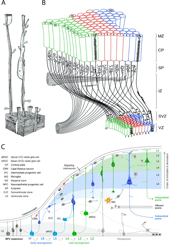
“C Illustration of the dynamics of major developmental events and diversity of progenitors involved in the development of primate cerebral neocortex based on studies of Rakic, with permission from Silver et al. [396].”
BackgroundMethodInfluentialSelf-citationRice, D., & Barone, S. (2000) · Environmental Health Perspectives · doi:10.1289/ehp.00108s3511 · Cited by 2,944
Zika Virus Associated with Microcephaly
Mlakar, J., Korva, M., Tul, N., Popović, M., Poljšak-Prijatelj, M., Mraz, J., Kolenc, M., Resman Rus, K., Vesnaver Vipotnik, T., Fabjan Vodušek, V., et al. (2016) · New England Journal of Medicine · doi:10.1056/nejmoa1600651 · Cited by 2,884
Berridge, C. W., & Waterhouse, B. D. (2003) · Brain Research Reviews · doi:10.1016/s0165-0173(03)00143-7 · Cited by 2,529
Excitatory actions of gaba during development: the nature of the nurture
Ben-Ari, Y. (2002) · Nature Reviews Neuroscience · doi:10.1038/nrn920 · Cited by 2,448
Mode of cell migration to the superficial layers of fetal monkey neocortex
Rakic, P. (1972) · Journal of Comparative Neurology · doi:10.1002/cne.901450105 · Cited by 2,170 · cited 6×
“C Illustration of the dynamics of major developmental events and diversity of progenitors involved in the development of primate cerebral neocortex based on studies of Rakic, with permission from Silver et al. [396].”
BackgroundInfluentialSelf-citation
Cited by
Opioid Receptor-Mediated Regulation of Neurotransmission in the Brain
Reeves, K. C., Shah, N., Muñoz, B., & Atwood, B. K. (2022) · Frontiers in Molecular Neuroscience · doi:10.3389/fnmol.2022.919773 · Cited by 156
Kringelbach, M. L., Perl, Y. S., Tagliazucchi, E., & Deco, G. (2023) · Science Advances · doi:10.1126/sciadv.ade6049 · Cited by 105
Habay, J., Uylenbroeck, R., Droogenbroeck, R. V., Wachter, J. D., Proost, M., Tassignon, B., Pauw, K. D., MEEUSEN, R. R., Pattyn, N., Cutsem, J. V., et al. (2023) · Sports Medicine - Open · doi:10.1186/s40798-023-00559-7 · Cited by 82
Reorganization of adolescent prefrontal cortex circuitry is required for mouse cognitive maturation
Pöpplau, J. A., Schwarze, T., Dorofeikova, M., Pochinok, I., Günther, A., Marquardt, A., & Hanganu‐Opatz, I. L. (2023) · Neuron · doi:10.1016/j.neuron.2023.10.024 · Cited by 67
“1,2,68 Moreover, pruning deficits during this time were proposed to precede the onset of mental disorders.”
Peters, K. Z., & Naneix, F. (2022) · Frontiers in Neural Circuits · doi:10.3389/fncir.2022.939235 · Cited by 85
“At the neurobiological level, adolescence is associated with neurophysiological changes, an increase in white matter volume and inverted U-shaped changes in gray matter volume related to synaptic overproduction and subsequent pruning (Lenroot and Giedd, 2006; Caballero et al., 2016; Drzewiecki and Juraska, 2020; Kolk…”
Metadata
- Authors
- Kolk, S. M., & Rakic, P.
- Affiliations
- Radboud University Nijmegen (NL)
- Yale University (US)
- Corresponding author
- Kolk, Sharon M.
- Rakic, Pasko
- Venue
- Neuropsychopharmacology
- Published
- 2021-10-13
- Volume
- 47
- Issue
- 1
- Pages
- 41-57
- ISSN
- 0893-133X (ISSN-L)
- 1740-634X
- PMC
- PMC8511863
- OpenAlex
- W3207566007
- Cited by
- 378
- References
- 406
- Citations by year
- 2026: 85 · 2025: 119 · 2024: 87 · 2023: 58 · 2022: 28 · 2021: 1
- FWCI
- 6.56
- Type
- journal article
- Language
- en
- Open access
- hybrid
- doi.org
- Licence
- cc-by
- creativecommons.org/licenses/by/4.0 (tdm)
- creativecommons.org/licenses/by/4.0 (vor)
- Locations
- Neuropsychopharmacology (publishedVersion, cc-by)
- PubMed Central (publishedVersion, cc-by)
- PubMed (publishedVersion)
- Radboud Repository (Radboud University) (submittedVersion)
- Radboud Repository (Radboud University) (submittedVersion, other-oa)
- Funders
- Nederlandse Organisatie voor Wetenschappelijk Onderzoek (NWA.1160.18.320)
- U.S. Department of Health & Human Services | National Institutes of Health (DA023999)
- National Institutes of Health (USA) NIDA
- Topics
- Life Sciences › Neuroscience › Developmental Neuroscience › Neurogenesis and neuroplasticity mechanisms
- Life Sciences › Biochemistry, Genetics and Molecular Biology › Genetics › Genetics and Neurodevelopmental Disorders
- Life Sciences › Neuroscience › Cellular and Molecular Neuroscience › Neuroscience and Neuropharmacology Research
- Keywords
- prefrontal cortex development, neuronal migration, synaptogenesis, neuronal differentiation, neurotransmitter systems, gene expression regulation, cognitive development, intellectual disability, neuropsychiatric disorders, cerebral cortex evolution, synaptic pruning
- MeSH terms
- Animals, Cerebral Cortex, Humans, Neurons, Synapses, Prefrontal Cortex, Neurotransmitter Agents
About these checks
Registry, retraction, Cites and statistics results are computed, not judged, with one exception: a printed reference that no index lists is read once by a model before it counts as a citation error. Claims are extracted by a model and kept only when their quote appears verbatim in the text; each cited source is then checked, with a model’s help, for whether it says what the claim relies on. Those verdicts can be wrong — read the claim’s quote and the cited source before relying on one.
The claim verdicts this report counts are experimental: their accuracy is not yet measured.
A language model drew the claims from the paper on 2026-10-09, under version 3 of the claim definition (model deepseek-v4.1-flash, prompt extract-v3).
A second model labelled each claim on 2026-10-09 (model jev-1.13-20260917, definition revision gold-guideline-v0.2).
Cited sources are checked with a language model’s help (gpt-6-sol).
Statistics and arithmetic are recomputed, not judged.
Confidence bands are shown only for fields calibrated on our annotated test set. None is calibrated yet, so no band is shown.
What counts as a claim ›How we measure confidence ›How we check this ›
Are you an author, or do you see a problem with this page? Request a review
How to cite
Cite CoChat for the checks on this page, and the paper for its own findings.
- This page
- CoChat. (2026). Citation check: Development of prefrontal cortex. https://app.cochat.ai/papers/10.1038/s41386-021-01137-9
- The paper
- Kolk, S. M., & Rakic, P. (2022). Development of prefrontal cortex. Neuropsychopharmacology. https://doi.org/10.1038/s41386-021-01137-9
Data sources
Bibliographic data: Semantic Scholar, OpenAlex, Crossref.
Contains information from the Semantic Scholar Academic Graph Datasets, made available under the ODC Attribution License (ODC-BY 1.0).
Semantic Scholar’s data is described in: Kinney, R., et al. (2023). The Semantic Scholar Open Data Platform. arXiv:2301.10140.
