

The Basics of Brain Development
Open accessThe DOI matches its Crossref record; 128 of the 132 references it cites were found in a registry or an index.
Abstract
Over the past several decades, significant advances have been made in our understanding of the basic stages and mechanisms of mammalian brain development. Studies elucidating the neurobiology of brain development span the levels of neural organization from the macroanatomic, to the cellular, to the molecular. Together this large body of work provides a picture of brain development as the product of a complex series of dynamic and adaptive processes operating within a highly constrained, genetically organized but constantly changing context. The view of brain development that has emerged from the developmental neurobiology literature presents both challenges and opportunities to psychologists seeking to understand the fundamental processes that underlie social and cognitive development, and the neural systems that mediate them. This chapter is intended to provide an overview of some very basic principles of brain development, drawn from contemporary developmental neurobiology, that may be of use to investigators from a wide range of disciplines.
Overview
The claims and statistics of this paper haven’t been checked yet.
Free: it reads the open-access text and checks the first 5 claims that cite a source we can check. Create a free account to check every claim.
Integrity signals
Registry
Cites
Statistics
Figures





Verification
Claims the paper makes
The claims and statistics of this paper haven’t been checked yet.
Free: it reads the open-access text and checks the first 5 claims that cite a source we can check. Create a free account to check every claim.
Claims other papers make about this one
Each is checked against this paper’s own text: does it say what the citing paper says it does?
The source backs part of the claim, not all of it — or backs it for a different population, condition or comparison.
In the citing paper
“We now know from recent studies that it is the birth and migration of neurons derived from oRGCs that play a role in the development of the primary sulci (superior frontal, inferior frontal and precentral) in week 25–26 of gestation [23, 76].”
From Development of prefrontal cortex (2022) · cites this paper as [23]
Experimental — accuracy not yet measuredIn the citing paper
“This intricate structural-functional interplay underscores that the ontogeny of functional networks likely reflects programmed neurodevelopmental events (e.g. neurogenesis, cell death, myelination, pruning, synaptic plasticity, and glial development) (Innocenti and Price, 2005; Stiles and Jernigan, 2010)”
From Development of large-scale functional networks from birth to adulthood: A guide to the neuroimaging literature (2017) · cites this paper as Stiles and Jernigan, 2010
In the citing paper
“The brain’s major fiber pathways become consolidated through myelination, though their presence is largely established prenatally (Stiles and Jernigan, 2010).”
From Development of large-scale functional networks from birth to adulthood: A guide to the neuroimaging literature (2017) · cites this paper as Stiles and Jernigan, 2010
Self-check
Percentages, sums and reported differences re-computed from the paper’s own numbers.
The self-check needs the paper’s full text.
Citations
Cites
Philosophical transactions of the Royal Society of London. B
Press, H. (1886) · Cited by 10,080
MR diffusion tensor spectroscopy and imaging
Basser, P. J., Mattiello, J., & LeBihan, D. (1994) · Biophysical Journal · doi:10.1016/s0006-3495(94)80775-1 · Cited by 5,970
Dynamic mapping of human cortical development during childhood through early adulthood
Gogtay, N., Giedd, J. N., Lusk, L., Hayashi, K. M., Greenstein, D., Vaituzis, A. C., Nugent, T. F., Herman, D. H., Clasen, L. S., Toga, A. W., et al. (2004) · Proceedings of the National Academy of Sciences · doi:10.1073/pnas.0402680101 · Cited by 5,604
(2005) · Choice Reviews Online · doi:10.5860/choice.43-0626 · Cited by 4,674
Neurotrophins: Roles in Neuronal Development and Function
Huang, E. J., & Reichardt, L. F. (2001) · Annual Review of Neuroscience · doi:10.1146/annurev.neuro.24.1.677 · Cited by 4,394
Ferrier lecture. Functional architecture of macaque monkey visual cortex
Hubel, D. H., & Wiesel, T. N. (1977) · Proceedings of the Royal Society of London. Series B. Biological Sciences · doi:10.1098/rspb.1977.0085 · Cited by 2,608 · cited 1×
“The seminal studies of Hubel and Weisel (Hubel and Wiesel 1977; Hubel et al. 1977) showed that monocular visual deprivation in the early postnatal period can substantially alter basic patterns of organization within primary visual cortex (PVC).”
Background(1995) · Choice Reviews Online · doi:10.5860/choice.32-5931 · Cited by 2,311
Mode of cell migration to the superficial layers of fetal monkey neocortex
Rakic, P. (1972) · Journal of Comparative Neurology · doi:10.1002/cne.901450105 · Cited by 2,170 · cited 1×
“Because of the greater distances, neurons require what was originally identified as a special population of cells within the VZ called “radial glial guides” to support their migration (Rakic 1972).”
BackgroundRegional Development of the Brain in Early Life
(1968) · Archives of Disease in Childhood · doi:10.1136/adc.43.229.388 · Cited by 2,090
Neurons derived from radial glial cells establish radial units in neocortex
Noctor, S. C., Flint, A. C., Weissman, T. A., Dammerman, R. S., & Kriegstein, A. R. (2001) · Nature · doi:10.1038/35055553 · Cited by 2,016 · cited 2×
“…radial glial guides were originally thought to be a special, transient population of cells, it has recently been discovered that the cells that provide the scaffolding are actually the neural progenitor cells (Noctor et al. 2001; Noctor et al. 2002; Parnavelas et al. 2002; Weissman et al. 2003).”
Background
Research works printed but not linked: 1
- 56. Lebel, C., & Beaulieu, C. (2009). Longitudinal diffusion tensor imaging of healthy brain development in children. Honolulu: ISMRM Annual Meeting.
Cited by
The central nervous system and the gut microbiome
Sharon, G., Sampson, T. R., Geschwind, D. H., & Mazmanian, S. K. (2016) · Cell · doi:10.1016/j.cell.2016.10.027 · Cited by 1,513
“In the cerebral cortex, which is most elaborated in humans (Geschwind and Rakic, 2013), neural progenitor cells (NPCs; also known as radial glial cells) proliferate in the ventricular zone.”
BackgroundInfluentialMaternal immune activation and abnormal brain development across CNS disorders
Knuesel, I., Chicha, L., Britschgi, M., Schobel, S. A., Bodmer, M., Hellings, J. A., Toovey, S., & Prinssen, E. P. (2014) · Nature Reviews Neurology · doi:10.1038/nrneurol.2014.187 · Cited by 880
Dubois, J., Dehaene-Lambertz, G., Kulikova, S., Poupon, C., Hüppi, P. S., & Hertz-Pannier, L. (2014) · Neuroscience · doi:10.1016/j.neuroscience.2013.12.044 · Cited by 851
“All these processes of overproduction/elimination are essential to sustain the functional networks plasticity (Stiles and Jernigan 2010).”
BackgroundThe Cellular and Molecular Landscapes of the Developing Human Central Nervous System
Silbereis, J. C., Pochareddy, S., Zhu, Y., Li, M., & Sestan, N. (2016) · Neuron · doi:10.1016/j.neuron.2015.12.008 · Cited by 818
“…progression of myelination, and tractography over the same ages have been noted in certain neurological and psychiatric disorders (Lee et al., 2014; Stiles and Jernigan, 2010; Tau and Peterson, 2010), highlighting the importance of the appropriate regulation of these prolonged human…”
InfluentialFilipello, F., Morini, R., Corradini, I., Zerbi, V., Canzi, A., Michalski, B., Erreni, M., Markicevic, M., Starvaggi-Cucuzza, C., Otero, K., et al. (2018) · Immunity · doi:10.1016/j.immuni.2018.04.016 · Cited by 695
“Patients included in the age window between 5 and 23 years (i.e., the developmental period coinciding with synaptic pruning in humans; Stiles and Jernigan, 2010) displayed a significant reduction of TREM2 protein expression relative to healthy subjects (Figures 7A and 7B).”
BackgroundA deep learning approach for Parkinson’s disease diagnosis from EEG signals
Oh, S. L., Hagiwara, Y., Raghavendra, U., Yuvaraj, R., Arunkumar, N., Murugappan, M., & Acharya, U. R. (2020) · Neural Computing and Applications · doi:10.1007/s00521-018-3689-5 · Cited by 627
The development of brain white matter microstructure
Lebel, C., & Deoni, S. (2018) · NeuroImage · doi:10.1016/j.neuroimage.2017.12.097 · Cited by 602
“Infancy and early childhood encompass the period of peak brain growth, but brain structure continues to be refined and remodeled throughout childhood, adolescence, and into young adulthood in response to genetics and environmental conditions (Rice and Barone, 2000; Stiles and Jernigan, 2010).”
BackgroundDo children really recover better? Neurobehavioural plasticity after early brain insult
Anderson, V., Spencer-Smith, M., & Wood, A. (2011) · Brain · doi:10.1093/brain/awr103 · Cited by 584
“For more detailed discussion of these issues see recent reviews by Stiles and Jernigan (2010) and Spencer-Smith and Anderson (2009).”
Bershteyn, M., Nowakowski, T. J., Pollen, A. A., Di Lullo, E., Nene, A., Wynshaw-Boris, A., & Kriegstein, A. R. (2017) · Cell Stem Cell · doi:10.1016/j.stem.2016.12.007 · Cited by 566
“Although brain folding in the human does not begin until the end of the second trimester (after gestation week 23 (GW23)) (Chi et al., 1977; Martin et al., 1988; Hansen et al., 1993; Armstrong et al., 1995), many of the key cellular events that influence this process, including expansion of the progenitor population a…”
BackgroundInfant gut microbiome associated with cognitive development
Carlson, A. L., Xia, K., Azcarate-Peril, M. A., Goldman, B. D., Ahn, M., Styner, M. A., Thompson, A. L., Geng, X., Gilmore, J. H., & Knickmeyer, R. C. (2018) · Biological Psychiatry · doi:10.1016/j.biopsych.2017.06.021 · Cited by 541
“migration, and apoptosis of glia, myelination of axons, and synaptogenesis (29).”
BackgroundGenetic insights into the neurodevelopmental origins of schizophrenia
Birnbaum, R., & Weinberger, D. R. (2017) · Nature Reviews Neuroscience · doi:10.1038/nrn.2017.125 · Cited by 494
Generation and assembly of human brain region-specific three-dimensional cultures
Sloan, S. A., Andersen, J., Pașca, A. M., Birey, F., & Pașca, S. P. (2018) · Nature Protocols · doi:10.1038/s41596-018-0032-7 · Cited by 490
Environmental influences on the pace of brain development
Tooley, U. A., Bassett, D. S., & Mackey, A. P. (2021) · Nature reviews. Neuroscience · doi:10.1038/s41583-021-00457-5 · Cited by 488
National Strength and Conditioning Association Position Statement on Long-Term Athletic Development
Lloyd, R. S., Cronin, J. B., Faigenbaum, A. D., Haff, G. G., Howard, R., Kraemer, W. J., Micheli, L. J., Myer, G. D., & Oliver, J. L. (2016) · Journal of Strength and Conditioning Research · doi:10.1519/jsc.0000000000001387 · Cited by 485
“This stage of development involves the process of pruning and an overall strengthening of the synaptic pathways (148,237,238,241) and provides an opportunity to take advantage of the motor skill potential of children.”
BackgroundInfluence of maternal thyroid hormones during gestation on fetal brain development
Moog, N. K., Entringer, S., Heim, C., Wadhwa, P. D., Kathmann, N., & Buss, C. (2017) · Neuroscience · doi:10.1016/j.neuroscience.2015.09.070 · Cited by 474
“The neurons, however, begin a process of migration that results in the anatomical development of the cortex, which primarily occurs during the fetal period (9 weeks until birth) (Stiles and Jernigan, 2010).”
BackgroundChanges in Sleep as a Function of Adolescent Development
Colrain, I. M., & Baker, F. C. (2011) · Neuropsychology Review · doi:10.1007/s11065-010-9155-5 · Cited by 444
“Several recent reviews cover structural MRI and diffusion tensor imaging (DTI) changes in adolescence (Bava and Tapert 2010; Giedd et al. 2010; Paus 2010; Schmithorst and Yuan 2010) and more generally from gestation to adulthood (Stiles and Jernigan 2010).”
BackgroundAtzil, S., Gao, W., Fradkin, I., & Barrett, L. F. (2018) · Nature Human Behaviour · doi:10.1038/s41562-018-0384-6 · Cited by 415
Grayson, D. S., & Fair, D. A. (2017) · NeuroImage · doi:10.1016/j.neuroimage.2017.01.079 · Cited by 411
“…events (e.g. neurogenesis, cell death, myelination, pruning, synaptic plasticity, and glial development) (Innocenti and Price, 2005; Stiles and Jernigan, 2010) and that FC aberrations may point towards the etiological bases of neuropsychiatric disorders (e.g. (Swartz and Monk,…”
BackgroundEffect of alcohol use on the adolescent brain and behavior
Lees, B., Meredith, L. R., Kirkland, A. E., Bryant, B. E., & Squeglia, L. M. (2020) · Pharmacology Biochemistry and Behavior · doi:10.1016/j.pbb.2020.172906 · Cited by 411
“Various regions of the brain have time-varying developmental trajectories, with lower order sensorimotor regions maturing first, followed by limbic regions important for processing rewards, and frontal regions associated with higher order cognitive functioning developing later in adolescence and young adulthood (15, 1…”
BackgroundRappazzo, K., Coffman, E., & Hines, E. (2017) · International Journal of Environmental Research and Public Health · doi:10.3390/ijerph14070691 · Cited by 405
“The brain and nervous system development is a dynamic process, occurring over various lifestages with important programming and vulnerability at each stage of development [26,27].”
BackgroundSex differences in the developing brain: insights from multimodal neuroimaging
Kaczkurkin, A. N., Raznahan, A., & Satterthwaite, T. D. (2019) · Neuropsychopharmacology · doi:10.1038/s41386-018-0111-z · Cited by 403
The Neonatal Connectome During Preterm Brain Development
van den Heuvel, M. P., Kersbergen, K. J., de Reus, M. A., Keunen, K., Kahn, R. S., Groenendaal, F., de Vries, L. S., & Benders, M. J. N. L. (2015) · Cerebral Cortex · doi:10.1093/cercor/bhu095 · Cited by 386
“…formed, migrate to their final position, and start to grow their axonal and dendritic projections from which they connect to other neurons, with a steady, but relative slow growth of synaptic connections (Lagercrantz 1998; Innocenti and Price 2005; Stiles and Jernigan 2010; Kostovic et al. 2014).”
BackgroundResearch Review: What have we learned about adolescent substance use?
Gray, K. M., & Squeglia, L. M. (2018) · Journal of Child Psychology and Psychiatry · doi:10.1111/jcpp.12783 · Cited by 383
“…mostly nerve cell bodies and dendrites, tends to decrease during adolescence via synaptic pruning and changes in the extracellular matrix (Gogtay et al., 2004; Paus, 2005; Petanjek et al., 2011; Raznahan et al., 2014; Sowell, Thompson, Holmes, Jernigan, & Toga, 1999; Stiles & Jernigan, 2010).”
Development of prefrontal cortex
Kolk, S. M., & Rakic, P. (2022) · Neuropsychopharmacology · doi:10.1038/s41386-021-01137-9 · Cited by 378
“We now know from recent studies that it is the birth and migration of neurons derived from oRGCs that play a role in the development of the primary sulci (superior frontal, inferior frontal and precentral) in week 25–26 of gestation [23, 76].”
BackgroundMetabolites as signalling molecules
Baker, S. A., & Rutter, J. (2023) · Nature Reviews Molecular Cell Biology · doi:10.1038/s41580-022-00572-w · Cited by 368
Brain organoids for the study of human neurobiology at the interface of in vitro and in vivo
Chiaradia, I., & Lancaster, M. A. (2020) · Nature Neuroscience · doi:10.1038/s41593-020-00730-3 · Cited by 348
Teffer, K., & Semendeferi, K. (2012) · Progress in Brain Research · doi:10.1016/b978-0-444-53860-4.00009-x · Cited by 346
“The chief external landmarks of the PFC, its primary sulci (superior frontal, inferior frontal, and precentral), develop during gestational weeks 25–26 (Stiles and Jernigan, 2010).”
BackgroundBioprinting: From Tissue and Organ Development to in Vitro Models
Mota, C., Camarero-Espinosa, S., Baker, M. B., Wieringa, P., & Moroni, L. (2020) · Chemical Reviews · doi:10.1021/acs.chemrev.9b00789 · Cited by 345
Childhood maltreatment and its effect on neurocognitive functioning: timing and chronicity matter
Cowell, R. A., Cicchetti, D., Rogosch, F. A., & Toth, S. L. (2015) · Development and Psychopathology · doi:10.1017/s0954579415000139 · Cited by 307
“Developmental neuroscientists generally agree that the brain is more malleable at early stages of development, with the greatest amount of growth and change happening prenatally and during the first year of life (Fox, Levitt, & Nelson, 2010; Stiles & Jernigan, 2010; Thompson & Nelson, 2001).”
BackgroundGennatas, E. D., Avants, B. B., Wolf, D. H., Satterthwaite, T. D., Ruparel, K., Ciric, R., Hakonarson, H., Gur, R. E., & Gur, R. C. (2017) · The Journal of Neuroscience · doi:10.1523/jneurosci.3550-16.2017 · Cited by 302
“Note that only in CT the direction of the difference changes in frontal and occipital lobes as well as the bilateral insula from a male to a female advantage. tion, a progressive event, both of which are essential aspects of normal development (Stiles and Jernigan, 2010).”
BackgroundMethodInfluentialPregnancy as a period of risk, adaptation, and resilience for mothers and infants
Davis, E. P., & Narayan, A. J. (2020) · Development and Psychopathology · doi:10.1017/s0954579420001121 · Cited by 297
“Neurogenesis commences as early as 42 days after conception (Stiles & Jernigan, 2010) and by the end of the second trimester, 200 billion neurons have been produced (Bourgeois, 1997).”
BackgroundExploring the Role of Neuroplasticity in Development, Aging, and Neurodegeneration
Marzola, P., Melzer, T., Pavesi, E., Gil-Mohapel, J., & Brocardo, P. S. (2023) · Brain Sciences · doi:10.3390/brainsci13121610 · Cited by 281
“The process of differentiation of NPCs into neurons involves the successive expression of specific transcription factors and proteins, including Neurogenin (Ngn) and Mash 1 (Ascl1), doublecortin (DCX), β III-tubulin (Tuj1), and finally NeuN, a marker of mature neurons [34].”
BackgroundInfluentialFundamental Elements in Autism: From Neurogenesis and Neurite Growth to Synaptic Plasticity
Gilbert, J., & Man, H.-Y. (2017) · Frontiers in Cellular Neuroscience · doi:10.3389/fncel.2017.00359 · Cited by 280
CLIP in medical imaging: A survey
Zhao, Z., Liu, Y., Wu, H., Wang, M., Li, Y., Wang, S., Teng, L., Liu, D., Cui, Z., Wang, Q., et al. (2025) · Medical Image Analysis · doi:10.1016/j.media.2025.103551 · Cited by 281
Nutrition in school-age children: a rationale for revisiting priorities
Saavedra, J. M., & Prentice, A. M. (2022) · Nutrition Reviews · doi:10.1093/nutrit/nuac089 · Cited by 282
“Brain size increases by 4-fold during the preschool years, reaching approximately 90% of adult volume by age 6.(34) However, brain development will be a continuous process with age-specific phases until adulthood.”
BackgroundCross talk: the microbiota and neurodevelopmental disorders
Kelly, J. R., Minuto, C., Cryan, J. F., Clarke, G., & Dinan, T. G. (2017) · Frontiers in Neuroscience · doi:10.3389/fnins.2017.00490 · Cited by 271
Towards a unified analysis of brain maturation and aging across the entire lifespan: A MRI analysis
Coupé, P., Catheline, G., Lanuza, E., Manjón, J. V., & Initiative, F. T. A. D. N. (2017) · Human Brain Mapping · doi:10.1002/hbm.23743 · Cited by 271
“The steeper decrease of normalized volume for cortical GM in the 0–20 y period (compared to the almost linear dynamics of the deep GM) is probably due to the very high pruning rate of the exuberant connectivity generated in the cerebral cortex [Stiles and Jernigan, 2010] or is due to myelination of nearby subcortical…”
BackgroundResultInfluentialNeurotoxic Effects of Alcohol in Adolescence
Jacobus, J., & Tapert, S. F. (2013) · Annual Review of Clinical Psychology · doi:10.1146/annurev-clinpsy-050212-185610 · Cited by 267
Brain changes in a maternal immune activation model of neurodevelopmental brain disorders
Bergdolt, L., & Dunaevsky, A. (2019) · Progress in Neurobiology · doi:10.1016/j.pneurobio.2018.12.002 · Cited by 265
“Brain development is a complex organization of processes under genetic, environmental, and immune regulation, and consequently is vulnerable to a variety of insults (Garay and McAllister, 2010; Stiles and Jernigan, 2010).”
Microglia and the aging brain: are senescent microglia the key to neurodegeneration?
Angelova, D. M., & Brown, D. R. (2019) · Journal of Neurochemistry · doi:10.1111/jnc.14860 · Cited by 262
The fetal origins of mental illness
al-Haddad, B. J. S., Oler, E., Armistead, B., Elsayed, N. A., Weinberger, D. R., Bernier, R., Burd, I., Kapur, R., Jacobsson, B., Wang, C., et al. (2019) · American Journal of Obstetrics and Gynecology · doi:10.1016/j.ajog.2019.06.013 · Cited by 261
Effects of Cannabis on the Adolescent Brain
Jacobus, J., & Tapert, S. (2014) · Current Pharmaceutical Design · doi:10.2174/13816128113199990426 · Cited by 259
“, subtraction of overproduced or weaker synaptic connections) to some degree, whereas some subcortical structures such as the hippocampus and amygdala have been shown to increase with age [2, 31, 32].”
BackgroundPfefferbaum, A., Rohlfing, T., Rosenbloom, M. J., Chu, W., Colrain, I. M., & Sullivan, E. V. (2013) · NeuroImage · doi:10.1016/j.neuroimage.2012.10.008 · Cited by 256
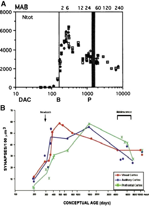
“Cross-sectional developmental studies focused on adolescence have revealed a curvilinear function of cortical gray matter with an increase from birth to about 10 years of age followed by a continuous decline through adulthood to old age (Blanton et al., 2001; Carne et al., 2006; Courchesne et al., 2000; Giedd et al.,…”
BackgroundThe development of brain network hubs
Oldham, S., & Fornito, A. (2019) · Developmental Cognitive Neuroscience · doi:10.1016/j.dcn.2018.12.005 · Cited by 250
“Key developmental events, such as the initial growth of axons during the second trimester, or the pruning of excess synapses during childhood and adolescence, are critical in sculpting the anatomical wiring that links spatially distributed neural elements (de GraafPeters and Hadders-Algra, 2006; Stiles and Jernigan, 2…”
Alcohol and Drug Use and the Developing Brain
Squeglia, L. M., & Gray, K. M. (2016) · Current Psychiatry Reports · doi:10.1007/s11920-016-0689-y · Cited by 249
Modesto, T., Tiemeier, H., Peeters, R. P., Jaddoe, V. W. V., Hofman, A., Verhulst, F. C., & Ghassabian, A. (2015) · JAMA Pediatrics · doi:10.1001/jamapediatrics.2015.0498 · Cited by 248
Innate immunity at the crossroads of healthy brain maturation and neurodevelopmental disorders
Zengeler, K. E., & Lukens, J. R. (2021) · Nature Reviews Immunology · doi:10.1038/s41577-020-00487-7 · Cited by 246
Structural Growth Trajectories and Rates of Change in the First 3 Months of Infant Brain Development
Holland, D., Chang, L., Ernst, T. M., Curran, M., Buchthal, S. D., Alicata, D., Skranes, J., Johansen, H., Hernandez, A., Yamakawa, R., et al. (2014) · JAMA Neurology · doi:10.1001/jamaneurol.2014.1638 · Cited by 242
Microglial Dynamics During Human Brain Development
Menassa, D. A., & Gomez-Nicola, D. (2018) · Frontiers in Immunology · doi:10.3389/fimmu.2018.01014 · Cited by 239
“The endoderm, forms the gut lining; the mesoderm generates the circulatory, cardiovascular and lymphatic systems; and the ectoderm generates both skin and the neuroectoderm, from which the nervous system develops (28, 29).”
BackgroundMaternal Nutrition and Neurodevelopment: A Scoping Review
Cortés-Albornoz, M. C., García-Guáqueta, D. P., Velez-van-Meerbeke, A., & Talero-Gutiérrez, C. (2021) · Nutrients · doi:10.3390/nu13103530 · Cited by 238
“Neurulation occurs early in embryonic life, beginning on day 18 after conception and continuing until day 28 [7].”
BackgroundYouth Mental Health Should Be a Top Priority for Health Care in Canada
Malla, A., Shah, J., Iyer, S., Boksa, P., Joober, R., Andersson, N., Lal, S., & Fuhrer, R. (2018) · The Canadian Journal of Psychiatry · doi:10.1177/0706743718758968 · Cited by 232
Moving toward a neuroplasticity view of bilingualism, executive control, and aging
BAUM, S., & TITONE, D. (2014) · Applied Psycholinguistics · doi:10.1017/s0142716414000174 · Cited by 230
“From before birth through adolescence, there is evidence of rapid growth of the brain in terms of synaptogenesis, neurogenesis, and neuronal migration, which yields increases in both gray and white matter density (for reviews, see Stiles & Jernigan, 2010; Tau & Peterson, 2010).”
BackgroundMRI of the Neonatal Brain: A Review of Methodological Challenges and Neuroscientific Advances
Dubois, J., Alison, M., Counsell, S. J., Hertz‐Pannier, L., Hüppi, P. S., & Benders, M. J. N. L. (2021) · Journal of Magnetic Resonance Imaging · doi:10.1002/jmri.27192 · Cited by 226
“The human brain develops slowly, from embryonic conception to early adulthood, based on multiple events occurring within a highly constrained but constantly changing context.(1,2) During gestation, the brain shows a sequential development of transient laminar compartments, from the center to the periphery: the prolife…”
BackgroundEarly nutrition influences developmental myelination and cognition in infants and young children
Deoni, S., Dean, D., Joelson, S., O'Regan, J., & Schneider, N. (2018) · NeuroImage · doi:10.1016/j.neuroimage.2017.12.056 · Cited by 221
“These adaptive processes are modulated by neural activity and are responsive to environmental, genetic, hormonal, and other influences (Stiles and Jernigan, 2010).”
Genetic topography of brain morphology
Chen, C.-H., Fiecas, M., Gutiérrez, E. D., Panizzon, M. S., Eyler, L. T., Vuoksimaa, E., Thompson, W. K., Fennema-Notestine, C., Hagler, D. J., Jernigan, T. L., et al. (2013) · Proceedings of the National Academy of Sciences · doi:10.1073/pnas.1308091110 · Cited by 217
“Sensorimotor region and frontal pole, temporal, and occipital poles mature before higher-order association areas (12, 39).”
BackgroundMethodSelf-citationCircadian and wakefulness-sleep modulation of cognition in humans
Wright, K. P., Lowry, C. A., & LeBourgeois, M. K. (2012) · Frontiers in Molecular Neuroscience · doi:10.3389/fnmol.2012.00050 · Cited by 209
Morton, P. D., Ishibashi, N., & Jonas, R. A. (2017) · Circulation Research · doi:10.1161/circresaha.116.309048 · Cited by 200
“Human brain development is a long process which begins in the third week of gestation and continues into early adulthood.(12) After birth, the brain increases over 100% in volume in the first year and another 15% by the end of the second year of life.”
Collin, G., & van den Heuvel, M. P. (2013) · The Neuroscientist · doi:10.1177/1073858413503712 · Cited by 198
Vértes, P. E., & Bullmore, E. T. (2015) · Journal of Child Psychology and Psychiatry · doi:10.1111/jcpp.12365 · Cited by 197
“The bulk of neurogenesis and neuronal migration is thought to be complete by about week 20 after conception (Stiles & Jernigan, 2010) at which point axons begin to form and the density of synapses increases steadily (by about 4% per week) until week 27 after conception (Collin & van den Heuvel,…”
BackgroundThe Role of Neurod Genes in Brain Development, Function, and Disease
Tutukova, S., Tarabykin, V., & Hernandez-Miranda, L. R. (2021) · Frontiers in Molecular Neuroscience · doi:10.3389/fnmol.2021.662774 · Cited by 193
“This can be particularly seen in disorders affecting the development of the cerebral cortex, which are frequently associated with seizures both in childhood and in adult life (Subramanian et al., 2019).”
BackgroundDNA Modifications: Function and Applications in Normal and Disease States
Liyanage, V., Jarmasz, J., Murugeshan, N., Del Bigio, M., Rastegar, M., & Davie, J. (2014) · Biology · doi:10.3390/biology3040670 · Cited by 150
“six, and reaching full adult weight in early teen years [200,201].”
BackgroundThe effect of alcohol use on human adolescent brain structures and systems
(2014) · Handbook of Clinical Neurology · doi:10.1016/b978-0-444-62619-6.00028-8 · Cited by 189
“Normal adolescent brain development involves substantial cortical thinning, which has been linked to elimination of weak synaptic connections (Giedd, 2004; Stiles and Jernigan, 2010).”
BackgroundWen, D. J., Poh, J. S., Ni, S. N., Chong, Y.-S., Chen, H., Kwek, K., Shek, L. P., Gluckman, P. D., Fortier, M. V., Meaney, M. J., et al. (2017) · Translational Psychiatry · doi:10.1038/tp.2017.74 · Cited by 186
Cytosine-5 RNA Methylation Regulates Neural Stem Cell Differentiation and Motility
Flores, J. V., Cordero-Espinoza, L., Oeztuerk-Winder, F., Andersson-Rolf, A., Selmi, T., Blanco, S., Tailor, J., Dietmann, S., & Frye, M. (2017) · Stem Cell Reports · doi:10.1016/j.stemcr.2016.11.014 · Cited by 186
“Human brain development begins with the differentiation of neural progenitor cells in the third gestational week and extends through to adolescence (Stiles and Jernigan, 2010).”
BackgroundOpposing Brain Differences in 16p11.2 Deletion and Duplication Carriers
Qureshi, A. Y., Mueller, S., Snyder, A. Z., Mukherjee, P., Berman, J. I., Roberts, T. P. L., Nagarajan, S. S., Spiro, J. E., Chung, W. K., Sherr, E. H., et al. (2014) · The Journal of Neuroscience · doi:10.1523/jneurosci.1366-14.2014 · Cited by 183
“Alterations in cortical thickness can occur either during embryonic formation or postnatally as a result of synaptic exuberance and pruning (Stiles and Jernigan, 2010).”
BackgroundPriming for Life: Early Life Nutrition and the Microbiota-Gut-Brain Axis
Ratsika, A., Codagnone, M. C., O’Mahony, S., Stanton, C., & Cryan, J. F. (2021) · Nutrients · doi:10.3390/nu13020423 · Cited by 182
“The brain is subjected to multiple structural and functional, time-specific changes during gestation such as axonal growth [76], synapse formation and dendritic and axonal arborization [77].”
BackgroundExploring early human brain development with structural and physiological neuroimaging
Vasung, L., Abaci Turk, E., Ferradal, S. L., Sutin, J., Stout, J. N., Ahtam, B., Lin, P.-Y., & Grant, P. E. (2019) · NeuroImage · doi:10.1016/j.neuroimage.2018.07.041 · Cited by 177
The emergence of functional architecture during early brain development
Keunen, K., Counsell, S. J., & Benders, M. J. N. L. (2017) · NeuroImage · doi:10.1016/j.neuroimage.2017.01.047 · Cited by 175
“eventually resulting in an abundance of connections (Huttenlocher and Courten de, 1987; Huttenlocher and Dabholkar, 1997; Innocenti and Price, 2005; Kostovic and Jovanov-Milosevic, 2006; Stiles and Jernigan, 2010).”
BackgroundInfluentialBrain Fuel Utilization in the Developing Brain
Steiner, P. (2019) · Annals of Nutrition and Metabolism · doi:10.1159/000508054 · Cited by 156
Fair, S. R., Julian, D., Hartlaub, A. M., Pusuluri, S. T., Malik, G., Summerfied, T. L., Zhao, G., Hester, A. B., Ackerman, W. E., Hollingsworth, E. W., et al. (2020) · Stem Cell Reports · doi:10.1016/j.stemcr.2020.08.017 · Cited by 174
“Although they have provided important fundamental knowledge, these tools are not amenable to experimental manipulation (Hartlaub et al., 2019; Stiles and Jernigan, 2010).”
BackgroundUnderstanding the genetic determinants of the brain with MOSTest
van der Meer, D., Frei, O., Kaufmann, T., Shadrin, A. A., Devor, A., Smeland, O. B., Thompson, W. K., Fan, C. C., Holland, D., Westlye, L. T., et al. (2020) · Nature Communications · doi:10.1038/s41467-020-17368-1 · Cited by 173
Palumbo, S., Mariotti, V., Iofrida, C., & Pellegrini, S. (2018) · Frontiers in Behavioral Neuroscience · doi:10.3389/fnbeh.2018.00117 · Cited by 111
“…animals (Mosaferi et al., 2015) and humans (McEwen et al., 2012) indicate that the environment, mostly during prenatal stage and infancy, impact significantly on neural development, as several critical periods with lasting consequences on behavior have been documented (Stiles and Jernigan, 2010).”
Genome integrity and disease prevention in the nervous system
McKinnon, P. J. (2017) · Genes & Development · doi:10.1101/gad.301325.117 · Cited by 157
“…other organ systems, this tissue has amultitude of levels of control, including homeostasis within the brain, and controlling roles in fundamental aspects of human physiology, including breathing, heart function, endocrine control, and integration of environmental cues (Stiles and Jernigan 2010).”
BackgroundDevelopmental origins of the human hypothalamic-pituitary-adrenal axis
Howland, M. A., Sandman, C. A., & Glynn, L. M. (2017) · Expert Review of Endocrinology & Metabolism · doi:10.1080/17446651.2017.1356222 · Cited by 168
“Prolific neurogenesis occurs, with the majority of the brain’s billions of neurons produced by mid-gestation [1].”
BackgroundNewborns are sensitive to multiple cues for word segmentation in continuous speech
Fló, A., Brusini, P., Macagno, F., Nespor, M., Mehler, J., & Ferry, A. L. (2019) · Developmental Science · doi:10.1111/desc.12802 · Cited by 167
Pfefferbaum, A., Rohlfing, T., Pohl, K. M., Lane, B., Chu, W., Kwon, D., Nolan Nichols, B., Brown, S. A., Tapert, S. F., Cummins, K., et al. (2016) · Cerebral Cortex · doi:10.1093/cercor/bhv205 · Cited by 165
“Underlying this complementary pattern of gray matter and white matter volume dynamics are regional differences in the timing of these changes (Bava et al. 2010; Raznahan et al. 2010) (for review, Toga et al. 2006; Stiles and Jernigan 2010; Giedd et al. 2014).”
BackgroundSelf-citationGut microbiota: a potential regulator of neurodevelopment
Tognini, P. (2017) · Frontiers in Cellular Neuroscience · doi:10.3389/fncel.2017.00025 · Cited by 163
“MINI REVIEW published: 07 February 2017 doi: 10.3389/fncel.2017.00025 Gut Microbiota: A Potential Regulator of Neurodevelopment Paola Tognini * Sassone-Corsi Laboratory, Center for Epigenetics and Metabolism, Department of Biological Chemistry, University of California Irvine, Irvine, CA, USA Edited by: Flavia Trettel…”
InfluentialGenetic Etiologies, Diagnosis, and Treatment of Tuberous Sclerosis Complex
Salussolia, C. L., Klonowska, K., Kwiatkowski, D. J., & Sahin, M. (2019) · Annual Review of Genomics and Human Genetics · doi:10.1146/annurev-genom-083118-015354 · Cited by 163
Vohr, B. R., Poggi Davis, E., Wanke, C. A., & Krebs, N. F. (2017) · Pediatrics · doi:10.1542/peds.2016-2828f · Cited by 163
Early-Life Exposure to Polycyclic Aromatic Hydrocarbons and ADHD Behavior Problems
Perera, F. P., Chang, H.-W., Tang, D., Roen, E. L., Herbstman, J., Margolis, A., Huang, T.-J., Miller, R. L., Wang, S., & Rauh, V. (2014) · PLoS ONE · doi:10.1371/journal.pone.0111670 · Cited by 163
“However, because brain development and growth occurs throughout childhood, postnatal exposures to environmental pollutants may also affect children’s neurodevelopment and behavior [83].”
BackgroundEGF receptor in organ development, tissue homeostasis and regeneration
Tito, C., Masciarelli, S., Colotti, G., & Fazi, F. (2025) · Journal of Biomedical Science · doi:10.1186/s12929-025-01119-9 · Cited by 164
“Here, neurons participate in the branching morphogenesis process, establishing connections (axons and dendrites) and forming an intracellular network for signals transmission [132, 133].”
Bal-Price, A., Pistollato, F., Sachana, M., Bopp, S. K., Munn, S., & Worth, A. (2018) · Toxicology and Applied Pharmacology · doi:10.1016/j.taap.2018.02.008 · Cited by 148
3D Bioprinting of Neural Tissues
Cadena, M., Ning, L., King, A. T., Hwang, B., Jin, L., Serpooshan, V., & Sloan, S. A. (2020) · Advanced Healthcare Materials · doi:10.1002/adhm.202001600 · Cited by 158
Perinatal Interactions between the Microbiome, Immunity, and Neurodevelopment
Pronovost, G. N., & Hsiao, E. Y. (2019) · Immunity · doi:10.1016/j.immuni.2018.11.016 · Cited by 149
“Following differentiation of neural stem cells in early gestation, neural progenitor cells (NPCs) from the ventricular zone proliferate and migrate to pattern the major structures of the brain (Kriegstein and Alvarez-Buylla, 2009; Stiles and Jernigan, 2010).”
Abıdın, Z. U., Naqvi, R. A., Haider, A., Kim, H. S., Jeong, D., & Lee, S. W. (2024) · Frontiers in Bioengineering and Biotechnology · doi:10.3389/fbioe.2024.1392807 · Cited by 156
Early life adversity: Lasting consequences for emotional learning
Krugers, H. J., Arp, J. M., Xiong, H., Kanatsou, S., Lesuis, S. L., Korosi, A., Joels, M., & Lucassen, P. J. (2017) · Neurobiology of Stress · doi:10.1016/j.ynstr.2016.11.005 · Cited by 131
“During the early postnatal developmental period, the connectivity in the brain is larger than in adults (Innocenti and Price, 2005; Stiles and Jernigan, 2010; Lopez-Larson et al., 2011) which is later fine-tuned via pruning processes (Stiles and Jernigan, 2010).”
BackgroundInfluentialCortical Malformations: Lessons in Human Brain Development
Subramanian, L., Calcagnotto, M. E., & Paredes, M. F. (2020) · Frontiers in Cellular Neuroscience · doi:10.3389/fncel.2019.00576 · Cited by 155
Regulation of cerebral cortical neurogenesis by the Pax6 transcription factor
Manuel, M. N., Mi, D., Mason, J. O., & Price, D. J. (2015) · Frontiers in Cellular Neuroscience · doi:10.3389/fncel.2015.00070 · Cited by 153
“Projection neurons in each layer tend to exhibit similar gene expression patterns, morphologies and organization of afferent and efferent connections (Stiles and Jernigan, 2010).”
Cornblath, E. J., Ashourvan, A., Kim, J. Z., Betzel, R. F., Ćirić, R., Adebimpe, A., Baum, G. L., He, X., Ruparel, K., Moore, T. M., et al. (2020) · Communications Biology · doi:10.1038/s42003-020-0961-x · Cited by 152
Docosahexaenoic Acid and Arachidonic Acid Nutrition in Early Development
Carlson, S. E., & Colombo, J. (2016) · Advances in Pediatrics · doi:10.1016/j.yapd.2016.04.011 · Cited by 135
“By the end of the embryonic period (ie, the first 8 prenatal weeks), the fundamental structures of the brain and central nervous system are fairly well defined [43], and neurogenesis has begun [44].”
Associations between Neighborhood SES and Functional Brain Network Development
Tooley, U. A., Mackey, A. P., Ciric, R., Ruparel, K., Moore, T. M., Gur, R. C., Gur, R. E., Satterthwaite, T. D., & Bassett, D. S. (2020) · Cerebral Cortex · doi:10.1093/cercor/bhz066 · Cited by 132
The Impact of Infection in Pregnancy on Placental Vascular Development and Adverse Birth Outcomes
Weckman, A. M., Ngai, M., Wright, J. K., McDonald, C. R., & Kain, K. C. (2019) · Frontiers in Microbiology · doi:10.3389/fmicb.2019.01924 · Cited by 146
Multiscale Structure–Function Gradients in the Neonatal Connectome
Larivière, S., Vos de Wael, R., Hong, S.-J., Paquola, C., Tavakol, S., Lowe, A. J., Schrader, D. V., & Bernhardt, B. C. (2020) · Cerebral Cortex · doi:10.1093/cercor/bhz069 · Cited by 139
Age, plasticity, and homeostasis in childhood brain disorders
Dennis, M., Spiegler, B. J., Juranek, J. J., Bigler, E. D., Snead, O. C., & Fletcher, J. M. (2013) · Neuroscience & Biobehavioral Reviews · doi:10.1016/j.neubiorev.2013.09.010 · Cited by 147
“…within a highly constrained, genetically organized but changing environment that has bidirectional effects on the brain; at each point in development, the child has a stable state and a plasticity history that limit which factors will influence its further development (Stiles & Jernigan, 2010).”
BackgroundPiezoelectric Effects of Materials on Bio-Interfaces
Marino, A., Genchi, G. G., Sinibaldi, E., & Ciofani, G. (2017) · ACS Applied Materials & Interfaces · doi:10.1021/acsami.7b04323 · Cited by 142
Sphingomyelin in Brain and Cognitive Development: Preliminary Data
Schneider, N., Hauser, J., de Oliveira, M. C., Cazaubon, E., Mottaz, S. C., O’Neill, B. V., Steiner, P., & Deoni, S. (2019) · eNeuro · doi:10.1523/eneuro.0421-18.2019 · Cited by 142
“Ensuring efficient transmission of nerve impulses along neuronal axons (Fields, 2008; Stiles and Jernigan, 2010), CNS myelination is important in the maturation of brain networks, coordinated information processing and ultimately cognitive performance in infants and children (O’Muircheartaigh et…”
BackgroundAppraising the Role of Iron in Brain Aging and Cognition: Promises and Limitations of MRI Methods
Daugherty, A. M., & Raz, N. (2015) · Neuropsychology Review · doi:10.1007/s11065-015-9292-y · Cited by 131
“Multiple factors shape the trajectory of brain change across the lifespan, but unlike development that follows a largely programmed albeit modifiable course (Stiles and Jernigan 2010), aging is unlikely to reflect the unfolding of a grand plan and appears more as a sum of overlapping time-dependent processes and influ…”
BackgroundTiming of therapies for Down syndrome: the sooner, the better
Stagni, F., Giacomini, A., Guidi, S., Ciani, E., & Bartesaghi, R. (2015) · Frontiers in Behavioral Neuroscience · doi:10.3389/fnbeh.2015.00265 · Cited by 124
“In the human forebrain neocortical neurons are generated during a restricted period that begins at approximately gestational week 6 and is largely completed by week 18 (Stiles and Jernigan, 2010) (Figure 4B).”
BackgroundDevelopments and Opportunities for 3D Bioprinted Organoids
Ren, Y., Yang, X., Ma, Z., Sun, X., Zhang, Y., Li, W., Yang, H., Qiang, L., Yang, Z., Liu, Y., et al. (2024) · International Journal of Bioprinting · doi:10.18063/ijb.v7i3.364 · Cited by 126
“Brain tissue is composed of tightly packed glial and neuronal cells, and the ability of cells to self-organize depends mostly on the influence of component gradients and intercellular interactions[70,71].”
BackgroundDifferential Responses of Human Fetal Brain Neural Stem Cells to Zika Virus Infection
McGrath, E. L., Rossi, S. L., Gao, J., Widen, S. G., Grant, A. C., Dunn, T. J., Azar, S. R., Roundy, C. M., Xiong, Y., Prusak, D. J., et al. (2017) · Stem Cell Reports · doi:10.1016/j.stemcr.2017.01.008 · Cited by 134
“First, it is known that there are no significant developmental milestones between gestational weeks 9 and 13 (Stiles and Jernigan, 2010).”
Background
Metadata
- Authors
- Stiles, J., & Jernigan, T. L.
- Affiliations
- University of California San Diego (US)
- Corresponding author
- Jernigan, Terry L.
- ORCID
- Jernigan, Terry L.
- Venue
- Neuropsychology Review
- Published
- 2010-11-03
- Volume
- 20
- Issue
- 4
- Pages
- 327-348
- ISSN
- 1040-7308 (ISSN-L)
- 1573-6660
- PMC
- PMC2989000
- OpenAlex
- W2115599658
- Cited by
- 2,009
- References
- 132
- Citations by year
- 2026: 142 · 2025: 184 · 2024: 199 · 2023: 209 · 2022: 184 · 2021: 197 · 2020: 195 · 2019: 155 · 2018: 158 · 2017: 122 · 2016: 92 · 2015: 64 · 2014: 49 · 2013: 24 · 2012: 23
- FWCI
- 1.43
- Type
- journal article
- Language
- en
- Open access
- hybrid
- doi.org
- Licence
- cc-by-nc
- Locations
- Neuropsychology Review (publishedVersion, cc-by-nc)
- Europe PMC (PubMed Central) (publishedVersion, cc-by-nc)
- PubMed Central (publishedVersion, cc-by-nc)
- PubMed (publishedVersion)
- eScholarship (California Digital Library) (submittedVersion, public-domain)
- eScholarship (California Digital Library) (submittedVersion)
- Funders
- Lundbeckfonden (R32‐A3161)
- H. Lundbeck A/S
- National Institutes of Health (5r01hd025077-10)
- U.S. Public Health Service (R32-A3161)
- University of California, San Diego (RC2DA029475)
- National Institute on Drug Abuse (RC2DA029475)
- National Institute of Child Health and Human Development
- Eunice Kennedy Shriver National Institute of Child Health and Human Development (R01HD060595, R01 HD 25077)
- Topics
- Life Sciences › Neuroscience › Developmental Neuroscience › Neurogenesis and neuroplasticity mechanisms
- Life Sciences › Biochemistry, Genetics and Molecular Biology › Genetics › Genetics and Neurodevelopmental Disorders
- Social Sciences › Psychology › Developmental and Educational Psychology › Child and Animal Learning Development
- Keywords
- brain development, developmental neuroscience, mammalian brain, neuronal organization, social development (developmental psychology), cognitive development, neural systems, genetic constraints, developmental mechanisms
- MeSH terms
- Animals, Brain, Cell Differentiation, Diagnostic Imaging, Humans, Nerve Net, Neural Pathways, Neurobiology, Neurons, Gene Expression Regulation, Developmental, Body Patterning, Neurogenesis
About these checks
Registry, retraction, Cites and statistics results are computed, not judged, with one exception: a printed reference that no index lists is read once by a model before it counts as a citation error. Claims are extracted by a model and kept only when their quote appears verbatim in the text; each cited source is then checked, with a model’s help, for whether it says what the claim relies on. Those verdicts can be wrong — read the claim’s quote and the cited source before relying on one.
Statistics and arithmetic are recomputed, not judged.
Confidence bands are shown only for fields calibrated on our annotated test set. None is calibrated yet, so no band is shown.
What counts as a claim ›How we measure confidence ›How we check this ›
Are you an author, or do you see a problem with this page? Request a review
Data sources
Bibliographic data: Semantic Scholar, OpenAlex, Crossref.
Contains information from the Semantic Scholar Academic Graph Datasets, made available under the ODC Attribution License (ODC-BY 1.0).
Semantic Scholar’s data is described in: Kinney, R., et al. (2023). The Semantic Scholar Open Data Platform. arXiv:2301.10140.
