

The DOI matches its Crossref record; 228 of the 228 references it cites were found in a registry or an index.
The potential of P2X7 receptors as a therapeutic target, including inflammation and tumour progression
Open accessAbstract
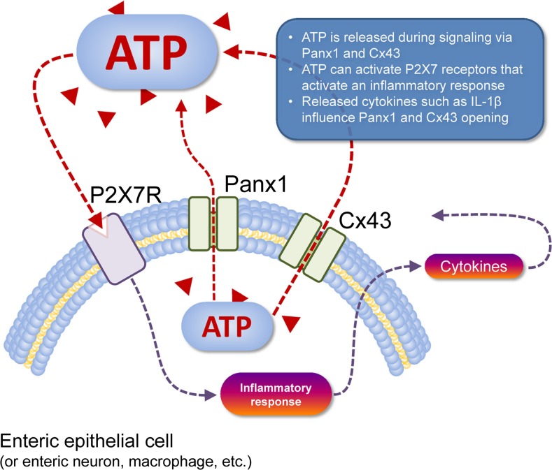
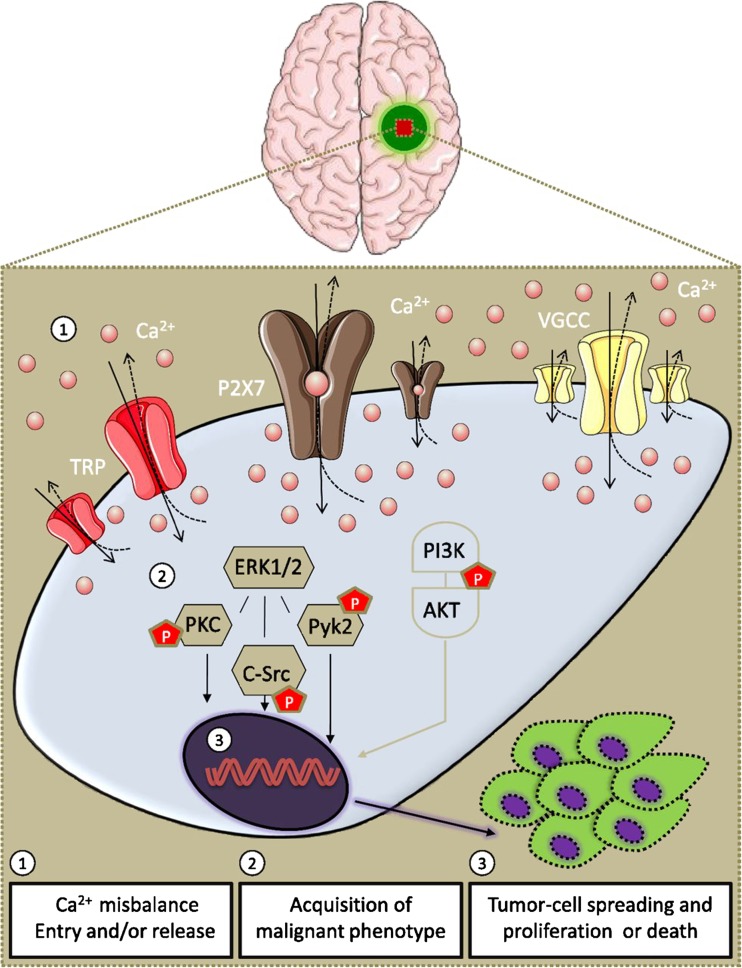
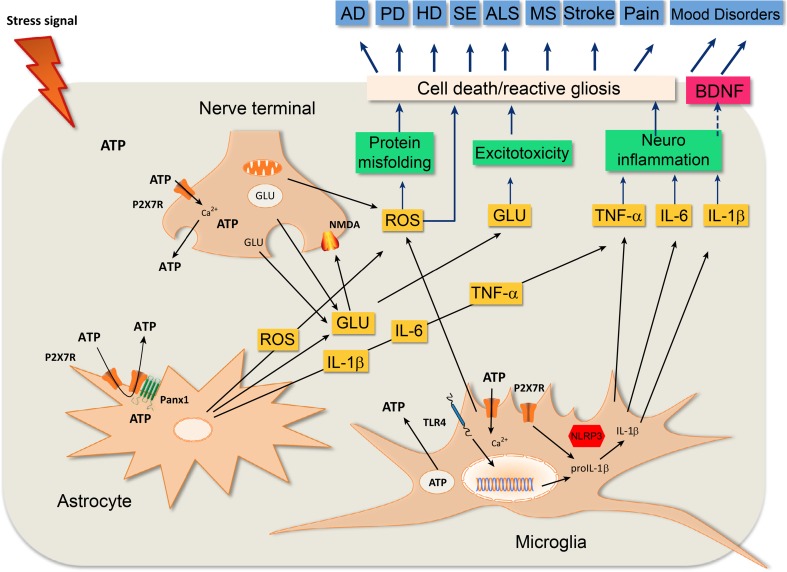
Seven P2X ion channel nucleotide receptor subtypes have been cloned and characterised. P2X7 receptors (P2X7R) are unusual in that there are extra amino acids in the intracellular C terminus. Low concentrations of ATP open cation channels sometimes leading to cell proliferation, whereas high concentrations of ATP open large pores that release inflammatory cytokines and can lead to apoptotic cell death. Since many diseases involve inflammation and immune responses, and the P2X7R regulates inflammation, there has been recent interest in the pathophysiological roles of P2X7R and the potential of P2X7R antagonists to treat a variety of diseases. These include neurodegenerative diseases, psychiatric disorders, epilepsy and a number of diseases of peripheral organs, including the cardiovascular, airways, kidney, liver, bladder, skin and musculoskeletal. The potential of P2X7R drugs to treat tumour progression is discussed.
Overview
The claims and statistics of this paper haven’t been checked yet.
Run the check — free, no account needed. It reads the open-access text and checks the sources it cites.
Integrity signals
Registry
Cites
Statistics
Figures
Verification
Claims the paper makes
The claims and statistics of this paper haven’t been checked yet.
Run the check — free, no account needed. It reads the open-access text and checks the sources it cites.
Claims other papers make about this one
Each is checked against this paper’s own text: does it say what the citing paper says it does?
In the citing paper
“P2X7R-mediated effects, which mainly occurs through activation of the neuroinflammatory response, have been involved in MDD, neurodegenerative diseases, schizophrenia, epilepsy, neuropathic pain and brain injury [28,66,67,68].”
From P2X7 Receptor Signaling in Stress and Depression (2019) · cites this paper as [66]
Self-check
Percentages, sums and reported differences re-computed from the paper’s own numbers.
The self-check needs the paper’s full text.
Citations
Cites
Receptors for Purines and Pyrimidines
Burnstock, G., & Verkhratsky, A. (2012) · Springer eBooks · doi:10.1007/978-3-642-28863-0_5 · Cited by 3,962
Self-citationReceptors for Purines and Pyrimidines
Ralevic, V., & Burnstock, G. (1998) · Pharmacological Reviews · doi:10.1016/s0031-6997(24)01373-5 · Cited by 2,028
Self-citationPhysiology and Pathophysiology of Purinergic Neurotransmission
Burnstock, G. (2007) · Physiological Reviews · doi:10.1152/physrev.00043.2006 · Cited by 1,609
Self-citationThe P2X7 Receptor in Infection and Inflammation
Di Virgilio, F., Dal Ben, D., Sarti, A. C., Giuliani, A. L., & Falzoni, S. (2017) · Immunity · doi:10.1016/j.immuni.2017.06.020 · Cited by 1,267
A P2X purinoceptor expressed by a subset of sensory neurons
Chen, C., Akopian, A. N., Sivilotti, L. G., Colquhoun, D., Burnstock, G., & Wood, J. N. (1995) · Nature · doi:10.1038/377428a0 · Cited by 1,016
Self-citationYang, D., He, Y., Muñoz-Planillo, R., Liu, Q., & Núñez, G. (2015) · Immunity · doi:10.1016/j.immuni.2015.10.009 · Cited by 622
Pathophysiology and Therapeutic Potential of Purinergic Signaling
Burnstock, G. (2006) · Pharmacological Reviews · doi:10.1124/pr.58.1.5 · Cited by 618
Self-citationExtracellular purines, purinergic receptors and tumor growth
Virgilio, F. D., & Adinolfi, E. (2016) · Oncogene · doi:10.1038/onc.2016.206 · Cited by 566
CD73 promotes anthracycline resistance and poor prognosis in triple negative breast cancer
Loi, S., Pommey, S., Haibe‐Kains, B., Beavis, P. A., Darcy, P. K., Smyth, M. J., & Stagg, J. (2013) · Proceedings of the National Academy of Sciences · doi:10.1073/pnas.1222251110 · Cited by 504
Abdulqawi, R., Dockry, R., Holt, K. J., Layton, G. R., McCarthy, B. G., Ford, A. P. D. W., & Smith, J. A. (2014) · The Lancet · doi:10.1016/s0140-6736(14)61255-1 · Cited by 488
Cited by
Sorted by citations
Extracellular ATP and P2 purinergic signalling in the tumour microenvironment
Virgilio, F. D., Sarti, A. C., Falzoni, S., Marchi, E. D., & Adinolfi, E. (2018) · Nature reviews. Cancer · doi:10.1038/s41568-018-0037-0 · Cited by 748
The Role of Inflammation in the Pathogenesis of Osteoarthritis
Chow, Y. Y., & Chin, K. (2020) · Mediators of Inflammation · doi:10.1155/2020/8293921 · Cited by 536
Grams, R. J., Santos, W. L., Scorei, I. R., Abad-García, A., Rosenblum, C. A., Biţă, A., Cerecetto, H., Viñas, C., & Soriano-Ursua, M. A. (2024) · Chemical Reviews · doi:10.1021/acs.chemrev.3c00663 · Cited by 389
Interleukin-1β is a potential therapeutic target for periodontitis: a narrative review
Cheng, R., Wu, Z., Li, M., Shao, M., & Hu, T. (2020) · International Journal of Oral Science · doi:10.1038/s41368-019-0068-8 · Cited by 304
Microglia in Alzheimer’s disease: pathogenesis, mechanisms, and therapeutic potentials
Miao, J., Ma, H., Yang, Y., Liao, Y., Lin, C., Zheng, J., Yu, M., & Lan, J. (2023) · Frontiers in Aging Neuroscience · doi:10.3389/fnagi.2023.1201982 · Cited by 293
Full-Length P2X7 Structures Reveal How Palmitoylation Prevents Channel Desensitization
McCarthy, A. E., Yoshioka, C., & Mansoor, S. (2019) · Cell · doi:10.1016/j.cell.2019.09.017 · Cited by 268
Thawkar, B. S., & Kaur, G. (2018) · Journal of Neuroimmunology · doi:10.1016/j.jneuroim.2018.11.010 · Cited by 254
P2X7 Interactions and Signaling – Making Head or Tail of It
Kopp, R., Krautloher, A., Ramírez-Fernández, A., & Nicke, A. (2019) · Frontiers in Molecular Neuroscience · doi:10.3389/fnmol.2019.00183 · Cited by 217
Targeting Adenosine Receptor Signaling in Cancer Immunotherapy
Sek, K., Mølck, C., Stewart, G. D., Kats, L. M., Darcy, P. K., & Beavis, P. A. (2018) · International Journal of Molecular Sciences · doi:10.3390/ijms19123837 · Cited by 206
Eisinger, S., Sarhan, D., Boura, V. F., Ibarlucea-Benitez, I., Tyystjärvi, S., Oliynyk, G., Arsenian‐Henriksson, M., Lane, D. P., Wickström, S. L., Kiessling, R. W., et al. (2020) · Proceedings of the National Academy of Sciences · doi:10.1073/pnas.2015343117 · Cited by 173
P2X7 Receptor Signaling in Stress and Depression
Ribeiro, D. E., Roncalho, A. L., Glaser, T., Ulrich, H., Wegener, G., & Joca, S. (2019) · International Journal of Molecular Sciences · doi:10.3390/ijms20112778 · Cited by 129
Wei, F., Wang, D., Wei, J., Tang, N., Tang, L. M., Xiong, F., Guo, C. J., Zhou, M., Li, X., Li, G., et al. (2020) · Cellular and Molecular Life Sciences · doi:10.1007/s00018-020-03581-0 · Cited by 128
Mechanosensitive ion channels push cancer progression
Pethő, Z., Najder, K., Bulk, E., & Schwab, A. (2019) · Cell Calcium · doi:10.1016/j.ceca.2019.03.007 · Cited by 125
Animal models of pain: Diversity and benefits
Abboud, C., Duveau, A., Bouali‐Benazzouz, R., Massé, K. L., Mattar, J., Brochoire, L., Fossat, P., Boué‐Grabot, É., Hleihel, W., & Landry, M. (2020) · Journal of Neuroscience Methods · doi:10.1016/j.jneumeth.2020.108997 · Cited by 114
Wang, H., Wang, X., Xu, L., Zhang, J., & Cao, H. (2020) · Purinergic Signalling · doi:10.1007/s11302-020-09711-4 · Cited by 104
CD73 Downregulation Decreases In Vitro and In Vivo Glioblastoma Growth
Azambuja, J. H., Gelsleichter, N. E., Beckenkamp, L. R., Iser, I. C., Fernandes, M. D. C., Figueiró, F., Battastini, A. M. O., Scholl, J. N., de Oliveira, F. H., Spanevello, R. M., et al. (2018) · Molecular Neurobiology · doi:10.1007/s12035-018-1240-4 · Cited by 96
P2RX7 Purinoceptor as a Therapeutic Target—The Second Coming?
Young, C., & Górecki, D. C. (2018) · Frontiers in Chemistry · doi:10.3389/fchem.2018.00248 · Cited by 97
Rotondo, J. C., Mazziotta, C., Lanzillotti, C., Stefani, C., Badiale, G., Campione, G., Martini, F., & Tognon, M. G. (2022) · Cancers · doi:10.3390/cancers14051116 · Cited by 98
The P2X7 Receptor in Microglial Cells Modulates the Endolysosomal Axis, Autophagy, and Phagocytosis
Campagno, K. E., & Mitchell, C. H. (2021) · Frontiers in Cellular Neuroscience · doi:10.3389/fncel.2021.645244 · Cited by 94
Extracellular ATP and Adenosine in Cancer Pathogenesis and Treatment
Chiarella, A. M., Ryu, Y. K., Manji, G. A., & Rustgi, A. K. (2021) · Trends in cancer · doi:10.1016/j.trecan.2021.04.008 · Cited by 87
Inherent P2X7 Receptors Regulate Macrophage Functions during Inflammatory Diseases
Ren, W.-J., Rubini, P., Tang, Y., Engel, T., & Illés, P. (2021) · International Journal of Molecular Sciences · doi:10.3390/ijms23010232 · Cited by 70
Blocking P2X7-Mediated Macrophage Polarization Overcomes Treatment Resistance in Lung Cancer
Qin, J., Zhang, X., Tan, B., Zhang, S., Yin, C., Xue, Q., Zhang, Z., Ren, H., Chen, J., Liu, M., et al. (2020) · Cancer Immunology Research · doi:10.1158/2326-6066.cir-20-0123 · Cited by 82
Nociceptive signaling mediated by P2X3, P2X4 and P2X7 receptors
Inoue, K., & Tsuda, M. (2021) · Biochemical Pharmacology · doi:10.1016/j.bcp.2020.114309 · Cited by 78
Pathological ATPergic Signaling in Major Depression and Bipolar Disorder
Illés, P., Verkhratsky, A., & Tang, Y. (2020) · Frontiers in Molecular Neuroscience · doi:10.3389/fnmol.2019.00331 · Cited by 78
Campagno, K. E., Lu, W., Jassim, A. H., Albalawi, F., Cenaj, A., Tso, H., Clark, S. P., Sripinun, P., Gómez, N. M., & Mitchell, C. H. (2021) · Journal of Neuroinflammation · doi:10.1186/s12974-021-02251-7 · Cited by 68
The role and pharmacological properties of the P2X7 receptor in neuropathic pain
Zhang, W., Zhu, J., & Liu, Z.-X. (2019) · Brain Research Bulletin · doi:10.1016/j.brainresbull.2019.11.006 · Cited by 67
Structural and Functional Basis for Understanding the Biological Significance of P2X7 Receptor
Martínez-Cuesta, M. Á., Blanch‐Ruiz, M. A., Ortega‐Luna, R., Sánchez‐López, A., & Álvarez, Á. (2020) · International Journal of Molecular Sciences · doi:10.3390/ijms21228454 · Cited by 68
The pharmacological and pharmacokinetic properties of esculin: A comprehensive review
Li, C., Li, J.-C., Lai, J., & Liu, Y. (2022) · Phytotherapy Research · doi:10.1002/ptr.7470 · Cited by 61
Guan, S., Shen, Y., Ge, H., Xiong, W., He, L., Liu, L., Yin, C., Wei, X., & Gao, Y. (2019) · Frontiers in Psychiatry · doi:10.3389/fpsyt.2019.00770 · Cited by 60
Irradiation causes senescence, ATP release, and P2X7 receptor isoform switch in glioblastoma
Zanoni, M., Sarti, A. C., Zamagni, A., Cortesi, M., Pignatta, S., Arienti, C., Tebaldi, M., Sarnelli, A., Romeo, A., Bartolini, D., et al. (2022) · Cell Death and Disease · doi:10.1038/s41419-022-04526-0 · Cited by 62
Price, G. W., Chadjichristos, C. E., Kavvadas, P., Tang, S. C. W., Yiu, W. H., Green, C. R., Potter, J. A., Siamantouras, E., Squires, P. E., & Hills, C. E. (2020) · Cell Communication and Signaling · doi:10.1186/s12964-020-00558-1 · Cited by 58
Orchestrated modulation of rheumatoid arthritis via crosstalking intracellular signaling pathways
Ibrahim, S. S. A., & Huttunen, K. M. (2021) · Inflammopharmacology · doi:10.1007/s10787-021-00800-3 · Cited by 56
Antagonism of the ATP-gated P2X7 receptor: a potential therapeutic strategy for cancer
Drill, M., Jones, N. C., Hunn, M., O'Brien, T. J., & Monif, M. (2021) · Purinergic Signalling · doi:10.1007/s11302-021-09776-9 · Cited by 56
Sidoryk‐Wȩgrzynowicz, M., & Strużyńska, L. (2021) · International Journal of Molecular Sciences · doi:10.3390/ijms22168404 · Cited by 51
P2X7 receptor in inflammation and pain
Hu, S.-Q., Hu, J., Zou, F.-L., Liu, J., Luo, H., Hu, D.-X., Wu, L., & Zhang, W. (2022) · Brain Research Bulletin · doi:10.1016/j.brainresbull.2022.07.006 · Cited by 52
Victoni, T., Barreto, E., Lagente, V., & Carvalho, V. F. (2021) · Oxidative Medicine and Cellular Longevity · doi:10.1155/2021/6646923 · Cited by 54
The progress and prospects of targeting the adenosine pathway in cancer immunotherapy
Yang, Y., Zhu, L., Xu, Y., Liang, L., Liu, L.-Z., Chen, X., Li, H., & Liu, H. (2025) · Biomarker Research · doi:10.1186/s40364-025-00784-0 · Cited by 55
Yang, C., Shi, S., Su, Y., Tong, J., & Li, L. (2020) · Journal of Cellular and Molecular Medicine · doi:10.1111/jcmm.15708 · Cited by 51
Role of the P2X7 receptor in the pathogenesis of type 2 diabetes and its microvascular complications
Solini, A., & Novak, I. (2019) · Current Opinion in Pharmacology · doi:10.1016/j.coph.2019.02.009 · Cited by 48
Williams, B. M., Cliff, C. L., Lee, K., Squires, P. E., & Hills, C. E. (2022) · Frontiers in Physiology · doi:10.3389/fphys.2022.907504 · Cited by 47
Novel biologics targeting the P2X7 ion channel
Koch‐Nolte, F., Eichhoff, A. M., Pinto-Espinoza, C., Schwarz, N., Schäfer, T., Menzel, S., Haag, F., Demeules, M., Gondé, H., & Adriouch, S. (2019) · Current Opinion in Pharmacology · doi:10.1016/j.coph.2019.03.001 · Cited by 51
P2X7R antagonists in chronic stress-based depression models: a review
von Mücke-Heim, I.-A., Ries, C., Urbina-Treviño, L., & Deussing, J. M. (2021) · European Archives of Psychiatry and Clinical Neuroscience · doi:10.1007/s00406-021-01306-3 · Cited by 47
Li, Y., Chen, J., Bolinger, A. A., Chen, H., Liu, Z., Cong, Y., Brasier, A. R., Pinchuk, I. V., Tian, B., & Zhou, J. (2021) · Inflammatory Bowel Diseases · doi:10.1093/ibd/izab190 · Cited by 45
da Silva, G. B., Manica, D., da Silva, A. P., Kosvoski, G. C., Hanauer, M. C., Assmann, C. E., Simões, J. L. B., Pillat, M. M., de Lara, J. D., Marafon, F., et al. (2022) · Journal of Molecular Medicine · doi:10.1007/s00109-022-02185-4 · Cited by 46
P2X7 promotes the progression of MLL-AF9 induced acute myeloid leukemia by upregulation of Pbx3
Feng, W., Yang, X. Q., Wang, L., Wang, R., Yang, F., Wang, H., Liu, X., Ren, Q., Zhang, Y., Zhu, X., et al. (2020) · Haematologica · doi:10.3324/haematol.2019.243360 · Cited by 44
P2X7 receptor antagonism inhibits tumour growth in human high-grade gliomas
Kan, L. K., Seneviratne, S. O., Drummond, K. J., Williams, D. A., O'Brien, T. J., & Monif, M. (2020) · Purinergic Signalling · doi:10.1007/s11302-020-09705-2 · Cited by 46
P2X7 receptor: a critical regulator and potential target for breast cancer
Zhu, X., Li, Q., Song, W., Peng, X., & Zhao, R. (2021) · Journal of Molecular Medicine · doi:10.1007/s00109-021-02041-x · Cited by 43
The Role of ATP Receptors in Pain Signaling
Inoue, K. (2022) · Neurochemical Research · doi:10.1007/s11064-021-03516-6 · Cited by 42
The P2X7 Receptor Is Shed Into Circulation: Correlation With C-Reactive Protein Levels
Giuliani, A. L., Berchan, M., Sanz, J. M., Passaro, A., Pizzicotti, S., Vultaggio-Poma, V., Sarti, A. C., & Virgilio, F. D. (2019) · Frontiers in Immunology · doi:10.3389/fimmu.2019.00793 · Cited by 42
Molecular Pharmacology of P2X Receptors: Exploring Druggable Domains Revealed by Structural Biology
Oken, A. C., Krishnamurthy, I., Savage, J. C., Lisi, N. E., Godsey, M., & Mansoor, S. (2022) · Frontiers in Pharmacology · doi:10.3389/fphar.2022.925880 · Cited by 41
P2X receptors in cancer growth and progression
Virgilio, F. D., Vultaggio-Poma, V., & Sarti, A. C. (2020) · Biochemical Pharmacology · doi:10.1016/j.bcp.2020.114350 · Cited by 43
Purinergic signaling and tumor microenvironment in cervical Cancer
Pfaffenzeller, M. S., Franciosi, M. L. M., & Cardoso, A. M. (2020) · Purinergic Signalling · doi:10.1007/s11302-020-09693-3 · Cited by 41
P2 Purinergic Signaling in the Distal Lung in Health and Disease
Wirsching, E., Fauler, M., Fois, G., & Frick, M. (2020) · International Journal of Molecular Sciences · doi:10.3390/ijms21144973 · Cited by 41
Cytokines and Their Genetic Polymorphisms Related to Periodontal Disease
Kozak, M., Dąbrowska‐Żamojcin, E., Mazurek−Mochol, M., & Pawlik, A. (2020) · Journal of Clinical Medicine · doi:10.3390/jcm9124045 · Cited by 42
Bernardazzi, C., Castelo-Branco, M. T. L., Pêgo, B., Ribeiro, B. E., Rosas, S. L. B., Santana, P. T., Machado, J. C., Leal, C. V., Thompson, F. L., Coutinho‐Silva, R., et al. (2022) · International Journal of Molecular Sciences · doi:10.3390/ijms23094616 · Cited by 39
Purinergic P2X7 Receptor: A Cation Channel Sensitive to Tumor Microenvironment
Scarpellino, G., Genova, T., & Munaron, L. (2019) · Recent Patents on Anti-Cancer Drug Discovery · doi:10.2174/1574892814666190116122256 · Cited by 36
The role of microglia and P2X7 receptors in gliomas
Kan, L. K., Williams, D. J., Drummond, K. J., O'Brien, T. J., & Monif, M. (2019) · Journal of Neuroimmunology · doi:10.1016/j.jneuroim.2019.04.010 · Cited by 37
The P2X7 purinergic receptor: a potential therapeutic target for lung cancer
Li, Q., Zhu, X., Song, W., Peng, X., & Zhao, R. (2020) · Journal of Cancer Research and Clinical Oncology · doi:10.1007/s00432-020-03379-4 · Cited by 36
To Inhibit or Enhance? Is There a Benefit to Positive Allosteric Modulation of P2X Receptors?
Stokes, L., Bidula, S. M., Bibič, L., & Allum, E. (2020) · Frontiers in Pharmacology · doi:10.3389/fphar.2020.00627 · Cited by 37
Possible role of purinergic signaling in COVID-19
Franciosi, M. L. M., Lima, M. D. M., Schetinger, M. R. C., & Cardoso, A. M. (2021) · Molecular and Cellular Biochemistry · doi:10.1007/s11010-021-04130-4 · Cited by 38
FTY720 in CNS injuries: Molecular mechanisms and therapeutic potential
Zhang, L., & Wang, H. (2020) · Brain Research Bulletin · doi:10.1016/j.brainresbull.2020.08.013 · Cited by 34
Cui, Z., Wang, G., Zhang, J., Song, J., Jiang, Y., Dou, J., Lian, L., Nan, J., & Wu, Y. (2021) · Phytotherapy Research · doi:10.1002/ptr.7214 · Cited by 34
Ernest James Phillips, T., & Maguire, E. (2021) · Frontiers in Cellular Neuroscience · doi:10.3389/fncel.2021.652593 · Cited by 33
Loss of P2X7 receptor function dampens whole body energy expenditure and fatty acid oxidation
Giacovazzo, G., Apolloni, S., & Coccurello, R. (2018) · Purinergic Signalling · doi:10.1007/s11302-018-9610-y · Cited by 34
Liu, X., Xie, Z., Li, S., He, J., Cao, S., & Xiao, Z. (2021) · International Journal of Biological Sciences · doi:10.7150/ijbs.59032 · Cited by 33
Zhang, W. (2021) · Purinergic Signalling · doi:10.1007/s11302-020-09761-8 · Cited by 31
Zhan, T., Tang, S., Du, J., Liu, J., Yu, B., Yang, Y., Xie, Y., Qiu, Y., Li, G., & Gao, Y. (2024) · International Journal of Molecular Sciences · doi:10.3390/ijms25021186 · Cited by 31
P2RX7 at the Host-Pathogen Interface of Infectious Diseases
Soare, A. Y., Freeman, T. L., Min, A. K., Malik, H. S., Osota, E. O., & Swartz, T. H. (2021) · Microbiology and Molecular Biology Reviews · doi:10.1128/mmbr.00055-20 · Cited by 31
Cancer Metabostemness and Metabolic Reprogramming via P2X7 Receptor
Rabelo, I. L. A., Arnaud-Sampaio, V. F., Adinolfi, E., Ulrich, H., & Lameu, C. (2021) · Cells · doi:10.3390/cells10071782 · Cited by 30
A promising independent prognostic biomarker in colorectal cancer: P2X7 receptor.
Çalık, İ., Çalık, M., Turken, G., & Özercan, İ. H. (2020) · PubMed Central · Cited by 29
Solini, A., Rossi, C., Santini, E., Giuntini, M., Raggi, F., Parolini, F., Biancalana, E., Prete, E. D., Bonuccelli, U., & Ceravolo, R. (2021) · European Journal of Neurology · doi:10.1111/ene.14918 · Cited by 32
P2 Receptors as Therapeutic Targets in the Salivary Gland: From Physiology to Dysfunction
Khalafalla, M. G., Woods, L. T., Jasmer, K. J., Forti, K. M., Camden, J. M., Jensen, J. L., Limesand, K. H., Galtung, H. K., & Weisman, G. A. (2020) · Frontiers in Pharmacology · doi:10.3389/fphar.2020.00222 · Cited by 28
Potential Therapeutic Role of Purinergic Receptors in Cardiovascular Disease Mediated by SARS‐CoV‐2
dos Anjos, F., Simões, J. L. B., Assmann, C. E., Carvalho, F. B., & Bagatini, M. D. (2020) · Journal of Immunology Research · doi:10.1155/2020/8632048 · Cited by 32
Spotlight on P2X7 Receptor PET Imaging: A Bright Target or a Failing Star?
Schmidt, S., Isaak, A., & Junker, A. (2023) · International Journal of Molecular Sciences · doi:10.3390/ijms24021374 · Cited by 28
P2X7 purinoceptor as a therapeutic target in muscular dystrophies
Górecki, D. C. (2019) · Current Opinion in Pharmacology · doi:10.1016/j.coph.2019.02.003 · Cited by 27
Purinergic Calcium Signals in Tumor-Derived Endothelium
Scarpellino, G., Genova, T., Avanzato, D., Bernardini, M., Bianco, S., Petrillo, S., Tolosano, E., Vieira, J. R. D. A., Bussolati, B., Fiorio, A., et al. (2019) · Cancers · doi:10.3390/cancers11060766 · Cited by 28
Oken, A. C., Lisi, N. E., Krishnamurthy, I., McCarthy, A. E., Godsey, M., Glasfeld, A., & Mansoor, S. (2024) · Nature Communications · doi:10.1038/s41467-024-50771-6 · Cited by 26
Sun, H. J., Xin, Y., Wu, J., ShuQin, Z., GuiLian, Z., Wu, H., Zhen, G., HongWei, R., & Yongnan, L. (2020) · Neurochemistry International · doi:10.1016/j.neuint.2020.104773 · Cited by 27
Strassheim, D., Karoor, V., Stenmark, K. R., Verin, A. D., & Gerasimovskaya, E. V. (2018) · Vessel Plus · doi:10.20517/2574-1209.2018.44 · Cited by 26
Ca2+ as a therapeutic target in cancer
Gross, S., Mallu, P., Joshi, H., Schultz, B. M., Go, C. K., & Soboloff, J. (2020) · Advances in cancer research · doi:10.1016/bs.acr.2020.05.003 · Cited by 29
Li, P., Zhang, Q., Xiao, Z., Yu, S., Yan, Y., & Qin, Y. (2018) · Molecular Pain · doi:10.1177/1744806918803039 · Cited by 26
Doǧan, E., Aygün, H., Arslan, G., Rzayev, E., Avcı, B., Ayyıldız, M., & Ağar, E. (2020) · Frontiers in Neuroscience · doi:10.3389/fnins.2020.00414 · Cited by 26
Bando, K., Kuroishi, T., Tada, H., Oizumi, T., Tanaka, Y., Takahashi, T., Mizoguchi, I., Sugawara, S., & Endo, Y. (2020) · Journal of Bone and Mineral Research · doi:10.1002/jbmr.4384 · Cited by 24
Wang, K., Sun, M., Juan, Z., Zhang, J., Sun, Y., Wang, G., Wang, C., Li, Y., Kong, W., Fan, L., et al. (2022) · Behavioural Neurology · doi:10.1155/2022/3777351 · Cited by 24
Purinergic Signaling in Pancreas—From Physiology to Therapeutic Strategies in Pancreatic Cancer
Novak, I., Yu, H., Magni, L., & Deshar, G. (2020) · International Journal of Molecular Sciences · doi:10.3390/ijms21228781 · Cited by 25
Photobiomodulation inhibits inflammation in the temporomandibular joint of rats
Pereira, E. D. S. B. M., Basting, R. T., Abdalla, H. B., Garcez, A. S., Napimoga, M. H., & Clemente‐Napimoga, J. T. (2021) · Journal of Photochemistry and Photobiology B Biology · doi:10.1016/j.jphotobiol.2021.112281 · Cited by 24
P2X 7 receptors exhibit at least three modes of allosteric antagonism
Oken, A. C., Ditter, I. A., Lisi, N. E., Krishnamurthy, I., Godsey, M., & Mansoor, S. (2024) · Science Advances · doi:10.1126/sciadv.ado5084 · Cited by 21
eATP/P2X7R Axis: An Orchestrated Pathway Triggering Inflammasome Activation in Muscle Diseases
Panicucci, C., Raffaghello, L., Bruzzone, S., Baratto, S., Principi, E., Minetti, C., Gazzerro, E., & Bruno, C. (2020) · International Journal of Molecular Sciences · doi:10.3390/ijms21175963 · Cited by 28
P2RX7 promotes osteosarcoma progression and glucose metabolism by enhancing c-Myc stabilization
Sheng, G., Gao, Y., Ding, Q., Zhang, R., Wang, T., Jing, S., Zhao, H., Ma, T., Wu, H. K., & Yang, Y. (2023) · Journal of Translational Medicine · doi:10.1186/s12967-023-03985-z · Cited by 24
P2X7R as an independent prognostic indicator in gastric cancer
Çalık, İ., Çalık, M., Sarıkaya, B., Özercan, İ. H., Arslan, R., Artaş, G., & Dağlı, A. F. (2020) · Bosnian Journal of Basic Medical Sciences · doi:10.17305/bjbms.2020.4620 · Cited by 23
Santana, P. T., de Lima, I. S., Souza, K. C. D. S. E., Barbosa, P. H. S., & de Souza, H. S. P. (2024) · International Journal of Molecular Sciences · doi:10.3390/ijms252010874 · Cited by 25
Emerging Roles of Cells and Molecules of Innate Immunity in Alzheimer’s Disease
Tamburini, B., Badami, G. D., Manna, M. P. L., Azgomi, M. S., Caccamo, N., & Dieli, F. (2023) · International Journal of Molecular Sciences · doi:10.3390/ijms241511922 · Cited by 22
P2X7 Receptor: an Emerging Target in Alzheimer’s Disease
Huang, Q., Ying, J., Yu, W., Dong, Y., Xiong, H., Zhang, Y., Liu, J., Wang, X., & Hua, F. (2023) · Molecular Neurobiology · doi:10.1007/s12035-023-03699-9 · Cited by 26
Arylboronic acids inhibit P2X7 receptor function and the acute inflammatory response
Faria, R. X., Hiller, N. D. J., Salles, J. P., Resende, J. A. L. C., Diogo, R. T., von Ranke, N. L., Bello, M. L., Rodrigues, C. R., Castro, H. C., & Martins, D. D. L. (2019) · Journal of Bioenergetics and Biomembranes · doi:10.1007/s10863-019-09802-x · Cited by 22
Radioligands targeting purinergic P2X7 receptor
Zheng, Q. (2020) · Bioorganic & Medicinal Chemistry Letters · doi:10.1016/j.bmcl.2020.127169 · Cited by 22
Bijelić, D. D., Milicevic, K. D., Lazarević, M. N., Miljković, D. M., Pristov, J. B., Savić, D. Z., Petković, B. B., Anđjus, P. R., Momčilović, M. B., & Nikolic, L. M. (2020) · Journal of Neuroscience Research · doi:10.1002/jnr.24699 · Cited by 24
Pandey, S. N., Babu, M. A., Goyal, K., Menon, S. V., Ray, S., Kaur, M., Sharma, S., Rana, M., Rekha, A., Ali, H., et al. (2025) · Inflammopharmacology · doi:10.1007/s10787-025-01926-4 · Cited by 21
P2X7 receptor inhibition by 2-amino-3-aryl-1,4-naphthoquinones
Martins, D. D. L., Borges, A. A., Silva, N. A. D. A. E., Faria, J. V., Hoelz, L. V. B., de Souza, H. V. C. M., Bello, M. L., Boechat, N., Ferreira, V. F., & Faria, R. X. (2020) · Bioorganic Chemistry · doi:10.1016/j.bioorg.2020.104278 · Cited by 20
Mishra, A., Behura, A., Kumar, A., Ghosh, A., Naik, L., Mawatwal, S., Mohanty, S. S., Mishra, A., Saha, S., Bhutia, S. K., et al. (2020) · Biochimica et Biophysica Acta (BBA) - General Subjects · doi:10.1016/j.bbagen.2020.129806 · Cited by 21
Apolloni, S., Fabbrizio, P., Amadio, S., Napoli, G., Freschi, M., Sironi, F. M., Pevarello, P., Tarroni, P., Liberati, C., Bendotti, C., et al. (2021) · International Journal of Molecular Sciences · doi:10.3390/ijms221910649 · Cited by 21
Metadata
- Authors
- Burnstock, G., & Knight, G. E.
- Affiliations
- The University of Melbourne (AU)
- University College London (GB)
- Corresponding author
- Burnstock, Geoffrey
- ORCID
- Burnstock, Geoffrey
- Venue
- Purinergic Signalling
- Published
- 2017-11-21
- Volume
- 14
- Issue
- 1
- Pages
- 1-18
- ISSN
- 1573-9538 (ISSN-L)
- 1573-9546
- PMC
- PMC5842154
- OpenAlex
- W2768734810
- Semantic Scholar
- 1aa37d0f163277fbeb968b6bb8cea0a8afcbe622
- Cited by
- 248
- References
- 228
- Citations by year
- 2026: 13 · 2025: 20 · 2024: 22 · 2023: 25 · 2022: 28 · 2021: 48 · 2020: 52 · 2019: 27 · 2018: 13
- FWCI
- 2.65
- Type
- journal article
- Language
- en
- Open access
- hybrid
- doi.org
- Locations
- Purinergic Signalling (publishedVersion, cc-by)
- Europe PMC (PubMed Central) (publishedVersion, cc-by)
- PubMed Central (publishedVersion, cc-by)
- PubMed (publishedVersion)
- UCL Discovery (University College London) (submittedVersion)
- Minerva Access (University of Melbourne) (submittedVersion, cc-by)
- Funders
- University College London (UCL)
- Topics
- Life Sciences › Biochemistry, Genetics and Molecular Biology › Physiology › Adenosine and Purinergic Signaling
- Health Sciences › Medicine › Physiology (medical) › Pain Mechanisms and Treatments
- Life Sciences › Biochemistry, Genetics and Molecular Biology › Physiology › Calcium signaling and nucleotide metabolism
- Keywords
- P2X7 receptor, ATP, P2X7 receptor antagonists, inflammation, tumor progression, apoptosis, neurodegenerative diseases, mental illness, epilepsy, therapeutic target
- MeSH terms
- Animals, Humans, Inflammation, Neoplasms, Disease Progression, Receptors, Purinergic P2X7, Purinergic P2X Receptor Antagonists
About these checks
Registry, retraction, Cites and statistics results are computed, not judged, with one exception: a printed reference that no index lists is read once by a model before it counts as a citation error. Claims are extracted by a model and kept only when their quote appears verbatim in the text; each cited source is then checked, with a model’s help, for whether it says what the claim relies on. Those verdicts can be wrong — read the claim’s quote and the cited source before relying on one.
Statistics and arithmetic are recomputed, not judged.
Confidence bands are shown only for fields calibrated on our annotated test set. None is calibrated yet, so no band is shown.
What counts as a claim ›How we measure confidence ›How we check this ›
Are you an author, or do you see a problem with this page? Request a review
