

The DOI matches its Crossref record; 444 of the 445 references it cites were found in a registry or an index.
The Oxygen Paradox, the French Paradox, and age-related diseases
Open accessAbstract
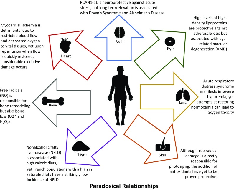
A paradox is a seemingly absurd or impossible concept, proposition, or theory that is often difficult to understand or explain, sometimes apparently self-contradictory, and yet ultimately correct or true. How is it possible, for example, that oxygen "a toxic environmental poison" could be also indispensable for life (Beckman and Ames Physiol Rev 78(2):547-81, 1998; Stadtman and Berlett Chem Res Toxicol 10(5):485-94, 1997)?: the so-called Oxygen Paradox (Davies and Ursini 1995; Davies Biochem Soc Symp 61:1-31, 1995). How can French people apparently disregard the rule that high dietary intakes of cholesterol and saturated fats (e.g., cheese and paté) will result in an early death from cardiovascular diseases (Renaud and de Lorgeril Lancet 339(8808):1523-6, 1992; Catalgol et al. Front Pharmacol 3:141, 2012; Eisenberg et al. Nat Med 22(12):1428-1438, 2016)?: the so-called, French Paradox. Doubtless, the truth is not a duality and epistemological bias probably generates apparently self-contradictory conclusions. Perhaps nowhere in biology are there so many apparently contradictory views, and even experimental results, affecting human physiology and pathology as in the fields of free radicals and oxidative stress, antioxidants, foods and drinks, and dietary recommendations; this is particularly true when issues such as disease-susceptibility or avoidance, "healthspan," "lifespan," and ageing are involved. Consider, for example, the apparently paradoxical observation that treatment with low doses of a substance that is toxic at high concentrations may actually induce transient adaptations that protect against a subsequent exposure to the same (or similar) toxin. This particular paradox is now mechanistically explained as "Adaptive Homeostasis" (Davies Mol Asp Med 49:1-7, 2016; Pomatto et al. 2017a; Lomeli et al. Clin Sci (Lond) 131(21):2573-2599, 2017; Pomatto and Davies 2017); the non-damaging process by which an apparent toxicant can activate biological signal transduction pathways to increase expression of protective genes, by mechanisms that are completely different from those by which the same agent induces toxicity at high concentrations. In this review, we explore the influences and effects of paradoxes such as the Oxygen Paradox and the French Paradox on the etiology, progression, and outcomes of many of the major human age-related diseases, as well as the basic biological phenomenon of ageing itself.
Overview
The claims and statistics of this paper haven’t been checked yet.
Run the check — free, no account needed. It reads the open-access text and checks the sources it cites.
Integrity signals
Registry
Cites
Statistics
Figures
Verification
Claims the paper makes
The claims and statistics of this paper haven’t been checked yet.
Run the check — free, no account needed. It reads the open-access text and checks the sources it cites.
Claims other papers make about this one
Each is checked against this paper’s own text: does it say what the citing paper says it does?
In the citing paper
“Under conditions of an imbalanced redox state, increased mitochondrial ROS production might lead to modifications of the arterial walls180, followed by an inflammatory environment181.”
From Mitochondrial and metabolic dysfunction in ageing and age-related diseases (2022) · cites this paper as [181]
Self-check
Percentages, sums and reported differences re-computed from the paper’s own numbers.
The self-check needs the paper’s full text.
Citations
Cites
1 retracted
Xiao, Y., Yan, W., Lu, L., Wang, Y., Lu, W., Cao, Y. L., & Cai, W. (2015) · Cell Cycle · doi:10.1080/15384101.2015.1026491 · Cited by 74
RetractedCancer incidence and mortality worldwide: Sources, methods and major patterns in GLOBOCAN 2012
Ferlay, J., Soerjomataram, I., Dikshit, R., Eser, S., Mathers, C., Rebelo, M., Parkin, D. M., Forman, D., & Bray, F. (2015) · International Journal of Cancer · doi:10.1002/ijc.29210 · Cited by 28,766
(2000) · New England Journal of Medicine · doi:10.1056/nejm200005043421801 · Cited by 12,941
The metabolic syndrome—a new worldwide definition
Alberti, K. G. M., Zimmet, P., & Shaw, J. (2005) · The Lancet · doi:10.1016/s0140-6736(05)67402-8 · Cited by 9,352 · cited 2×
“insulin resistance, and high blood pressure and blood triglycerides (Alberti et al. 2005).”
BackgroundGlutamate neurotoxicity and diseases of the nervous system
Choi, D. W. (1988) · Neuron · doi:10.1016/0896-6273(88)90162-6 · Cited by 8,188
Molecular Mechanisms of Stress-Responsive Changes in Collagen and Elastin Networks in Skin
Aziz, J., Shezali, H., Radzi, Z., Yahya, N. A., Abu Kassim, N. H., Czernuszka, J., & Rahman, M. T. (2016) · Skin Pharmacology and Physiology · doi:10.1159/000447017 · Cited by 7,323
The L-Arginine-Nitric Oxide Pathway
Epstein, F. H., Moncada, S., & Higgs, A. (1993) · New England Journal of Medicine · doi:10.1056/nejm199312303292706 · Cited by 6,678
Osteoclast differentiation and activation
Boyle, W. J., Simonet, W. S., & Lacey, D. L. (2003) · Nature · doi:10.1038/nature01658 · Cited by 6,670 · cited 1×
“If these two actions are not in balance, pathology ensues (Boyle et al. 2003; Nohl 1993; Wauquier et al. 2009; Vaananen et al. 2000).”
BackgroundHeart Disease and Stroke Statistics—2015 Update: A Report From the American Heart Association
Mozaffarian, D., Benjamin, E. J., Go, A. S., Arnett, D. K., Blaha, M. J., Cushman, M., de Ferranti, S., Després, J.-P., Fullerton, H. J., Howard, V. J., et al. (2015) · Circulation · doi:10.1161/cir.0000000000000152 · Cited by 5,797
Wong, W. L., Su, X., Li, X., Cheung, C. M. G., Klein, R., Cheng, C., & Wong, T. Y. (2014) · The Lancet Global Health · doi:10.1016/s2214-109x(13)70145-1 · Cited by 5,545
Research works printed but not linked: 4
- Garrod AB (1859) Treatise on nature of gout and rheumatic gout. Walton and Maberly, London
- Kurien BT, Mohan C, Scofield H (2012) Mechanisms of tissue damage-free radicals and fibrosis. In: Wallace DJ (ed) Dubois’ Lupus Erythematosus and Related Syndromes E-Book: Expert Consult-Online, 8th edn. Elsevier Health Sciences, London, p 175–189
- Rui Yin QC, Hamblin MR (2015) Skin aging and photoaging, in Skin photoaging. Morgan & Claypool, San Rafael, pp 1–4
- Yaar M, Gilchrest BA (2012) Aging of the skin, in Fitspatrick’s dermatology in general medicine. McGraw Hill, New York, pp 1213–1211
Cited by
Sorted by citations
Mitochondrial and metabolic dysfunction in ageing and age-related diseases
Amorim, J. A., Coppotelli, G., Rolo, A. P., Palmeira, C. M., Ross, J. M., & Sinclair, D. A. (2022) · Nature Reviews Endocrinology · doi:10.1038/s41574-021-00626-7 · Cited by 990
Charlton, N. C., Mastyugin, M., Török, B., & Török, M. (2023) · Molecules · doi:10.3390/molecules28031057 · Cited by 292
“The so-called “French Paradox” especially increased interest in dietary small molecule antioxidants after it was noted that despite high saturated fat and alcohol intake, Mediterranean populations are less likely to develop coronary heart disease [42].”
BackgroundOxInflammation: From Subclinical Condition to Pathological Biomarker
Valacchi, G., Virgili, F., Cervellati, C., & Pecorelli, A. (2018) · Frontiers in Physiology · doi:10.3389/fphys.2018.00858 · Cited by 212
“Possible leaks from this outpost can be then countered by further repair systems such as proteinases, lipases, DNA repair enzymes (Davies et al., 2017).”
BackgroundMelatonin: Clinical Perspectives in Neurodegeneration
Cardinali, D. P. (2019) · Frontiers in Endocrinology · doi:10.3389/fendo.2019.00480 · Cited by 210
“Although the regular intake of antioxidants has been proposed for prevention of neurodegeneration, its effectiveness, however, has been questioned (3).”
BackgroundMarcucci, G., Domazetovic, V., Nediani, C., Ruzzolini, J., Favre, C., & Brandi, M. L. (2023) · Antioxidants · doi:10.3390/antiox12020373 · Cited by 200
“It has also been demonstrated that ROS stimulates osteoclastogenesis through RANKL as reported in human stromal cells [26,81].”
BackgroundNeuroprotective Activities of Curcumin in Parkinson’s Disease: A Review of the Literature
Nebrisi, E. E. (2021) · International Journal of Molecular Sciences · doi:10.3390/ijms222011248 · Cited by 165
“Although oxygen is very fundamental for any living system, it is inherently harmful at the same time, a phenomenon known as “the oxygen paradox” [100,101].”
BackgroundInflammation in Neurological Disorders: The Thin Boundary Between Brain and Periphery
Cervellati, C., Trentini, A., Pecorelli, A., & Valacchi, G. (2020) · Antioxidants and Redox Signaling · doi:10.1089/ars.2020.8076 · Cited by 140
The Implication of Reactive Oxygen Species and Antioxidants in Knee Osteoarthritis
Tudorachi, N. B., Totu, E. E., Fifere, A., Ardeleanu, V., Mocanu, V., Mircea, C., Işıldak, İ., Smilkov, K., & Cărăușu, E. M. (2021) · Antioxidants · doi:10.3390/antiox10060985 · Cited by 137
“ROS could affect the musculoskeletal system, including multiple cellular structures at the bone level [64].”
BackgroundOxidative stress and antioxidants in the pathophysiology of malignant melanoma
Obrador, E., Liu-Smith, F., Dellinger, R. W., Salvador, R., Meyskens, F. L., & Estrela, J. M. (2019) · Biological Chemistry · doi:10.1515/hsz-2018-0327 · Cited by 119
“Of course, this balance depends on concentration as too high a concentration of the reactive species can be toxic (Davies et al., 2017).”
BackgroundAnti-aging properties of Dendrobium nobile Lindl.: From molecular mechanisms to potential treatments
Nie, X., Chen, Y., Li, W., & Lu, Y. (2020) · Journal of Ethnopharmacology · doi:10.1016/j.jep.2020.112839 · Cited by 112
Hussain, M. S., Gupta, G., Mishra, R., Patel, N. K., Gupta, S. K., Alzarea, S. I., Kazmi, I. A., Kumbhar, P., Disouza, J., Dureja, H., et al. (2024) · Pathology - Research and Practice · doi:10.1016/j.prp.2024.155157 · Cited by 120
Exercise, redox system and neurodegenerative diseases
Quan, H., Koltai, E., Suzuki, K., Aguiar, A. S., Pinho, R. A., Boldogh, I. R., Berkes, I., & Radak, Z. O. (2020) · Biochimica et Biophysica Acta (BBA) - Molecular Basis of Disease · doi:10.1016/j.bbadis.2020.165778 · Cited by 89
Lawrence, G. D. (2021) · Advances in Nutrition · doi:10.1093/advances/nmab013 · Cited by 88
“The wide array of ROS and lipid peroxidation products are implicated in oxidative stress, aging, and many diseases (29, 30).”
BackgroundOxidative stress as a possible pathogenic cofactor of post-menopausal osteoporosis
Bonaccorsi, G., Piva, I., Greco, P., & Cervellati, C. (2018) · The Indian Journal of Medical Research · doi:10.4103/ijmr.ijmr_524_18 · Cited by 81
Implications of Oxidative Stress and Cellular Senescence in Age-Related Thymus Involution
Barbouti, A., Vasileiou, P. V. S., Evangelou, K., Vlasis, K. G., Papoudou‐Bai, A., Gorgoulis, V. G., & Kanavaros, P. (2020) · Oxidative Medicine and Cellular Longevity · doi:10.1155/2020/7986071 · Cited by 86
“Aerobic organisms use additionally lipid- and water-soluble antioxidant “sacrificial” compounds such as vitamins C and E, carotenoids, and glutathione (GSH) that are preferentially oxidized to preserve more important biomolecules, as well as defense mechanisms that remove or repair oxidized cellular components [64].”
BackgroundInfluentialRanking Biomarkers of Aging by Citation Profiling and Effort Scoring
Hartmann, A., Hartmann, C., Secci, R., Hermann, A., Fuellen, G., & Walter, M. (2021) · Frontiers in Genetics · doi:10.3389/fgene.2021.686320 · Cited by 86
“Such hallmarks of aging as discussed here were observed in various organs and tissues (Stadtman and Berlett, 1997; Baraibar and Friguet, 2013; Davies et al., 2017).”
BackgroundHyperbaric oxygen therapy for healthy aging: From mechanisms to therapeutics
Fu, Q., Duan, R., Sun, Y., & Li, Q. (2022) · Redox Biology · doi:10.1016/j.redox.2022.102352 · Cited by 75
Melatonin as a Chronobiotic and Cytoprotective Agent in Parkinson’s Disease
Perez‐Lloret, S., & Cardinali, D. P. (2021) · Frontiers in Pharmacology · doi:10.3389/fphar.2021.650597 · Cited by 66
“Hence, the effectiveness of administering antioxidants for neurodegeneration prevention has been proposed but albeit questioned (Davies et al., 2017).”
BackgroundGeriatric Medicine and Gerontology
Zawada, E. T. (2019) · IntechOpen eBooks · doi:10.5772/intechopen.77654 · Cited by 64
Tian, W., Yang, L., Liu, Y., He, J., Yang, L., Zhang, Q., Liu, F., Li, J., Liu, J., Sumi, S., et al. (2019) · The Journal of Nutritional Biochemistry · doi:10.1016/j.jnutbio.2019.01.018 · Cited by 54
Resveratrol Action on Lipid Metabolism in Cancer
Kisková, T., & Kassayová, M. (2019) · International Journal of Molecular Sciences · doi:10.3390/ijms20112704 · Cited by 52
Lundquist, A. J., Parizher, J., Petzinger, G. M., & Jakowec, M. W. (2019) · Journal of Neuroscience Research · doi:10.1002/jnr.24430 · Cited by 50
“In the context of normal brain function, exercise can act as a mechanism for brain maintenance, allowing motor and cognitive learning to occur (Davies et al., 2017).”
BackgroundSelf-citationMolecular Targets in Alzheimer’s Disease
Bjørklund, G., Aaseth, J., Dadar, M., & Chirumbolo, S. (2019) · Molecular Neurobiology · doi:10.1007/s12035-019-1563-9 · Cited by 48
The role of NOX4 in pulmonary diseases
Li, Z., Xu, S., Feng, Y., Cheng, Y., Xiong, J., Zhou, Y., & Guan, C. (2020) · Journal of Cellular Physiology · doi:10.1002/jcp.30005 · Cited by 43
Self-citationEvaluating the link between Paraoxonase-1 levels and Alzheimer's disease development
Cervellati, C., Valacchi, G., Tisato, V., Zuliani, G., & Marsillach, J. (2019) · Minerva Medica · doi:10.23736/s0026-4806.18.05875-5 · Cited by 40
Salvagno, M., Coppalini, G., Taccone, F. S., Strapazzon, G., Mrakic‐Sposta, S., Rocco, M., Khalifé, M., & Balestra, C. (2022) · International Journal of Molecular Sciences · doi:10.3390/ijms24010082 · Cited by 43
“New evidence about oncologic progression has shown that cancer cells may evolve into a hypermetabolic state, which can be sustained even in the presence of a limited oxygen supply [76,77].”
BackgroundSystemic and skin-targeting beneficial effects of lycopene-enriched ice cream: A pilot study
Chernyshova, M. P., Pristensky, D. V., Lozbiakova, M. V., Chalyk, N. E., Bandaletova, T. Y., & Petyaev, I. M. (2018) · Journal of Dairy Science · doi:10.3168/jds.2018-15282 · Cited by 39
“Lycopene concentration in serum samples was measured using HPLC (Diwadkar-Navsariwala et al., 2003) with modifications.”
MethodNègre‐Salvayre, A., & Salvayre, R. (2022) · Antioxidants · doi:10.3390/antiox11112281 · Cited by 39
Nutritional Barriers to the Adherence to the Mediterranean Diet in Non-Mediterranean Populations
Sam-Yellowe, T. Y. (2024) · Foods · doi:10.3390/foods13111750 · Cited by 34
“The relationship between eating patterns, in particular the association of dietary fats and the risk of chronic non-infectious diseases was established by these major studies [7,8].”
BackgroundMethodBorbély, É., Payrits, M., Hunyady, Á., Mező, G., & Pintér, E. (2019) · GeroScience · doi:10.1007/s11357-019-00083-1 · Cited by 31
4-Hydroxynonenal Contributes to Fibroblast Senescence in Skin Photoaging Evoked by UV-A Radiation
Swiader, A., Camaré, C., Guerby, P., Salvayre, R., & Nègre‐Salvayre, A. (2021) · Antioxidants · doi:10.3390/antiox10030365 · Cited by 32
Are melatonin doses employed clinically adequate for melatonin-induced cytoprotection?
Cardinali, D. P. (2019) · Melatonin Research · doi:10.32794/mr11250025 · Cited by 29
Glycerylphytate compounds with tunable ion affinity and osteogenic properties
Mora‐Boza, A., López-Donaire, M. L., Saldaña, L., Vilaboa, N. E., Vázquez‐Lasa, B., & Román, J. S. (2019) · Scientific Reports · doi:10.1038/s41598-019-48015-5 · Cited by 28
Galaup, C., Picard, C., Couderc, F., Gilard, V., & Collin, F. (2021) · FEBS Journal · doi:10.1111/febs.15859 · Cited by 28
Melanoma in the liver: Oxidative stress and the mechanisms of metastatic cell survival
Obrador, E., Salvador, R., López‐Blanch, R., Jihad-Jebbar, A., Alcácer, J., Benlloch, M., Pellicer, J. A., & Estrela, J. M. (2020) · Seminars in Cancer Biology · doi:10.1016/j.semcancer.2020.05.001 · Cited by 27
Geltinger, F., Tevini, J., Briza, P., Geiser, A., Bischof, J., Richter, K., Felder, T., & Rinnerthaler, M. (2020) · GeroScience · doi:10.1007/s11357-019-00103-0 · Cited by 26
Nirmal, N. P., Mahale, K. R., Rathod, N. B., Siddiqui, S. A., & Dhar, B. K. (2024) · Process Safety and Environmental Protection · doi:10.1016/j.psep.2024.11.106 · Cited by 26
Oxidative Stress in Urolithiasis
Boonla, C. (2018) · InTech eBooks · doi:10.5772/intechopen.75366 · Cited by 24
Fratantonio, D., Speciale, A., Molonia, M. S., Bashllari, R., Palumbo, M. A., Saija, A., Cimino, F., Monastra, G., & Virgili, F. (2018) · Archives of Biochemistry and Biophysics · doi:10.1016/j.abb.2018.08.003 · Cited by 26
Szegeczki, V., Bauer, B., Jüngling, A., Fulop, B. D., Vágó, J., Perényi, H., Tarantini, S., Tamás, A., Zákány, R., Reglődi, D., et al. (2019) · GeroScience · doi:10.1007/s11357-019-00097-9 · Cited by 22
Therapeutic potential of melatonin and its derivatives in aging and neurodegenerative diseases
Verma, A. K., Singh, S., & Rizvi, S. I. (2022) · Biogerontology · doi:10.1007/s10522-022-10006-x · Cited by 23
Cardiovascular Adaptive Homeostasis in Exercise
Davies, K. J. A. (2018) · Frontiers in Physiology · doi:10.3389/fphys.2018.00369 · Cited by 24
“…including yeast, various mammalian/human cells, Caenorhabditis elegans nematode worms, Drosophila melanogaster flies, mice, rats, and human beings (Pickering et al., 2012; Zhang et al., 2015; Davies et al., 2017; Pomatto and Davies, 2017; Pomatto et al., 2017c, 2018a,b; Raynes et al., 2017).”
BackgroundSelf-citationBatinac, T., Batičić, L., Kršek, A., Knežević, D., Marcucci, E., Tokmadžić, V. S., & Ćurko‐Cofek, B. (2024) · Journal of Cardiovascular Development and Disease · doi:10.3390/jcdd11120408 · Cited by 21
“The difference in the effects and outcomes obtained after HBOT is explained by the concept of hormesis and is dose-dependent [101], suggesting that treatment with subtoxic doses of a toxicant could lead to adaptations to prevent damage [14,101,102].”
Villarreal-Leal, R. A., Cooke, J. P., & Corradetti, B. (2021) · Nanomedicine Nanotechnology Biology and Medicine · doi:10.1016/j.nano.2021.102385 · Cited by 21
α-Arbutin ameliorates UVA-induced photoaging through regulation of the SIRT3/PGC-1α pathway
Lu, F. Y., Zhou, Q., Liang, M., Liang, H., Yu, Y., Yang, L., Zhang, Y., Lu, L., Zheng, Y., Hao, J., et al. (2024) · Frontiers in Pharmacology · doi:10.3389/fphar.2024.1413530 · Cited by 17
Soussi, D., Danion, J., Baulier, E., Favreau, F. D., Sauvageon, Y., Bossard, V., Matillon, X., Turpin, F., Belgsir, E. M., Thuillier, R., et al. (2019) · International Journal of Molecular Sciences · doi:10.3390/ijms20092268 · Cited by 16
“Resveratrol is a polyphenol found in numerous plants, among which things such as the well-documented health benefits of grapes have been linked to the French Paradox [15].”
BackgroundNew role for an old molecule: The 2,3-diphosphoglycerate case
Tellone, E., Barreca, D., Russo, A., Galtieri, A., & Ficarra, S. (2019) · Biochimica et Biophysica Acta (BBA) - General Subjects · doi:10.1016/j.bbagen.2019.07.002 · Cited by 9
Homeostasis, Homeodynamics and Aging
Rattan, S. I. S. (2019) · Elsevier eBooks · doi:10.1016/b978-0-12-801238-3.11425-4 · Cited by 9
Qiu, C., Zhang, Z., Wang, H., Liu, N., Li, R., Wei, Z., Wang, B., & Zhang, N. (2025) · Scientific Reports · doi:10.1038/s41598-025-11667-7 · Cited by 8
“…annotation and Kyoto Encyclopedia of Genes and Genomes (KEGG) enrichment of the candidate genes, we utilized R packages “clusterProfiler”(4.2.2; h t t p s : / / b i o c o n d u c t o r . o r g / p a c k a g e s / r e l e a s e / b i o c / h t m l / c l u s t e r P r o fi l e r . h t m l) 17 .”
InfluentialKotur‐Stevuljević, J., Savić, J. S., Simić, M. R., & Ivanišević, J. (2023) · Arhiv za farmaciju · doi:10.5937/arhfarm73-45369 · Cited by 8
“These observations underscore the importance of an antioxidant-rich diet as a potential strategy to reduce the risk of cardiovascular disease (22).”
BackgroundPinker, S. A., & Ullman, M. T. (2002) · Cited by 8
Kashirina, D. N., Pastushkova, L. K., Goncharova, A. G., & Larina, I. M. (2024) · Frontiers in Physiology · doi:10.3389/fphys.2024.1375929 · Cited by 9
Podlutsky, A. J. (2019) · GeroScience · doi:10.1007/s11357-019-00060-8 · Cited by 5
Popova, O. V., Kashirina, D. N., Pastushkova, L. K., Гончарова, А. Г., & Larina, I. M. (2024) · Bulletin of Experimental Biology and Medicine · doi:10.1007/s10517-024-06197-7 · Cited by 3
Polygenic risk score for disability and insights into disability-related molecular mechanisms
Kulminski, A. M., Kang, C., Kolpakov, S. A., Loika, Y., Nazarian, A., Yashin, A. I., Stallard, E., & Culminskaya, I. V. (2019) · GeroScience · doi:10.1007/s11357-019-00125-8 · Cited by 3
“Oxidative stress is associated with aging and various pathological conditions including cardiovascular and neurodegenerative diseases, macular degeneration, autoimmune disorders, and cancer (Davies et al. 2017; Liguori et al. 2018; Martin et al. 1996; Pham-Huy et al. 2008; Xiong et al. 2011).”
BackgroundOlive polyphenols and chronic alcohol protection
Petrella, C., Ferraguti, G., Tarani, L., Chaldakov, G. N., Ceccanti, M., Greco, A., Ralli, M., & Fiore, M. (2021) · Elsevier eBooks · doi:10.1016/b978-0-12-819528-4.00003-1 · Cited by 3
Risk factors and health status among older adults in Europe: a socioeconomic analysis
Tur‐Sinai, A., & Becker, N. (2021) · Journal of Biosocial Science · doi:10.1017/s0021932021000432 · Cited by 3
Melatonin in Aging and Aging-Related Disorders
Süzen, S. (2023) · Springer eBooks · doi:10.1007/978-981-19-7443-4_9 · Cited by 3
Modeling oxidative stress on neuronal dendrites and dendritic spines
Katsenos, A. P., Kotoula, E., Sereti, S., Zoidi, D., Tzavellas, N. P., Polyzou, A., Leondaritis, G., Kanellos, F. S., Bellou, S., Bellos, S., et al. (2025) · Neuroscience · doi:10.1016/j.neuroscience.2025.11.037 · Cited by 2
Trust Mukudzei Pfukwa, Motlhalamme, T. Y., Setati, M. E., Fawole, O. A., Manley, M., Heerden, C. J. V., & Mapiye, C. (2023) · Food Bioscience · doi:10.1016/j.fbio.2023.103464 · Cited by 2
Khalifé, M., Salvagno, M., Sosnowski, M., & Balestra, C. (2024) · Journal of Anesthesia Analgesia and Critical Care · doi:10.1186/s44158-024-00179-x · Cited by 2
Bhattacharya, A., Pulliam, D. A., Liu, Y., & Salmon, A. B. (2020) · Aging Pathobiology and Therapeutics · doi:10.31491/apt.2020.03.012 · Cited by 2
“On the other hand, aging is a biological process, and there is clear interest in understanding how age-related changes in health (and not just lifespan) might be regulated by processes such as mitochondrial oxidative stress [28, 29].”
BackgroundLi, S., Ma, X., Wei, J., Cheng, L., Chen, H., Yan, J., Chen, M., Zhang, Q. R., Tang, J., Zhang, Y., et al. (2025) · Food Chemistry · doi:10.1016/j.foodchem.2025.146907 · Cited by 1
Circadian rhythms, NRF2 signalling and redox homeostasis: A holy grail for a healthy life course?
Sutton, E., & Pekovic‐Vaughan, V. (2026) · Free Radical Biology and Medicine · doi:10.1016/j.freeradbiomed.2026.05.318 · Cited by 2
Dominguez, L. J., & Barbagallo, M. (2019) · Elsevier eBooks · doi:10.1016/b978-0-12-801238-3.11370-4 · Cited by 2
Unravelling the Photoprotection Capacity of Resveratrol on Histidine Oxidation
Recky, J. R. N., Dántola, M. L., & Lorente, C. (2021) · Photochem · doi:10.3390/photochem1020012 · Cited by 1
Role of oxidative stress in the pathogenesis of postmenopausal osteoporosis (literature review)
Bulgakova, S. V., & Melikova, A. V. (2020) · Terapevt (General Physician) · doi:10.33920/med-12-2010-03 · Cited by 1
Kinetic study of peroxidase enzyme in the presence of purine alkaloids
darvazi, T. R., & Sariri, R. (2022) · Nova Biologica Reperta · doi:10.52547/nbr.8.4.279 · Cited by 1
Microorganisms as a Potential Source of Antioxidants
Hernández‐Almanza, A., Ramírez‐Guzmán, N., Martínez-Medina, G. A., Loredo‐Treviño, A., Verma, D. K., & Aguilar, C. N. (2021) · doi:10.1002/9781119717317.ch11 · Cited by 1
Always enough but never too much: the how and why of downregulating tissue oxygenation
Olson, K. R. (2023) · American Journal of Physiology-Heart and Circulatory Physiology · doi:10.1152/ajpheart.00449.2023 · Cited by 1
Primary Prevention of Alzheimer’s Disease (AD)
Bergamini, E., & Cavallini, G. (2019) · IntechOpen eBooks · doi:10.5772/intechopen.85418
FUNCIÓN PROTECTORA DE LA MELATONINA DURANTE EL EJERCICIO FÍSICO
GARCÍA, J. J. G., MARÍN, A. F., CAPÓN, E. M. M., Alcalde, I. G., LAPIEDRA, M. P. A., Zubero, E. E., & Pingarrón, L. L. (2021) · Dykinson eBooks · doi:10.2307/j.ctv2gz3t0r.22
The Role of Oxidative Stress in the Pathogenesis of Eye Diseases
Bissell, M., Fetian, I. R., & Mozaffarieh, M. (2022) · doi:10.36000/hbt.2022.06.002
Cortés-Palma, F., Tehozol-Limón, D., & Juárez, R. G. (2023) · Casos y Revisiones de Salud · doi:10.22201/fesz.26831422e.2023.5.2.6
Identification of Potential Therapeutic Drugs for Diabetic Cardiomyopathy
You, Z., Wang, Y., & Huang, L. (2024) · Journal of Biomedical Nanotechnology · doi:10.1166/jbn.2024.3827
Lv, X., Ji, Q., Liu, Q., Zhao, Q., Xu, Y., Meng, Y., Zhou, L., Zhao, H., Wang, N., & Zhan, Y. (2025) · eFood · doi:10.1002/efd2.70062
Multi-species genomic study of oxidative stress response and adaptation
Thomès, L. (2020) · theses.fr (ABES) · doi:10.70675/9321ae18zf7e1z454azaabdzd4f01a10941b
Ma, W., Jiang, M., Zhang, J., Liu, W., Dan, Y., Sun, W., Ma, L., Leihong, X., & Ren, J. (2026) · Journal of Photochemistry and Photobiology B Biology · doi:10.1016/j.jphotobiol.2026.113451 · Cited by 1
Hu, J., Huang, S., Yang, H., Gao, F., Liu, Q., & Jiang, M. (2026) · Molecular Neurobiology · doi:10.1007/s12035-026-06154-7
Bagyinszky, E., & An, S. S. A. (2026) · Current Issues in Molecular Biology · doi:10.3390/cimb48101081
Metadata
- Authors
- Davies, J. M. S., Cillard, J., Friguet, B., Cadenas, E., Cadet, J., Cayce, R., Fishmann, A., Liao, D., Bulteau, A.-L., Derbré, F., Rébillard, A., Burstein, S., Hirsch, E., Kloner, R. A., Jakowec, M., Petzinger, G., Sauce, D., Sennlaub, F., Limon, I., Ursini, F., Maiorino, M., Economides, C., Pike, C. J., Cohen, P., Salvayre, A. N., Halliday, M. R., Lundquist, A. J., Jakowec, N. A., Mechta-Grigoriou, F., Mericskay, M., Mariani, J., Li, Z., Huang, D., Grant, E., Forman, H. J., Finch, C. E., Sun, P. Y., Pomatto, L. C. D., Agbulut, O., Warburton, D., Neri, C., Rouis, M., Cillard, P., Capeau, J., Rosenbaum, J., & Davies, K. J. A.
- Affiliations
- University of Southern California (US)
- Université de Rennes (FR)
- Centre National de la Recherche Scientifique (FR)
- Université de Sherbrooke (CA)
- Good Samaritan Hospital (US)
- École Normale Supérieure de Lyon (FR)
- Université Rennes 2 (FR)
- Huntington Medical Research Institutes (US)
- Inserm (FR)
- Institut de la Vision (FR)
- University of Padua (IT)
- Université Paris-Sud (FR)
- Corresponding author
- Davies, Kelvin J. A.
- ORCID
- Friguet, Bertrand
- Cadenas, Enrique
- Cadet, Jean
- Bulteau, Anne-Laure
- Rébillard, Amélie
- Kloner, Robert A.
- Jakowec, Michael
- Petzinger, Giselle
- Sauce, Delphine
- Sennlaub, Florian
- Limon, Isabelle
- Ursini, Fulvio
- Maiorino, Matilde
- Pike, Christian J.
- Cohen, Pinchas
- Halliday, Matthew R.
- Lundquist, Adam J.
- Jakowec, Nicolaus A.
- Mericskay, Mathias
- Mariani, Jean
- Li, Zhenlin
- Grant, Ellsworth
- Forman, Henry J.
- Finch, Caleb E.
- Sun, Patrick Y.
- Pomatto, Laura C. D.
- Agbulut, Onnik
- Warburton, David
- Rouis, Mustapha
- Capeau, Jacqueline
- Rosenbaum, Jean
- Davies, Kelvin J. A.
- Venue
- GeroScience
- Published
- 2017-12
- Volume
- 39
- Issue
- 5-6
- Pages
- 499-550
- ISSN
- 2152-4041 (ISSN-L)
- 2509-2715
- 2509-2723
- PMC
- PMC5745211
- OpenAlex
- W2779963304
- Cited by
- 82
- References
- 445
- Citations by year
- 2026: 4 · 2025: 4 · 2024: 9 · 2023: 8 · 2022: 7 · 2021: 13 · 2020: 10 · 2019: 19 · 2018: 7
- FWCI
- 1.13
- Type
- journal article
- Language
- en
- Open access
- hybrid
- doi.org
- Licence
- Locations
- GeroScience (publishedVersion, cc-by)
- PubMed Central (publishedVersion, cc-by)
- PubMed (publishedVersion)
- HAL (submittedVersion, cc-by)
- eScholarship (California Digital Library) (submittedVersion)
- eScholarship (California Digital Library) (submittedVersion, cc-by)
- Padua Research Archive (University of Padova) (submittedVersion, cc-by)
- Funders
- National Science Foundation (#DGE-1418060)
- National Institute of Environmental Health Sciences (#ES023864, #R01ES020812, #D43ES022862, #ES003598)
- Institut National de la Santé et de la Recherche Médicale
- National Institute on Aging (# AG-040753, # AG058068, # P01AG034906, # R01AG034430, # AG052374)
- Agence Nationale de la Recherche
- Université Pierre et Marie Curie
- Université Toulouse III - Paul Sabatier
- Conseil Régional de Bretagne
- Ligue Contre le Cancer
- Fondation de France
- Centre National de la Recherche Scientifique
- Assistance Publique - Hôpitaux de Paris
- U.S. Department of Defense (DOD-PC160353U)
- American Foundation for Aging Research
- Don Roberto Gonzalez and Family Foundation
- National Parkinson Foundation
- National Heart, Lung, and Blood Institute (#U01HL122681)
- Pasadena Guild
- Children's Hospital Los Angeles
- the webb foundation
- The Garland Foundation
- Topics
- Health Sciences › Medicine › Biochemistry (medical) › Antioxidant Activity and Oxidative Stress
- Health Sciences › Medicine › Biochemistry (medical) › Phytochemicals and Antioxidant Activities
- Health Sciences › Medicine › Public Health, Environmental and Occupational Health › Nutritional Studies and Diet
- Keywords
- oxygen paradox, French paradox, free radicals, oxidative stress, antioxidants, cardiovascular disease, age-related diseases, healthspan, lifespan, aging, adaptive homeostasis, hormesis, epigenetics, epigenetic reprogramming, epigenetic drift, epigenetic plasticity, epigenetic memory, epigenetic inheritance, epigenetic noise
- MeSH terms
- Diet, High-Protein, Adaptation, Physiological, Aged, Aged, 80 and over, Aging, Female, France, Free Radicals, Humans, Hypercholesterolemia, Male, Middle Aged, Oxygen, Geriatric Assessment, Oxidative Stress, Risk Assessment
About these checks
Registry, retraction, Cites and statistics results are computed, not judged, with one exception: a printed reference that no index lists is read once by a model before it counts as a citation error. Claims are extracted by a model and kept only when their quote appears verbatim in the text; each cited source is then checked, with a model’s help, for whether it says what the claim relies on. Those verdicts can be wrong — read the claim’s quote and the cited source before relying on one.
Statistics and arithmetic are recomputed, not judged.
Confidence bands are shown only for fields calibrated on our annotated test set. None is calibrated yet, so no band is shown.
What counts as a claim ›How we measure confidence ›How we check this ›
Are you an author, or do you see a problem with this page? Request a review
