

The DOI matches its Crossref record; 146 of the 146 references it cites were found in a registry or an index.
Role of Chemokines in the Development and Progression of Alzheimer’s Disease
Open accessAbstract
Alzheimer’s disease (AD) is a progressive neurogenerative disorder manifested by gradual memory loss and cognitive decline due to profound damage of cholinergic neurons. The neuropathological hallmarks of AD are intracellular deposits of neurofibrillary tangles (NFTs) and extracellular aggregates of amyloid β (Aβ). Mounting evidence indicates that intensified neuroinflammatory processes play a pivotal role in the pathogenesis of AD. Chemokines serve as signaling molecules in immune cells but also in nerve cells. Under normal conditions, neuroinflammation plays a neuroprotective role against various harmful factors. However, overexpression of chemokines initiates disruption of the integrity of the blood–brain barrier, facilitating immune cells infiltration into the brain. Then activated adjacent glial cells–astrocytes and microglia, release massive amounts of chemokines. Prolonged inflammation loses its protective role and drives an increase in Aβ production and aggregation, impairment of its clearance, or enhancement of tau hyperphosphorylation, contributing to neuronal loss and exacerbation of AD. Moreover, chemokines can be further released in response to growing deposits of toxic forms of Aβ. On the other hand, chemokines seem to exert multidimensional effects on brain functioning, including regulation of neurogenesis and synaptic plasticity in regions responsible for memory and cognitive abilities. Therefore, underexpression or complete genetic ablation of some chemokines can worsen the course of AD. This review covers the current state of knowledge on the role of particular chemokines and their receptors in the development and progression of AD. Special emphasis is given to their impact on forming Aβ and NFTs in humans and in transgenic murine models of AD.
Overview
The claims and statistics of this paper haven’t been checked yet.
Run the check — free, no account needed. It reads the open-access text and checks the sources it cites.
Integrity signals
Registry
Cites
Statistics
Figures
Verification
Claims the paper makes
The claims and statistics of this paper haven’t been checked yet.
Run the check — free, no account needed. It reads the open-access text and checks the sources it cites.
Claims other papers make about this one
Each is checked against this paper’s own text: does it say what the citing paper says it does?
The cited source says what the claim attributes to it.
In the citing paper
“consistent with the known capacity of CCL2 to break down the blood brain barrier and recruit peripheral immune cells to sites of damage.58–62”
From TET2-mutant myeloid cells mitigate Alzheimer’s disease progression via CNS infiltration and enhanced phagocytosis in mice (2025) · cites this paper as [60]
Self-check
Percentages, sums and reported differences re-computed from the paper’s own numbers.
The self-check needs the paper’s full text.
Citations
Cites
McKhann, G. M., Knopman, D. S., Chertkow, H., Hyman, B. T., Jack, C. R., Kawas, C. H., Klunk, W. E., Koroshetz, W. J., Manly, J. J., Mayeux, R., et al. (2011) · Alzheimer's & Dementia · doi:10.1016/j.jalz.2011.03.005 · Cited by 18,657 · cited 1×
“As the disease progresses, preclinical AD transits to mild cognitive impairment (MCI) in which cognition deficits do not affect patients’ performance at work or regular daily activities, but eventually may develop into AD (McKhann et al. 2011; Saido 2013).”
BackgroundA new class of membrane-bound chemokine with a CX3C motif
Bazan, J. F., Bacon, K. B., Hardiman, G., Wang, W., Soo, K., Rossi, D., Greaves, D. R., Zlotnik, A., & Schall, T. J. (1997) · Nature · doi:10.1038/385640a0 · Cited by 2,013
The cholinergic system in the pathophysiology and treatment of Alzheimer’s disease
Hampel, H., Mesulam, M.-M., Cuello, A. C., Farlow, M. R., Giacobini, E., Grossberg, G. T., Khachaturian, A. S., Vergallo, A., Cavedo, E., Snyder, P. J., et al. (2018) · Brain · doi:10.1093/brain/awy132 · Cited by 1,918
The ageing systemic milieu negatively regulates neurogenesis and cognitive function
Villeda, S. A., Luo, J., Mosher, K. I., Zou, B., Britschgi, M., Bieri, G., Stan, T. M., Fainberg, N., Ding, Z., Eggel, A., et al. (2011) · Nature · doi:10.1038/nature10357 · Cited by 1,763
A guide to chemokines and their receptors
Hughes, C. E., & Nibbs, R. J. B. (2018) · FEBS Journal · doi:10.1111/febs.14466 · Cited by 1,510 · cited 2×
“To date, six CXC receptors have been identified, named from CXCR1 to CXCR6, and eleven CC receptors, from CCR1 to CCR11, along with a single receptor for CX3CL1 and one for lymphotactin α/β, called CX3CR1 and XCR1, respectively (Hughes and Nibbs 2018).”
BackgroundZhao, Q.-F., Tan, L., Wang, H.-F., Jiang, T., Tan, M.-S., Tan, L., Xu, W., Li, J.-Q., Wang, J., Lai, T.-J., et al. (2016) · Journal of Affective Disorders · doi:10.1016/j.jad.2015.09.069 · Cited by 916
El Khoury, J., Toft, M., Hickman, S. E., Means, T. K., Terada, K., Geula, C., & Luster, A. D. (2007) · Nature Medicine · doi:10.1038/nm1555 · Cited by 915
Risk factors for Alzheimer’s disease
Armstrong, R. A. (2019) · Folia Neuropathologica · doi:10.5114/fn.2019.85929 · Cited by 679
Hippocampus and its involvement in Alzheimer’s disease: a review
Rao, Y. L., Ganaraja, B., Murlimanju, B. V., Joy, T., Krıshnamurthy, A., & Agrawal, A. (2022) · 3 Biotech · doi:10.1007/s13205-022-03123-4 · Cited by 646
Neurotactin, a membrane-anchored chemokine upregulated in brain inflammation
Pan, Y., Lloyd, C. M., Zhou, H., Dolich, S., Deeds, J., Gonzalo, J.-Á., Vath, J., Gosselin, M., Ma, J., Dussault, B. J., et al. (1997) · Nature · doi:10.1038/42491 · Cited by 643 · cited 1×
“This chemokine exists in both soluble and membrane-bound forms (Bajetto et al. 2002; Bazan et al. 1997; Pan et al. 1997).”
Background
Research works printed but not linked: 1
- Gutiérrez SE, Allison EK, Coleman U, Kingery-Gallagher ND, El Khoury J (2019b) Heterozygous CX3CR1 deficiency in microglia restores neuronal β-amyloid clearance pathways and slows progression of Alzheimer’s like-disease in PS1-APP mice. Front Immunol 10:2780. https://doi.org/10.3389/fimmu.2019.02780 · DOI · Paper page
Cited by
Sorted by citations
Jorfi, M., Park, J., Hall, C. K., Lin, C.-C. J., Chen, M., von Maydell, D., Kruskop, J. M., Kang, B., Choi, Y., Prokopenko, D., et al. (2023) · Nature Neuroscience · doi:10.1038/s41593-023-01415-3 · Cited by 214
Molecular Mechanisms of Neuroinflammation in Aging and Alzheimer’s Disease Progression
Andronie-Cioară, F. L., Ardelean, A. I., Nistor-Cseppento, C. D., Jurcău, A., Jurcău, M. C., Pașcalău, N., & Marcu, F. M. (2023) · International Journal of Molecular Sciences · doi:10.3390/ijms24031869 · Cited by 170
Obesity-induced inflammation: connecting the periphery to the brain
Le Thuc, O., & García-Cáceres, C. (2024) · Nature Metabolism · doi:10.1038/s42255-024-01079-8 · Cited by 125
Kamila, P., Kar, K., Chowdhury, S., Chakraborty, P., Dutta, R., S, S., Singh S, A., & Prajapati, B. G. (2025) · IBRO Neuroscience Reports · doi:10.1016/j.ibneur.2025.05.005 · Cited by 90
Zeng, X., Lafferty, T. K., Sehrawat, A., Chen, Y., Ferreira, P. C. L., Bellaver, B., Povala, G., Kamboh, M. I., Klunk, W. E., Cohen, A. D., et al. (2024) · Molecular Neurodegeneration · doi:10.1186/s13024-024-00753-5 · Cited by 91
Adaptive immune changes associate with clinical progression of Alzheimer’s disease
van Olst, L., Kamermans, A., Halters, S., van der Pol, S. M. A., Rodríguez, E., Verberk, I. M. W., Verberk, S. G. S., Wessels, D. W. R., Rodriguez-Mogeda, C., Verhoeff, J., et al. (2024) · Molecular Neurodegeneration · doi:10.1186/s13024-024-00726-8 · Cited by 61
“CCL2 is a key factor in lymphocyte recruitment [33] and is highly produced by Aβ-associated microglia and astrocytes [34, 35].”
BackgroundSex Differences in the Inflammatory Profile in the Brain of Young and Aged Mice
Cyr, B. S., & Vaccari, J. P. D. R. (2023) · Cells · doi:10.3390/cells12101372 · Cited by 42
“CCL3 and CCL4, along with CXCL1, have been found to be upregulated in the brain due to age and AD [32,43].”
BackgroundResultNeuroinflammation of Microglial Regulation in Alzheimer’s Disease: Therapeutic Approaches
Chen, H., Zeng, Y., Wang, D., Li, Y., Xing, J., Zeng, Y., Liu, Z., Zhou, X., & Fan, H. J. (2024) · Molecules · doi:10.3390/molecules29071478 · Cited by 41
Badr, A., Daily, K. P., Eltobgy, M. M., Estfanous, S. Z. K., Tan, M. H., Kuo, J. C., Whitham, O. D., Carafice, C., Gupta, G., Amer, H. M., et al. (2024) · Brain Behavior and Immunity · doi:10.1016/j.bbi.2024.05.006 · Cited by 37
Wang, C., Wang, J., Zhu, Z., Hu, J., & Lin, Y. (2024) · Frontiers in Immunology · doi:10.3389/fimmu.2024.1421076 · Cited by 35
Kumari, S., Dhapola, R., Sharma, P., Paidlewar, M., Vellingiri, B., Medhi, B., & HariKrishnaReddy, D. (2025) · Ageing Research Reviews · doi:10.1016/j.arr.2025.102883 · Cited by 35
Arfaei, R., Mikaeili, N., Daj, F., Boroumand, A., Kheyri, A., Yaraghi, P., Shirzad, Z., Keshavarz, M., Hassanshahi, G., Jafarzadeh, A., et al. (2024) · International Immunopharmacology · doi:10.1016/j.intimp.2024.112328 · Cited by 27
Shafi, A., Akmal, M., Sethi, A., & Chauhdary, Z. (2025) · Inflammopharmacology · doi:10.1007/s10787-025-02041-0 · Cited by 28
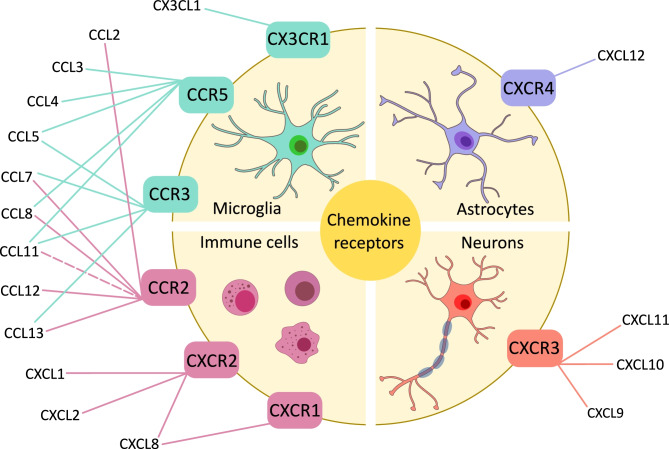
“Microglia, astrocyte, and neurons can all release chemokines and each of these cell types has the ability to express specific chemokine receptors (Wojcieszak et al. 2022).”
IL-8 (CXCL8) Correlations with Psychoneuroimmunological Processes and Neuropsychiatric Conditions
Shkundin, A., & Halaris, A. E. (2024) · Journal of Personalized Medicine · doi:10.3390/jpm14050488 · Cited by 28
“On the other hand, the overexpression of chemokines can disrupt the integrity of the brain barrier and allow immune cells to infiltrate the brain [7,147].”
BackgroundTraumatic brain injury: Bridging pathophysiological insights and precision treatment strategies
Lu, Y., Jin, J., Zhang, H., Lu, Q., Zhang, Y., Liu, C., Liang, Y., Tian, S., Zhao, Y., & Fan, H. (2025) · Neural Regeneration Research · doi:10.4103/nrr.nrr-d-24-01398 · Cited by 27
Matatall, K. A., Wathan, T. K., Nguyen, M., Chen, H., McDonald, A., Qi, G., Belk, J. A., Florez, M. A., Le, D. T., Olarinde, T., et al. (2025) · Cell Stem Cell · doi:10.1016/j.stem.2025.06.006 · Cited by 23
Investigating interleukin-8 in Alzheimer's disease: A comprehensive review
Righi, D., Manco, C., Pardini, M., Stufano, A., Schino, V., Pelagotti, V., Massa, F., Stefano, N. D., & Plantone, D. (2024) · Journal of Alzheimer s Disease · doi:10.1177/13872877241298973 · Cited by 21
“29 – 31 Pro-in fl ammatory cytokine signalling plays various roles in both neurodegeneration and neuroprotection.”
Temmerman, J., Engelborghs, S., Bjerke, M., & D’haeseleer, M. (2023) · Frontiers in Immunology · doi:10.3389/fimmu.2023.1162340 · Cited by 23
“In the early stages, it may enhance accumulation of microglia and clearance of Ab, whereas in the late stage, it may cause exaggerated inflammatory responses and the development of insoluble Ab within microglia (81).”
BackgroundEllakwa, D. E., Rashed, L. A., Ali, O. S. M., & El-Sabbagh, N. A. (2025) · Future Science OA · doi:10.1080/20565623.2025.2458434 · Cited by 22
“…age” for aD [8]. increasing evidence points to the importance of increased neuroinflammatory processes in the emergence of alzheimer’s disease [7]. there is evidence that the pathophysiology and neuronal damage of aD patients are significantly influenced by high levels of MDa and poor levels…”
Sleep deprivation-induced shifts in gut microbiota: Implications for neurological disorders
Wankhede, N. L., Kale, M. B., Kyada, A., Rekha, M. M., Chaudhary, K., Naidu, K. S., Rahangdale, S. R., Shende, P. V., Taksande, B. G., Khalid, M., et al. (2024) · Neuroscience · doi:10.1016/j.neuroscience.2024.11.070 · Cited by 21
Role of chemokines in aging and age-related diseases
Chaudhary, J. K., Danga, A. K., Kumari, A., Bhardwaj, A., & Rath, P. C. (2024) · Mechanisms of Ageing and Development · doi:10.1016/j.mad.2024.112009 · Cited by 24
Yan, D.-K., Qu, X., Chen, M., Wang, J., Li, X., Zhang, Z., Liu, Y., Kong, L., Yu, Y., Ju, R.-J., et al. (2023) · Journal of Drug Delivery Science and Technology · doi:10.1016/j.jddst.2023.104633 · Cited by 20
Adipose tissue as a therapeutic target for vascular damage in Alzheimer's disease
Bettinetti‐Luque, M., Trujillo‐Estrada, L., García‐Fuentes, E., Andreo‐Lopez, J., Sanchez-Varo, R. M., Garrido‐Sánchez, L., Mediavilla, A. G., López, M. G., García‐Caballero, M., Gutiérrez, A., et al. (2023) · British Journal of Pharmacology · doi:10.1111/bph.16243 · Cited by 21
“Within the brain, chemokines may be released by neurons, astrocytes or microglia and contribute to neurogenesis, synaptic plasticity and also to neuroinflammation (Wojcieszak et al., 2022).”
BackgroundImmune response of BV-2 microglial cells is impacted by peroxisomal beta-oxidation
Tawbeh, A., Raas, Q., Tahri-Joutey, M., Keime, C., Kaiser, R., Trompier, D., Nasser, B., Bellanger, E., Dessard, M., Hamon, Y., et al. (2023) · Frontiers in Molecular Neuroscience · doi:10.3389/fnmol.2023.1299314 · Cited by 18
“These chemokines are small cytokines that play a key role in the regulation of immune cell migration and actively participate in neurodegenerative processes (Wojcieszak et al., 2022).”
BackgroundTremendous Fidelity of Vitamin D3 in Age-related Neurological Disorders
Skv, M., Abraham, S. M., Eshwari, O., Golla, K., Jhelum, P., Maity, S., & Komal, P. (2024) · Molecular Neurobiology · doi:10.1007/s12035-024-03989-w · Cited by 18
Involvement of inflammasomes in the pathogenesis of Alzheimer's disease
Beder, N., Belkhelfa, M., & Leklou, H. (2024) · Journal of Alzheimer s Disease · doi:10.1177/13872877241283677 · Cited by 20
“71,72 When A β binds to CD36, toll-like receptors 4 (TLR4), and TLR6, it triggers the activation of microglia, which subsequently begin producing proin-fl ammatory cytokines and chemokines such as CCL2, CXCL10, CCL3, and CX3CL1.”
Guo, X., Liu, H. N., Song, Y., Wang, J., Liu, D., Zheng, Z., Li, J., Li, B., Song, A., He, W., et al. (2026) · MedComm · doi:10.1002/mco2.70497 · Cited by 26
Chemokines in patients with Alzheimer's disease: A meta-analysis
Wang, H., Zong, Y., Zhu, L., Wang, W., & Han, Y. (2023) · Frontiers in Aging Neuroscience · doi:10.3389/fnagi.2023.1047810 · Cited by 19
“Therefore, chemokines and their receptors were involved in the occurrence and progression of AD (Liu C. et al., 2014; Wojcieszak et al., 2022).”
BackgroundWang, P., Anderson, D. E., & Ye, Y. (2023) · Cell & Bioscience · doi:10.1186/s13578-023-01128-x · Cited by 20
The role of monocytes and macrophages in the progression of Alzheimer’s disease
Nie, W., Yue, Y., & Hu, J. (2025) · Frontiers in Immunology · doi:10.3389/fimmu.2025.1590909 · Cited by 15
Yang, Q., Yang, C., Lv, H., Zheng, X., Mao, S., Liu, N., Mo, S., Liao, B., Yang, M., Lü, Z., et al. (2024) · Inflammation · doi:10.1007/s10753-024-02043-0 · Cited by 12
Gaunt, J. R., Zainolabidin, N., Yip, A. K. K., Tan, J. M., Low, A. Y. T., Chen, A. I., & Ch’ng, T. H. (2023) · Journal of Neuroinflammation · doi:10.1186/s12974-023-02913-8 · Cited by 12
“The largest changes in expression were observed for CCL3 and CCL4, both of which are chemokines that are heavily expressed in inflamed tissues and upregulated in AD [57].”
BackgroundPalmitic Acid Modulates Microglial Cell Response to Metabolic Endotoxemia in an In Vitro Study
Chmielarz, M., Sobieszczańska, B., Teisseyre, A., Wawrzyńska, M., Bożemska, E., & Środa-Pomianek, K. (2023) · Nutrients · doi:10.3390/nu15153463 · Cited by 12
Cardamone, A., Coppoletta, A., Macrì, R., Nucera, S., Ruga, S., Scarano, F., Mollace, R., Mollace, A., Maurotti, S., Micotti, E., et al. (2024) · Pharmacological Research · doi:10.1016/j.phrs.2024.107417 · Cited by 12
mosGraphGPT: a foundation model for multi-omic signaling graphs using generative AIPreprint
Zhang, H., Huang, D., Chen, E., Cao, D., Xu, T., Dizdar, B., Li, G., Chen, Y., Payne, P., Province, M. A., et al. (2024) · bioRxiv (Cold Spring Harbor Laboratory) · doi:10.1101/2024.08.01.606222 · Cited by 14
“…and their receptors, part of neuroactive ligand-receptor interactions, have a dual role in AD: promoting neuroprotection and synaptic plasticity under normal conditions but leading to chronic inflammation when overexpressed, further contributing to Aβ aggregation and tau hyperphosphorylation 79 .”
BackgroundKoo, J., Jeong, B., Baek, J. Y., Lee, W. S., Gong, J., Park, S., Hong, J., Sim, Y., Kim, D., Kim, S. R., et al. (2025) · Nanoscale · doi:10.1039/d5nr00638d · Cited by 10
Pillat, M. M., Ayupe, A. C., Juvenal, G. D. A., Meinerz, C., Glaser, T., Pellegrina, D. V. D. S., Paiva, D. D. S., Mello, C. F., Longo, B. M., Reis, E. M. R., et al. (2023) · Stem Cell Reviews and Reports · doi:10.1007/s12015-023-10542-0 · Cited by 8
“Unlike many other chemokines, CX3CL1 interacts with only with the CX3CR1 expressed on microglia, thus playing a vital role in neuron-microglia communication [35].”
BackgroundT Cells Trafficking into the Brain in Aging and Alzheimer’s Disease
Ma, Y., Cao, J., Zhang, Y., Su, X.-M., Jing, Y., & Gao, L. (2024) · Journal of Neuroimmune Pharmacology · doi:10.1007/s11481-024-10147-5 · Cited by 11
“These chemokines are involved in the pathogenesis of AD (Wojcieszak et al. 2022 ).”
Bridging the Gap: The Neuro-immune Axis as a Key Player in Neurodegenerative Disorders
Liu, T., Wu, H., & Wei, J. (2025) · Neuroscience Bulletin · doi:10.1007/s12264-025-01439-8 · Cited by 9
“Cytokines and chemokines regulate the activity of immune cells, such as promoting the activation, proliferation, and migration of microglia and peripheral immune cells [56].”
InfluentialAfrakoti, L. G. M. P., Moghadam, S. D., & Hadinezhad, P. (2025) · Journal of Alzheimer s Disease Reports · doi:10.1177/25424823251329188 · Cited by 12
Glial Cells in Alzheimer’s Disease: Pathogenic Mechanisms and Therapeutic Frontiers
Sil, M., Mukherjee, N., Chatterjee, I., Ghosh, A., & Goswami, A. (2025) · Journal of Molecular Neuroscience · doi:10.1007/s12031-025-02379-8 · Cited by 9
Zhu, Q., Song, Y., Qian, Y., Zhang, R., Xue, J., & Hou, Y. (2025) · Ageing and Neurodegenerative Diseases · doi:10.20517/and.2025.25 · Cited by 6
Upadhayay, S. (2026) · International Immunopharmacology · doi:10.1016/j.intimp.2026.116285 · Cited by 7
Bello, E., Long, K. R., Iwama, S., Steer, J., Cooper, S., Alasoo, K., Kumasaka, N., Schwartzentruber, J., Panousis, N. I., & Bassett, A. R. (2026) · iScience · doi:10.1016/j.isci.2026.115688 · Cited by 7
“They are known to recruit peripheral immune cells through the blood-brain barrier as well as activate brain resident macrophages and microglia.”
Wei, C., Liu, J., Wu, B., Shen, T., Fan, J. J., Lin, Y., Li, L., Guo, Y., Shang, Y., Zhou, B., et al. (2025) · Brain Behavior and Immunity · doi:10.1016/j.bbi.2025.04.034 · Cited by 9
Singh, A. S., Naqvi, A. R., & Chanu, M. T. (2026) · Journal of Psychiatric Research · doi:10.1016/j.jpsychires.2026.01.050 · Cited by 7
Microglia protein profiles in CSF across Alzheimer’s disease clinical stages
Blujdea, E. R., van Bokhoven, P., Adami, P. V. M., Marshe, V. S., Vromen, E., Hok‐A‐Hin, Y. S., Boiten, W. A., Irwin, D. J., Chen‐Plotkin, A. S., Lemstra, A. W., et al. (2026) · Nature Aging · doi:10.1038/s43587-026-01088-0 · Cited by 7
“This potential decline in immune regulation, coupled with ongoing immune cell infiltration at the site of injury, suggests that initial neuroinflammation increases may confer protection, but, as this inflammation continues to be exacerbated over time, it becomes neurotoxic 59,64,65 .”
Ma, X., Zhiming, H., Mingwen, S., Hong, W., Tao, Y., Jun, X., & Gong, N. (2025) · Scientific Reports · doi:10.1038/s41598-025-94861-x · Cited by 5
“Prolonged inflammation loses its protective effect and contributes to Aβ accumulation, impaired clearance, enhanced tau hyperphorylation, neuronal loss, and AD deterioration 57–61 .”
Rice, M., Nuovo, G. J., Sawant, D. B., Mishra, A., & Tili, E. (2023) · Molecular Neurobiology · doi:10.1007/s12035-023-03822-w · Cited by 4
The glia-neutrophil axis: an understudied crosstalk in bacteria-induced neuroinflammation
Dunphy, A. M., & Marriott, I. (2026) · Frontiers in Neurology · doi:10.3389/fneur.2026.1765892 · Cited by 3
Heo, C., Kwak, H.-J., Ngo, L. H., Woo, R.-S., & Lee, S.-J. (2023) · Journal of Neuroscience Methods · doi:10.1016/j.jneumeth.2023.110029 · Cited by 4
Cytomegalovirus-induced T cell responses accelerate Alzheimer’s disease progression in mice
Marsden, M., McLaren, J. E., Bevan, R. J., Penn-Ripley, D., Somerville, M. S., Lauder, S. N., Jones, M. H., Maros, L.-B., McGurk, M. R., Gallimore, A., et al. (2026) · Brain · doi:10.1093/brain/awag043 · Cited by 2
“However, the preferential enrichment of virus-specific effector memory CD8 + T cells suggests that a selective mechanism, potentially involving inflammatory chemokines, 49 governs immune cell recruitment into the brains of 3xTg-AD mice.”
Comparison of the ApoE allelic variants in the formation of intracerebral Aβ deposits
Xu, G. M., Angelle, C., Huilgol, D., Sacilotto, P., McFarland, K. N., Fromholt, S., Lopez, A., Vo, Q., Rivasplata, A., Brkic, S., et al. (2025) · Neurobiology of Disease · doi:10.1016/j.nbd.2025.107105 · Cited by 2
Cognitive and transcriptomic effects of Epigallocatechin gallate in fetal alcohol spectrum disorders
Ramos-Triguero, A., Astals, M., Navarro‐Tapia, E., Vieiros, M., Bastons-Compta, A., Martínez, L., Andreu-Férnández, V., & García‐Algar, Ó. (2026) · Scientific Reports · doi:10.1038/s41598-025-34576-1 · Cited by 2
“PAE activates the immune system through TLR and NLR receptors in CNS 36 .”
InfluentialCao, Y., Wang, J., Li, X., Chen, X., Xiong, J., Wang, T., Sun, H., & Wang, Z. (2026) · Journal of Neuroinflammation · doi:10.1186/s12974-026-03852-w · Cited by 3
“Binding of Ccl4 to Ccr5 promotes immune cell migration; however, under pathological conditions, aberrant activation of this pathway may enhance microglial phagocytic activity and compromise synaptic integrity [15, 16].”
Targeting chemokine-receptor mediated molecular signaling by ethnopharmacological approaches
Naidu, G., Tripathi, D. K., Nagar, N., Mishra, A., & Poluri, K. M. (2024) · Journal of Ethnopharmacology · doi:10.1016/j.jep.2024.117837 · Cited by 2
An immune signature of postoperative cognitive decline in elderly patientsPreprint
Verdonk, F., Cambriel, A. I. F., Hedou, J. J., Ganio, E. A., Bellan, G., Gaudilliere, D. K., Einhaus, J. F., Sabayev, M., Stelzer, I. A., Feyaerts, D., et al. (2024) · bioRxiv (Cold Spring Harbor Laboratory) · doi:10.1101/2024.03.02.582845 · Cited by 2
“In addition, the model included a single plasma proteomic feature, chemokine CXCL1 (0.43 -log2 fold difference in median expression), a potent chemoattractant known to be implicated in neuro-240 inflammatory processes such as Alzheimer’s disease( 44 ).”
MethodInfluentialmosGraphFlow: a novel integrative graph AI model mining disease targets from multi-omic dataPreprint
Zhang, H., Cao, D., Xu, T., Chen, E., Li, G., Chen, Y., Payne, P., Province, M. A., & Li, F. (2024) · bioRxiv (Cold Spring Harbor Laboratory) · doi:10.1101/2024.08.01.606219 · Cited by 2
de Miranda, A. S., Vieira, É. L. M., Teixeira, A. L., & Bauer, M. E. (2026) · NeuroImmunoModulation · doi:10.1159/000550781 · Cited by 1
Wang, R., Feng, Y., Zhou, Z., Jiang, J., Zhang, R., Zou, W., Yang, H., Lv, W., & Shen, Y. (2026) · Frontiers in Aging Neuroscience · doi:10.3389/fnagi.2026.1748418 · Cited by 2
“Furthermore, the chemokine network forms a positive feedback loop with A β and tau protein pathology: A β oligomers stimulate microglia to release CCL3 and CCL5, which in turn exacerbate the hyperphosphorylation of tau protein within neurons (Jorda et al., 2020; Wojcieszak et al., 2022).”
Presto, P., Cardenas, J., Bustamante, C., Kisby, B. R., Ji, G., Ponomareva, O., Neugebauer, V., & Ponomarev, I. (2026) · Cells · doi:10.3390/cells15090775 · Cited by 1
“CXCL12 primarily signals through its receptors CXCR4 and, in the adult brain, it modulates neurotransmission, enhances synaptic plasticity, and generally provides neuroprotective effects against various insults [50].”
CXCL10 contributes to female-specific pathological progression in tauopathy model mice
Uenishi, R., Kawata, R., Manabe, T., Matsuba, Y., Mihira, N., Takeo, T., Saido, T. C., Hijioka, M., & Saito, T. (2026) · Journal of Neuroinflammation · doi:10.1186/s12974-026-03989-8 · Cited by 1
“While altered chemokine levels have been reported in the cerebrospinal fluid (CSF) and brain tissues of patients with AD [14], the specific molecular mediators linking glial-derived chemokine signaling to T cell infiltration and tau pathology—especially in a sex-dependent manner—remain largely…”
Sura, S., Jagadeesan, S., Moklas, M. A. M., Masrudin, S. S., Dandala, K. C. R., Judson, J. P., & Nor, N. H. M. (2026) · Molecular Biology Reports · doi:10.1007/s11033-026-12578-0 · Cited by 2
Nguyen, H. D., Jo, W. H., Cha, J. O., Hoang, N. M. H., & Kim, M.-S. (2024) · Toxicological Research · doi:10.1007/s43188-024-00228-1 · Cited by 1
Juvenal, G. D. A., Meinerz, C., Ayupe, A. C., Campos, H. C., Reis, E. M. R., Longo, B. M., Pillat, M. M., & Ulrich, H. (2024) · Cell & Bioscience · doi:10.1186/s13578-024-01251-3 · Cited by 1
“The chemokines CCL2, CCL3, CCL4, CCL5, CXCL10, and CXCL5 and the chemokine receptor CX3CR1 orchestrate neuroinflammation by attracting and activating microglia and play an important role in cell-to-cell communication in the CNS [31].”
BackgroundRico, E. M. G., Joseph, P., Noutsos, C., & Poon, K. (2024) · Neuroscience · doi:10.1016/j.neuroscience.2024.07.007 · Cited by 1
Wojcieszak, J. (2023) · Farmacja Polska · doi:10.32383/farmpol/166243 · Cited by 1
Self-citationNAD+ hydrolase Sarm1 is a key driver of synapse degeneration and memory loss in Alzheimer’s disease
Fan, F., Joshi, P., Liu, X., Kotturu, S. K., Abiola, M., Liu, Q., & Lou, X. (2025) · doi:10.64898/2025.12.21.695860
SARS-CoV-2 neurotropism-induced anxiety/depression-like behaviors require Microglia activationPreprint
Ge, Q., Zhou, S., Porras, J., Fu, P., Wang, T., Du, J., & Li, K. (2023) · bioRxiv (Cold Spring Harbor Laboratory) · doi:10.1101/2023.10.02.560570 · Cited by 1
“…studies support the idea that elevated cytokines like IL-1β, IL-6, CCL2 and TNF-α associated with the development of anxiety and depression-like behaviors in mice (65-68), underscoring the contribution of neuroinflammation to the behavioral manifestations observed following SARS-CoV-2 infection.”
BackgroundWang, P., Anderson, D. E., & Ye, Y. (2023) · Figshare · doi:10.6084/m9.figshare.24208974
Wang, P., Anderson, D. E., & Ye, Y. (2023) · Figshare · doi:10.6084/m9.figshare.24208992
Wang, P., Anderson, D. E., & Ye, Y. (2023) · Figshare · doi:10.6084/m9.figshare.24208983
Wang, P., Anderson, D. E., & Ye, Y. (2023) · Figshare · doi:10.6084/m9.figshare.24208980
Wang, P., Anderson, D. E., & Ye, Y. (2023) · Figshare · doi:10.6084/m9.figshare.24208971
Wang, P., Anderson, D. E., & Ye, Y. (2023) · Figshare · doi:10.6084/m9.figshare.24208977
Wang, P., Anderson, D. E., & Ye, Y. (2023) · Figshare · doi:10.6084/m9.figshare.24208989
Early cognitive dysfunction development immunological predictors in the ischemic stroke acute period
Tynterova, A. M. (2024) · Russian neurological Journal · doi:10.30629/2658-7947-2024-29-2-24-33
Catalão, C. H. R., Beeghly, C., Iwunze, N., Barichello, T., & Giridharan, V. V. (2025) · Neuromethods · doi:10.1007/978-1-0716-4474-4_17
Huang, L., Liu, Q., Ma, Z., Xue, J., Tian, X., Zhao, X., Zheng, Y., Wei, X., Yu, X., Feng, Y., et al. (2025) · Food Science and Human Wellness · doi:10.26599/fshw.2025.9250594
Changes of Microglial Cells in the Progression of Neurodegeneration in Natural Prion Disease
Monzón, M., Pérez, J. S., Hernández, R. S., Sarasa, R., Garcés, M., Bartolomé, M. J. L., & Badiola, J. J. (2025) · Frontiers in Bioscience-Landmark · doi:10.31083/fbl47074
“Thus, the modulation of these chemokines could also represent therapeutic strategies against neurodegeneration [43].”
Glial Cells, Cytokines, and Inflammasomes in Neurodegeneration
Patil, V. S., Desai, A., & Meena, H. (2025) · Springer eBooks · doi:10.1007/978-981-95-3945-1_3
van Olst, L., Kamermans, A., Halters, S., van der Pol, S. M. A., Rodríguez, E., Verberk, I. M. W., Verberk, S. G. S., Wessels, D. W. R., Rodriguez-Mogeda, C., Verhoeff, J., et al. (2024) · Figshare · doi:10.6084/m9.figshare.26711651
van Olst, L., Kamermans, A., Halters, S., van der Pol, S. M. A., Rodríguez, E., Verberk, I. M. W., Verberk, S. G. S., Wessels, D. W. R., Rodriguez-Mogeda, C., Verhoeff, J., et al. (2024) · Figshare · doi:10.6084/m9.figshare.26711621
van Olst, L., Kamermans, A., Halters, S., van der Pol, S. M. A., Rodríguez, E., Verberk, I. M. W., Verberk, S. G. S., Wessels, D. W. R., Rodriguez-Mogeda, C., Verhoeff, J., et al. (2024) · Figshare · doi:10.6084/m9.figshare.26711639
van Olst, L., Kamermans, A., Halters, S., van der Pol, S. M. A., Rodríguez, E., Verberk, I. M. W., Verberk, S. G. S., Wessels, D. W. R., Rodriguez-Mogeda, C., Verhoeff, J., et al. (2024) · Figshare · doi:10.6084/m9.figshare.26711654
van Olst, L., Kamermans, A., Halters, S., van der Pol, S. M. A., Rodríguez, E., Verberk, I. M. W., Verberk, S. G. S., Wessels, D. W. R., Rodriguez-Mogeda, C., Verhoeff, J., et al. (2024) · Figshare · doi:10.6084/m9.figshare.26711624
van Olst, L., Kamermans, A., Halters, S., van der Pol, S. M. A., Rodríguez, E., Verberk, I. M. W., Verberk, S. G. S., Wessels, D. W. R., Rodriguez-Mogeda, C., Verhoeff, J., et al. (2024) · Figshare · doi:10.6084/m9.figshare.26711672
Wang, P., Anderson, D. E., & Ye, Y. (2023) · Figshare · doi:10.6084/m9.figshare.24208986
Juvenal, G. D. A., Meinerz, C., Ayupe, A. C., Campos, H. C., Reis, E. M., Longo, B. M., Pillat, M. M., & Ulrich, H. (2024) · Figshare · doi:10.6084/m9.figshare.26061505
van Olst, L., Kamermans, A., Halters, S., van der Pol, S. M. A., Rodríguez, E., Verberk, I. M. W., Verberk, S. G. S., Wessels, D. W. R., Rodriguez-Mogeda, C., Verhoeff, J., et al. (2024) · Figshare · doi:10.6084/m9.figshare.26711636
van Olst, L., Kamermans, A., Halters, S., van der Pol, S. M. A., Rodríguez, E., Verberk, I. M. W., Verberk, S. G. S., Wessels, D. W. R., Rodriguez-Mogeda, C., Verhoeff, J., et al. (2024) · Figshare · doi:10.6084/m9.figshare.26711642
van Olst, L., Kamermans, A., Halters, S., van der Pol, S. M. A., Rodríguez, E., Verberk, I. M. W., Verberk, S. G. S., Wessels, D. W. R., Rodriguez-Mogeda, C., Verhoeff, J., et al. (2024) · Figshare · doi:10.6084/m9.figshare.26711657
Juvenal, G. D. A., Meinerz, C., Ayupe, A. C., Campos, H. C., Reis, E. M., Longo, B. M., Pillat, M. M., & Ulrich, H. (2024) · Figshare · doi:10.6084/m9.figshare.26735733
van Olst, L., Kamermans, A., Halters, S., van der Pol, S. M. A., Rodríguez, E., Verberk, I. M. W., Verberk, S. G. S., Wessels, D. W. R., Rodriguez-Mogeda, C., Verhoeff, J., et al. (2024) · Figshare · doi:10.6084/m9.figshare.26711663
Juvenal, G. D. A., Meinerz, C., Ayupe, A. C., Campos, H. C., Reis, E. M., Longo, B. M., Pillat, M. M., & Ulrich, H. (2024) · Figshare · doi:10.6084/m9.figshare.26735739
van Olst, L., Kamermans, A., Halters, S., van der Pol, S. M. A., Rodríguez, E., Verberk, I. M. W., Verberk, S. G. S., Wessels, D. W. R., Rodriguez-Mogeda, C., Verhoeff, J., et al. (2024) · Figshare · doi:10.6084/m9.figshare.26711630
van Olst, L., Kamermans, A., Halters, S., van der Pol, S. M. A., Rodríguez, E., Verberk, I. M. W., Verberk, S. G. S., Wessels, D. W. R., Rodriguez-Mogeda, C., Verhoeff, J., et al. (2024) · Figshare · doi:10.6084/m9.figshare.26711648
Juvenal, G. D. A., Meinerz, C., Ayupe, A. C., Campos, H. C., Reis, E. M., Longo, B. M., Pillat, M. M., & Ulrich, H. (2024) · Figshare · doi:10.6084/m9.figshare.26735730
van Olst, L., Kamermans, A., Halters, S., van der Pol, S. M. A., Rodríguez, E., Verberk, I. M. W., Verberk, S. G. S., Wessels, D. W. R., Rodriguez-Mogeda, C., Verhoeff, J., et al. (2024) · Figshare · doi:10.6084/m9.figshare.25687615
Juvenal, G. D. A., Meinerz, C., Ayupe, A. C., Campos, H. C., Reis, E. M., Longo, B. M., Pillat, M. M., & Ulrich, H. (2024) · Figshare · doi:10.6084/m9.figshare.26735724
Metadata
- Authors
- Wojcieszak, J., Kuczyńska, K., & Zawilska, J. B.
- Affiliations
- Karolinska Institutet (SE)
- Medical University of Lodz (PL)
- Corresponding author
- Zawilska, Jolanta B.
- Venue
- Journal of Molecular Neuroscience
- Published
- 2022-07-12
- Volume
- 72
- Issue
- 9
- Pages
- 1929-1951
- ISSN
- 0895-8696 (ISSN-L)
- 1559-1166
- PMC
- PMC9392685
- OpenAlex
- W4285089500
- Cited by
- 97
- References
- 146
- Citations by year
- 2026: 25 · 2025: 20 · 2024: 35 · 2023: 17
- FWCI
- 3.37
- Type
- journal article
- Language
- en
- Open access
- hybrid
- doi.org
- Locations
- Journal of Molecular Neuroscience (publishedVersion, cc-by)
- PubMed Central (publishedVersion, cc-by)
- PubMed (publishedVersion)
- Funders
- Uniwersytet Łódzki (503-31-001)
- Topics
- Life Sciences › Neuroscience › Cellular and Molecular Neuroscience › Alzheimer's disease research and treatments
- Life Sciences › Neuroscience › Neurology › Neuroinflammation and Neurodegeneration Mechanisms
- Health Sciences › Medicine › Psychiatry and Mental health › Dementia and Cognitive Impairment Research
- Keywords
- Alzheimer's disease, chemokines, chemokine receptors, neuroinflammation, amyloid-beta, neurofibrillary tangles, blood-brain barrier, microglia, astrocytes, tau hyperphosphorylation, neurogenesis, synaptic plasticity, cholinergic neurons, transgenic mouse model
- MeSH terms
- Alzheimer Disease, Animals, Humans, Amyloid beta-Peptides, Neurofibrillary Tangles, Microglia, Chemokines, Mice
About these checks
Registry, retraction, Cites and statistics results are computed, not judged, with one exception: a printed reference that no index lists is read once by a model before it counts as a citation error. Claims are extracted by a model and kept only when their quote appears verbatim in the text; each cited source is then checked, with a model’s help, for whether it says what the claim relies on. Those verdicts can be wrong — read the claim’s quote and the cited source before relying on one.
Statistics and arithmetic are recomputed, not judged.
Confidence bands are shown only for fields calibrated on our annotated test set. None is calibrated yet, so no band is shown.
What counts as a claim ›How we measure confidence ›How we check this ›
Are you an author, or do you see a problem with this page? Request a review
