

The DOI matches its Crossref record; 146 of the 146 references it cites were found in a registry or an index.
Role of Chemokines in the Development and Progression of Alzheimer’s Disease
Open accessAbstract
Alzheimer’s disease (AD) is a progressive neurogenerative disorder manifested by gradual memory loss and cognitive decline due to profound damage of cholinergic neurons. The neuropathological hallmarks of AD are intracellular deposits of neurofibrillary tangles (NFTs) and extracellular aggregates of amyloid β (Aβ). Mounting evidence indicates that intensified neuroinflammatory processes play a pivotal role in the pathogenesis of AD. Chemokines serve as signaling molecules in immune cells but also in nerve cells. Under normal conditions, neuroinflammation plays a neuroprotective role against various harmful factors. However, overexpression of chemokines initiates disruption of the integrity of the blood–brain barrier, facilitating immune cells infiltration into the brain. Then activated adjacent glial cells–astrocytes and microglia, release massive amounts of chemokines. Prolonged inflammation loses its protective role and drives an increase in Aβ production and aggregation, impairment of its clearance, or enhancement of tau hyperphosphorylation, contributing to neuronal loss and exacerbation of AD. Moreover, chemokines can be further released in response to growing deposits of toxic forms of Aβ. On the other hand, chemokines seem to exert multidimensional effects on brain functioning, including regulation of neurogenesis and synaptic plasticity in regions responsible for memory and cognitive abilities. Therefore, underexpression or complete genetic ablation of some chemokines can worsen the course of AD. This review covers the current state of knowledge on the role of particular chemokines and their receptors in the development and progression of AD. Special emphasis is given to their impact on forming Aβ and NFTs in humans and in transgenic murine models of AD.
Overview
The claims and statistics of this paper haven’t been checked yet.
Run the check — free, no account needed. It reads the open-access text and checks the sources it cites.
Integrity signals
Registry
Cites
Statistics
Figures
Verification
Claims the paper makes
The claims and statistics of this paper haven’t been checked yet.
Run the check — free, no account needed. It reads the open-access text and checks the sources it cites.
Claims other papers make about this one
Each is checked against this paper’s own text: does it say what the citing paper says it does?
The cited source says what the claim attributes to it.
In the citing paper
“consistent with the known capacity of CCL2 to break down the blood brain barrier and recruit peripheral immune cells to sites of damage.58–62”
From TET2-mutant myeloid cells mitigate Alzheimer’s disease progression via CNS infiltration and enhanced phagocytosis in mice (2025) · cites this paper as [60]
Self-check
Percentages, sums and reported differences re-computed from the paper’s own numbers.
The self-check needs the paper’s full text.
Citations
Cites
McKhann, G. M., Knopman, D. S., Chertkow, H., Hyman, B. T., Jack, C. R., Kawas, C. H., Klunk, W. E., Koroshetz, W. J., Manly, J. J., Mayeux, R., et al. (2011) · Alzheimer's & Dementia · doi:10.1016/j.jalz.2011.03.005 · Cited by 18,657 · cited 1×
“As the disease progresses, preclinical AD transits to mild cognitive impairment (MCI) in which cognition deficits do not affect patients’ performance at work or regular daily activities, but eventually may develop into AD (McKhann et al. 2011; Saido 2013).”
BackgroundA new class of membrane-bound chemokine with a CX3C motif
Bazan, J. F., Bacon, K. B., Hardiman, G., Wang, W., Soo, K., Rossi, D., Greaves, D. R., Zlotnik, A., & Schall, T. J. (1997) · Nature · doi:10.1038/385640a0 · Cited by 2,013
The cholinergic system in the pathophysiology and treatment of Alzheimer’s disease
Hampel, H., Mesulam, M.-M., Cuello, A. C., Farlow, M. R., Giacobini, E., Grossberg, G. T., Khachaturian, A. S., Vergallo, A., Cavedo, E., Snyder, P. J., et al. (2018) · Brain · doi:10.1093/brain/awy132 · Cited by 1,918
The ageing systemic milieu negatively regulates neurogenesis and cognitive function
Villeda, S. A., Luo, J., Mosher, K. I., Zou, B., Britschgi, M., Bieri, G., Stan, T. M., Fainberg, N., Ding, Z., Eggel, A., et al. (2011) · Nature · doi:10.1038/nature10357 · Cited by 1,763
A guide to chemokines and their receptors
Hughes, C. E., & Nibbs, R. J. B. (2018) · FEBS Journal · doi:10.1111/febs.14466 · Cited by 1,510 · cited 2×
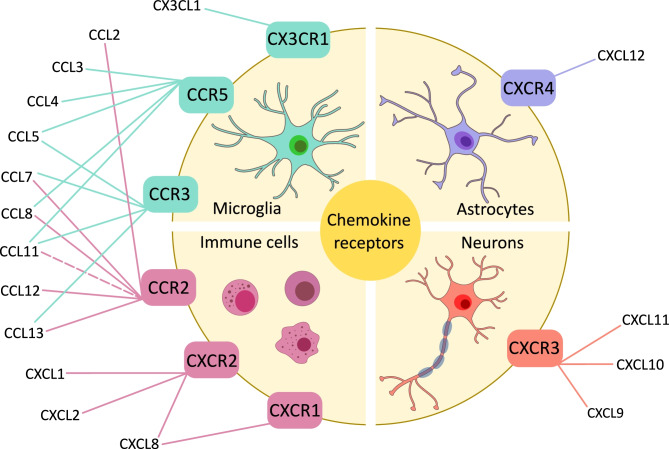
“To date, six CXC receptors have been identified, named from CXCR1 to CXCR6, and eleven CC receptors, from CCR1 to CCR11, along with a single receptor for CX3CL1 and one for lymphotactin α/β, called CX3CR1 and XCR1, respectively (Hughes and Nibbs 2018).”
BackgroundZhao, Q.-F., Tan, L., Wang, H.-F., Jiang, T., Tan, M.-S., Tan, L., Xu, W., Li, J.-Q., Wang, J., Lai, T.-J., et al. (2016) · Journal of Affective Disorders · doi:10.1016/j.jad.2015.09.069 · Cited by 916
El Khoury, J., Toft, M., Hickman, S. E., Means, T. K., Terada, K., Geula, C., & Luster, A. D. (2007) · Nature Medicine · doi:10.1038/nm1555 · Cited by 915
Risk factors for Alzheimer’s disease
Armstrong, R. A. (2019) · Folia Neuropathologica · doi:10.5114/fn.2019.85929 · Cited by 679
Hippocampus and its involvement in Alzheimer’s disease: a review
Rao, Y. L., Ganaraja, B., Murlimanju, B. V., Joy, T., Krıshnamurthy, A., & Agrawal, A. (2022) · 3 Biotech · doi:10.1007/s13205-022-03123-4 · Cited by 646
Neurotactin, a membrane-anchored chemokine upregulated in brain inflammation
Pan, Y., Lloyd, C. M., Zhou, H., Dolich, S., Deeds, J., Gonzalo, J.-Á., Vath, J., Gosselin, M., Ma, J., Dussault, B. J., et al. (1997) · Nature · doi:10.1038/42491 · Cited by 643 · cited 1×
“This chemokine exists in both soluble and membrane-bound forms (Bajetto et al. 2002; Bazan et al. 1997; Pan et al. 1997).”
BackgroundRegulation of Tau Pathology by the Microglial Fractalkine Receptor
Bhaskar, K., Konerth, M., Kokiko-Cochran, O. N., Cardona, A., Ransohoff, R. M., & Lamb, B. T. (2010) · Neuron · doi:10.1016/j.neuron.2010.08.023 · Cited by 639 · cited 1×
“Of note, these animals exhibited pronounced behavioral impairments (Bhaskar et al. 2010).”
BackgroundCX3CR1 deficiency leads to impairment of hippocampal cognitive function and synaptic plasticity
Rogers, J. T., Morganti, J. M., Bachstetter, A. D., Hudson, C. E., Peters, M. M., Grimmig, B. A., Weeber, E. J., Bickford, P. C., & Gemma, C. (2011) · The Journal of Neuroscience · doi:10.1523/jneurosci.3667-11.2011 · Cited by 624 · cited 3×
“Unlike many other chemokines, fractalkine interacts with only one receptor, CX3CR1, expressed on microglia, thus playing a vital role in neuron-microglia communication (Rogers et al. 2011).”
BackgroundThe Alzheimer’s Disease Clinical Spectrum
Atri, A. (2019) · Medical Clinics of North America · doi:10.1016/j.mcna.2018.10.009 · Cited by 623
Microglial Cx3cr1 knockout prevents neuron loss in a mouse model of Alzheimer's disease
Fuhrmann, M., Bittner, T., Jung, C. K. E., Burgold, S., Page, R. M., Mitteregger, G., Haass, C., LaFerla, F. M., Kretzschmar, H., & Herms, J. (2010) · Nature Neuroscience · doi:10.1038/nn.2511 · Cited by 562 · cited 1×
“On the other hand, knock-out of CX3CR1 in a 3xTg mice prevented neuronal loss, but not affected Aβ deposition and phagocytosis by microglia (Fuhrmann et al. 2010).”
BackgroundChitinase-3 like-protein-1 function and its role in diseases
Zhao, T., Su, Z., Li, Y., Zhang, X., & You, Q. (2020) · Signal Transduction and Targeted Therapy · doi:10.1038/s41392-020-00303-7 · Cited by 536
Hansen, R. A., Webb, A. P., Moore, C. G., Gartlehner, G., Jonas, D. E., & Morgan, L. C. (2020) · PubMed Central · doi:10.17615/ae8c-2p76 · Cited by 523
Lee, S., Varvel, N. H., Konerth, M. E., Xu, G., Cardona, A. E., Ransohoff, R. M., & Lamb, B. T. (2010) · The American Journal of Pathology · doi:10.2353/ajpath.2010.100265 · Cited by 522
CD36 Mediates the Innate Host Response to β-Amyloid
El Khoury, J. B., Moore, K. J., Means, T. K., Leung, J., Terada, K., Toft, M., Freeman, M. W., & Luster, A. D. (2003) · The Journal of Experimental Medicine · doi:10.1084/jem.20021546 · Cited by 483
The contribution of neuroinflammation to amyloid toxicity in Alzheimer's disease
Minter, M. R., Taylor, J. M., & Crack, P. J. (2016) · Journal of Neurochemistry · doi:10.1111/jnc.13411 · Cited by 451 · cited 2×
“, CCL2, CCL4, CCL11), which attract further microglia and astrocytes to an inflammatory site (Minter et al. 2016).”
BackgroundLai, K. S. P., Liu, C. S., Rau, A., Lanctôt, K. L., Köhler, C. A., Pakosh, M., Carvalho, A. F., & Herrmann, N. (2017) · Journal of Neurology, Neurosurgery & Psychiatry · doi:10.1136/jnnp-2017-316201 · Cited by 433 · cited 1×
“A meta-analysis revealed that plasma concentration of CXCL10 was significantly elevated in AD patients as compared to healthy controls (Lai et al. 2017).”
Dark microglia: A new phenotype predominantly associated with pathological states
Bisht, K., Sharma, K. P., Lecours, C., Gabriela Sánchez, M., El Hajj, H., Milior, G., Olmos‐Alonso, A., Gómez‐Nicola, D., Luheshi, G., Vallières, L., et al. (2016) · Glia · doi:10.1002/glia.22966 · Cited by 421
Fractalkine and CX3CR1 regulate hippocampal neurogenesis in adult and aged rats
Bachstetter, A. D., Morganti, J. M., Jernberg, J., Schlunk, A., Mitchell, S. H., Brewster, K. W., Hudson, C. E., Cole, M. J., Harrison, J. K., Bickford, P. C., et al. (2011) · Neurobiology of Aging · doi:10.1016/j.neurobiolaging.2009.11.022 · Cited by 364 · cited 1×
“In aged rats, a decrease of hippocampal neurogenesis is paralleled by reduction of CX3CL1 levels, while treatment with exogenous chemokine reverses this process (Bachstetter et al. 2011).”
McQuade, A., Kang, Y. J., Hasselmann, J., Jairaman, A., Sotelo, A., Coburn, M., Shabestari, S. K., Chadarevian, J. P., Fote, G., Tu, C. H., et al. (2020) · Nature Communications · doi:10.1038/s41467-020-19227-5 · Cited by 361
Mildner, A., Schlevogt, B., Kierdorf, K., Böttcher, C., Erny, D., Kummer, M. P., Quinn, M., Brück, W., Bechmann, I., Heneka, M. T., et al. (2011) · The Journal of Neuroscience · doi:10.1523/jneurosci.6209-10.2011 · Cited by 351 · cited 1×
“Furthermore, in Tg2576 mice, knock-out of CCR2 resulted in decreased clearance of Aβ by perivascular macrophages and accumulation of insoluble Aβ species (Mildner et al. 2011).”
BackgroundBajetto, A., Bonavia, R., Barbero, S., & Schettini, G. (2002) · Journal of Neurochemistry · doi:10.1046/j.1471-4159.2002.01091.x · Cited by 324 · cited 1×
“This chemokine exists in both soluble and membrane-bound forms (Bajetto et al. 2002; Bazan et al. 1997; Pan et al. 1997).”
BackgroundEarly-onset versus late-onset Alzheimer's disease: the case of the missing APOE ɛ4 allele
van der Flier, W. M., Pijnenburg, Y. A. L., Fox, N. C., & Scheltens, P. (2010) · The Lancet Neurology · doi:10.1016/s1474-4422(10)70306-9 · Cited by 321
Mouse models of Alzheimer's disease
Hall, A. M., & Roberson, E. D. (2012) · Brain Research Bulletin · doi:10.1016/j.brainresbull.2011.11.017 · Cited by 317 · cited 2×
“They usually harbor human mutations responsible for familial Alzheimer’s disease (FAD), including variants of APP, presenilin 1 (PS1), and microtubule-associated protein tau (MAPT) (Hall and Roberson 2012; Puzzo et al. 2015).”
BackgroundAging and Alzheimer's disease pathology
Sengoku, R. (2019) · Neuropathology · doi:10.1111/neup.12626 · Cited by 316 · cited 3×
“…changes in AD encompass formation and accumulation of pathological structures in the brain, namely extracellular senile plaques containing aggregates of amyloid β peptide (Aβ) and intracellular neurofibrillary tangles consisting of hyperphosphorylated tau protein (Saido 2013; Sengoku 2020).”
BackgroundMechanisms of secretion and spreading of pathological tau protein
Brunello, C. A., Merezhko, M., Uronen, R.-L., & Huttunen, H. J. (2020) · Cellular and Molecular Life Sciences · doi:10.1007/s00018-019-03349-1 · Cited by 304 · cited 3×
“Additionally, pathological tau can be secreted to extracellular space and enter surrounding neurons or glial cells further impairing axonal transport and causing neuronal synaptic loss thus contributing to progression of the disease (Brunello et al. 2019).”
BackgroundLaurent, C., Dorothée, G., Hunot, S., Martin, E., Monnet, Y., Duchamp, M., Dong, Y., Légeron, F.-P., Leboucher, A., Burnouf, S., et al. (2017) · Brain · doi:10.1093/brain/aww270 · Cited by 303 · cited 3×
“Overexpression of CCL3, CCL4, and CCL5, paralleled by infiltration of T lymphocytes into the hippocampus, was also observed in a Tg murine model of tauopathy, THY-Tau22 (Laurent et al.”
BackgroundXia, M., Qin, S., Wu, L., Mackay, C. R., & Hyman, B. T. (1998) · American Journal Of Pathology · doi:10.1016/s0002-9440(10)65542-3 · Cited by 300
Intrathecal Chemokine Synthesis in Mild Cognitive Impairment and Alzheimer Disease
Galimberti, D., Schoonenboom, N., Scheltens, P., Fenoglio, C., Bouwman, F., Venturelli, E., Guidi, I., Blankenstein, M. A., Bresolin, N., & Scarpini, E. (2006) · Archives of Neurology · doi:10.1001/archneur.63.4.538 · Cited by 289 · cited 17×
“Levels of CXCL8 were found to be significantly higher in samples, such as brain tissue, plasma, and CSF, obtained from AD patients as compared to healthy controls (Alsadany et al. 2013; Ashutosh et al. 2011; Corrêa et al. 2011; Galimberti et al. 2006; Sokolova et al. 2009).”
BackgroundResultInfluentialJouanne, M., Rault, S., & Voisin‐Chiret, A. S. (2017) · European Journal of Medicinal Chemistry · doi:10.1016/j.ejmech.2017.07.070 · Cited by 279
Westin, K., Buchhave, P., Nielsen, H., Minthon, L., Janciauskiene, S., & Hansson, O. (2012) · PLoS ONE · doi:10.1371/journal.pone.0030525 · Cited by 277 · cited 1×
“High levels of CCL2 in patients with prodromal AD were associated with a faster cognitive decline (Westin et al. 2012).”
BackgroundLiu, Z., Condello, C., Schain, A., Harb, R., & Grutzendler, J. (2010) · The Journal of Neuroscience · doi:10.1523/jneurosci.4403-10.2010 · Cited by 275
Glial activation and inflammation along the Alzheimer’s disease continuum
Nordengen, K., Kirsebom, B.-E., Henjum, K., Selnes, P., Gísladóttir, B., Wettergreen, M., Torsetnes, S. B., Grøntvedt, G. R., Waterloo, K. K., Aarsland, D., et al. (2019) · Journal of Neuroinflammation · doi:10.1186/s12974-019-1399-2 · Cited by 274
Xia, M. Q., Bacskai, B. J., Knowles, R. B., Qin, S. X., & Hyman, B. T. (2000) · Journal of Neuroimmunology · doi:10.1016/s0165-5728(00)00285-x · Cited by 269 · cited 1×
“Similarly, hippocampal astrocytes cultured in vitro produce increased amounts of CXCL10 in response to Aβ stimulation (Lai et al. 2013; Xia et al. 2000).”
BackgroundCho, S.-H., Sun, B., Zhou, Y., Kauppinen, T. M., Halabisky, B., Wes, P., Ransohoff, R. M., & Gan, L. (2011) · Journal of Biological Chemistry · doi:10.1074/jbc.m111.254268 · Cited by 268 · cited 5×
“Reduced levels of CX3CL1 were found in the hippocampus and frontal cortex of AD patients as compared to healthy controls (Cho et al. 2011), and in the CSF of patients with MCI and AD dementia (Perea et al.”
BackgroundInfluentialStuart, M. J., & Baune, B. T. (2014) · Neuroscience & Biobehavioral Reviews · doi:10.1016/j.neubiorev.2014.02.001 · Cited by 266 · cited 1×
“2019; Stuart and Baune 2014), and brain tissue, including critical structures for AD, such as the hippocampus, frontal, and temporal cortex of AD patients as compared to healthy controls (Liao et al.”
BackgroundSokolova, A., Hill, M. D., Rahimi, F., Warden, L. A., Halliday, G. M., & Shepherd, C. E. (2009) · Brain Pathology · doi:10.1111/j.1750-3639.2008.00188.x · Cited by 249
ISHIZUKA, K., KIMURA, T., IGATA‐YI, R., KATSURAGI, S., TAKAMATSU, J., & MIYAKAWA, T. (1997) · Psychiatry and Clinical Neurosciences · doi:10.1111/j.1440-1819.1997.tb02375.x · Cited by 245 · cited 1×
“Additionally, CCL2 is present in mature senile plaques and reactive microglia, where it is involved in microglial accumulation and phagocytosis of Aβ (Ishizuka et al. 1997; Sui et al. 2019).”
BackgroundAbsence of CX3CR1 impairs the internalization of Tau by microglia
Bolós, M., Llorens-Martín, M., Perea, J. R., Jurado-Arjona, J., Rábano, A., Hernández, F., & Avila, J. (2017) · Molecular Neurodegeneration · doi:10.1186/s13024-017-0200-1 · Cited by 231 · cited 3×
“2020), an observation suggesting that upregulation of this axis may play a compensatory role (Bolós et al. 2017).”
BackgroundEarly-Onset Alzheimer’s Disease: What Is Missing in Research?
Ayodele, T., Rogaeva, E. A., Kurup, J. T., Beecham, G. W., & Reitz, C. (2021) · Current Neurology and Neuroscience Reports · doi:10.1007/s11910-020-01090-y · Cited by 210
Naert, G., & Rivest, S. (2011) · The Journal of Neuroscience · doi:10.1523/jneurosci.0299-11.2011 · Cited by 207 · cited 2×
“Microglia of APP/PS1/ CCR2−/− mice exhibit increased expression of CX3CR1 which is involved in inhibition of microglial activation and phagocytosis (Naert and Rivest 2011).”
BackgroundLee, W., Liao, Y., Wang, Y.-F., Lin, I.-F., Wang, S., & Fuh, J. (2018) · Scientific Reports · doi:10.1038/s41598-018-19807-y · Cited by 197 · cited 16×
“Levels of CXCL8 were found to be significantly higher in samples, such as brain tissue, plasma, and CSF, obtained from AD patients as compared to healthy controls (Alsadany et al.”
BackgroundResultInfluentialDissociating Normal Aging from Alzheimer’s Disease: A View from Cognitive Neuroscience
Toepper, M. (2017) · Journal of Alzheimer s Disease · doi:10.3233/jad-161099 · Cited by 191 · cited 1×
“Moreover, linguistic abilities and verbal knowledge seems to be unaffected in normal aging processes (Sengoku 2020; Toepper 2017).”
BackgroundEotaxin is a natural antagonist for CCR2 and an agonist for CCR5
Ogilvie, P., Bardi, G., Clark‐Lewis, I., Baggiolini, M., & Uguccioni, M. (2001) · Blood · doi:10.1182/blood.v97.7.1920 · Cited by 188 · cited 14×
“CCL11 levels were increased in plasma of AD patients (Choi et al.”
BackgroundInfluentialNew role of P2X7 receptor in an Alzheimer’s disease mouse model
Martin, E., Amar, M., Dalle, C., Youssef, I., Boucher, C., Le Duigou, C., Brückner, M., Prigent, A., Sazdovitch, V., Halle, A., et al. (2019) · Molecular Psychiatry · doi:10.1038/s41380-018-0108-3 · Cited by 187 · cited 3×
“Similarly, levels of CXCL1 and CXCL2 were elevated in brains of APP/PS1 mice as compared to wild-type animals (Martin et al. 2019; Watson and Fan 2005), and treatment of mouse microglia with Aβ1-41 induced expression of CXCL2 (Ito et al.”
BackgroundRole of CCR2 in Inflammatory Conditions of the Central Nervous System
Chu, H. X., Arumugam, T. V., Gelderblom, M., Magnus, T. U., Drummond, G. R., & Sobey, C. G. (2014) · Journal of Cerebral Blood Flow & Metabolism · doi:10.1038/jcbfm.2014.120 · Cited by 179 · cited 1×
“Among them, CCL2 is of particular interest, as it is the most potent activator of CCR2 (Chu et al. 2014).”
BackgroundCCR5 is a suppressor for cortical plasticity and hippocampal learning and memory
Zhou, M., Greenhill, S., Huang, S., Silva, T. K., Sano, Y., Wu, S., Cai, Y., Nagaoka, Y., Sehgal, M., Cai, D. J., et al. (2016) · eLife · doi:10.7554/elife.20985 · Cited by 174
Fractalkine activates NRF2/NFE2L2 and heme oxygenase 1 to restrain tauopathy-induced microgliosis
Lastres-Becker, I., Innamorato, N. G., Jaworski, T., Rábano, A., Kügler, S., Van Leuven, F., & Cuadrado, A. (2014) · Brain · doi:10.1093/brain/awt323 · Cited by 160
Martin, E., Boucher, C., Fontaine, B., & Delarasse, C. (2017) · Aging Cell · doi:10.1111/acel.12522 · Cited by 156
Gene expression profiling of amyloid beta peptide-stimulated human post-mortem brain microglia
Walker, D. G. (2001) · Neurobiology of Aging · doi:10.1016/s0197-4580(01)00306-2 · Cited by 155
CXCR3 promotes plaque formation and behavioral deficits in an Alzheimer’s disease model
Krauthausen, M., Kummer, M. P., Zimmermann, J., Reyes-Irisarri, E., Terwel, D., Bulic, B., Heneka, M. T., & Müller, M. (2014) · Journal of Clinical Investigation · doi:10.1172/jci66771 · Cited by 152 · cited 2×
“2018) and APP/PS1 model of amyloidosis (Krauthausen et al. 2015).”
BackgroundAmyloid-β-induced chemokine production in primary human macrophages and astrocytes
Smits, H. A., Rijsmus, A., van Loon, J. H., Wat, J. W. Y., Verhoef, J., Boven, L. A., & Nottet, H. S. L. M. (2002) · Journal of Neuroimmunology · doi:10.1016/s0165-5728(02)00112-1 · Cited by 150 · cited 1×
“Importantly, it has been found that Aβ stimulates CCL2 release from astrocytes and microglia in vitro (El Khoury et al. 2003; Ito et al. 2006; Kiyota et al. 2013; Smits et al. 2002).”
RANTES upregulation in the Alzheimer's disease brain: A possible neuroprotective role
Tripathy, D., Thirumangalakudi, L., & Grammas, P. (2008) · Neurobiology of Aging · doi:10.1016/j.neurobiolaging.2008.03.009 · Cited by 150 · cited 1×
“However, levels of CCL5 did not correlate with cognitive scores (Tripathy et al. 2010; Vacinova et al. 2021).”
BackgroundThe Chemokine MIP-1α/CCL3 impairs mouse hippocampal synaptic transmission, plasticity and memory
Marciniak, E., Faivre, E., Dutar, P., Alves Pires, C., Demeyer, D., Caillierez, R., Laloux, C., Buée, L., Blum, D., & Humez, S. (2015) · Scientific Reports · doi:10.1038/srep15862 · Cited by 148 · cited 1×
“Similarly, in nonAD mice, subchronic intracerebroventricular injection of CCL3 resulted in inhibition of LTP and cognitive deficits, processes mediated by CCR5, as its antagonist abolished these effects (Marciniak et al. 2015).”
BackgroundYamamoto, M., Horiba, M., Buescher, J. L., Huang, D., Gendelman, H. E., Ransohoff, R. M., & Ikezu, T. (2005) · The American Journal of Pathology · doi:10.1016/s0002-9440(10)62364-4 · Cited by 137
Interleukin-8 receptor B immunoreactivity in brain and neuritic plaques of Alzheimer's disease.
Xia, M., Qin, S., McNamara, M. J., Mackay, C. R., & Hyman, B. T. (1997) · Europe PMC (PubMed Central) · Cited by 131
Man, S., Ma, Y., Shang, D., Zhao, W., Li, B., Guo, D., Fang, W., Zhu, L., & Chen, Y. (2006) · Neurobiology of Aging · doi:10.1016/j.neurobiolaging.2006.02.013 · Cited by 130
The efficacy and safety of memantine for the treatment of Alzheimer’s disease
Matsunaga, S., Kishi, T., Nomura, I., Sakuma, K., Okuya, M., Ikuta, T., & Iwata, N. (2018) · Expert Opinion on Drug Safety · doi:10.1080/14740338.2018.1524870 · Cited by 126 · cited 1×
“Although they stabilize the patient’s condition, slow exacerbation of symptoms, or improve cognitive functions, they do not stop the progression of the disease (Hansen et al. 2008; Matsunaga et al. 2018).”
BackgroundOpposing Effects of Membrane-Anchored CX3CL1 on Amyloid and Tau Pathologies via the p38 MAPK Pathway
Lee, S., Xu, G., Jay, T. R., Bhatta, S., Kim, K.-W., Jung, S., Landreth, G. E., Ransohoff, R. M., & Lamb, B. T. (2014) · The Journal of Neuroscience · doi:10.1523/jneurosci.0853-14.2014 · Cited by 123
Lee, J. K., Schuchman, E. H., Jin, H. K., & Bae, J. (2012) · Stem Cells · doi:10.1002/stem.1125 · Cited by 121
Li, M., Shang, D., Zhao, W., Li, T. X., Li, B., Fang, W., Zhu, L., Man, S., & Chen, Y. (2009) · The Journal of Immunology · doi:10.4049/jimmunol.0803013 · Cited by 121 · cited 12×
“These small-molecule proteins orchestrate the neuroinflammation as they are involved in attraction and activation of microglia and may induce a leakage of the blood–brain-barrier (BBB) to allow infiltration of peripheral immune cells.”
BackgroundInfluentialCCL5 induces a pro-inflammatory profile in microglia in vitro
Škuljec, J., Sun, H., Pul, R., Bénardais, K., Ragancokova, D., Moharregh-Khiabani, D., Kotsiari, A., Trebst, C., & Stangel, M. (2011) · Cellular Immunology · doi:10.1016/j.cellimm.2011.05.001 · Cited by 118 · cited 1×
“Agonists of CCR5 possess chemoattractant properties, promote infiltration of immune cells, their accumulation, and pro-inflammatory action (Laurent et al. 2017; Li et al. 2009; Skuljec et al. 2011).”
BackgroundThe keystone of Alzheimer pathogenesis might be sought in Aβ physiology
Puzzo, D., Gulisano, W., Arancio, O., & Palmeri, A. (2015) · Neuroscience · doi:10.1016/j.neuroscience.2015.08.039 · Cited by 116
CCL2 disrupts the adherens junction: implications for neuroinflammation
Roberts, T. K., Eugenin, E. A., Lopez, L., Romero, I. A., Weksler, B. B., Couraud, P.-O., & Berman, J. W. (2012) · Laboratory Investigation · doi:10.1038/labinvest.2012.80 · Cited by 115 · cited 1×
“However, when microglia and astrocytes fail to clear toxic forms of Aβ, chronic elevation of chemokines levels, resulting in prolonged activation of glial cells and neuroinflammation, may contribute to neuronal loss and a disease progression (Kiyota et al. 2009a; Li et al. 2009; Nordengen et al. 2019; Rezazadeh et al.…”
BackgroundKiyota, T., Yamamoto, M., Xiong, H., Lambert, M. P., Klein, W. L., Gendelman, H. E., Ransohoff, R. M., & Ikezu, T. (2009) · PLoS ONE · doi:10.1371/journal.pone.0006197 · Cited by 114
Coronel, R., Bernabeu-Zornoza, A., Palmer, C., Moreno, M. D. M. M., Zambrano, A., Cano, E., & Liste, I. (2018) · Molecular Neurobiology · doi:10.1007/s12035-018-0914-2 · Cited by 112
Fractalkine overexpression suppresses tau pathology in a mouse model of tauopathy
Nash, K. R., Lee, D. C., Hunt, J. B., Morganti, J. M., Selenica, M.-L., Moran, P., Reid, P., Brownlow, M., Guang-Yu Yang, C., Savalia, M., et al. (2013) · Neurobiology of Aging · doi:10.1016/j.neurobiolaging.2012.12.011 · Cited by 108
Joly‐Amado, A., Hunter, J., Quadri, Z., Zamudio, F., Rocha-Rangel, P., Chan, D., Kesarwani, A., Nash, K. R., Lee, D. C., Morgan, D., et al. (2020) · Frontiers in Immunology · doi:10.3389/fimmu.2020.00997 · Cited by 105
Xia, M., & Hyman, B. T. (2002) · Journal of Neuroimmunology · doi:10.1016/s0165-5728(01)00463-5 · Cited by 102 · cited 1×
“However, this chemokine increases tau phosphorylation, which may limit its neuroprotective activity (Xia and Hyman 2002).”
BackgroundNeurogranin: A Potential Biomarker of Neurological and Mental Diseases
Xiang, Y., Xin, J., Le, W., & Yang, Y. (2020) · Frontiers in Aging Neuroscience · doi:10.3389/fnagi.2020.584743 · Cited by 98
Microglial activation occurs late during preclinical Alzheimer's disease
Streit, W. J., Braak, H., Del Tredici, K., Leyh, J., Lier, J., Khoshbouei, H., Eisenlöffel, C., Müller, W., & Bechmann, I. (2018) · Glia · doi:10.1002/glia.23510 · Cited by 98 · cited 2×
“It is hypothesized that neuroinflammation occurs in the late stage of preclinical AD and determines its transition to symptomatic AD (Streit et al. 2018).”
BackgroundChidambaram, H., Das, R. R., & Chinnathambi, S. (2020) · Cell & Bioscience · doi:10.1186/s13578-020-00474-4 · Cited by 97 · cited 1×
“Interestingly, it was shown that tau competes with CX3CL1 for binding to its receptor (Chidambaram et al. 2020), an observation suggesting that upregulation of this axis may play a compensatory role (Bolós et al.”
BackgroundCXCR4 involvement in neurodegenerative diseases
Bonham, L. W., Karch, C. M. M., Fan, C. C., Tan, C. H., Geier, E. G., Wang, Y., Wen, N., Broce, I., Li, Y., Barkovich, M. J., et al. (2018) · Translational Psychiatry · doi:10.1038/s41398-017-0049-7 · Cited by 94
Axonal transport of APP and the spatial regulation of APP cleavage and function in neuronal cells
Brunholz, S., Sisodia, S., Lorenzo, A., Deyts, C., Kins, S., & Morfini, G. (2012) · Experimental Brain Research · doi:10.1007/s00221-011-2870-1 · Cited by 92 · cited 1×
“As γ-secretase cleaves CTF-β at various sites, Aβ is a heterogenous group of peptides, ranging from 38 to 49 amino acid residues in length (Brunholz et al. 2012; Coronel et al. 2018).”
BackgroundLiu, Y.-J., Guo, D.-W., Tian, L., Shang, D.-S., Zhao, W.-D., Li, B., Fang, W.-G., Zhu, L., & Chen, Y.-H. (2010) · Neurobiology of Aging · doi:10.1016/j.neurobiolaging.2008.03.024 · Cited by 92
Kiyota, T., Gendelman, H. E., Weir, R. A., Higgins, E. E., Zhang, G., & Jain, M. (2012) · Neurobiology of Aging · doi:10.1016/j.neurobiolaging.2012.08.009 · Cited by 92 · cited 4×
“Therefore, it can be concluded that the interaction of familial AD mutations and CCL2 deficiency results in impaired neuronal proliferation and differentiation, which can further contribute to the disease progression (Kiyota et al. 2013).”
BackgroundInfluentialZhang, K., Li, T. X., Liu, L., Feng, Y., Dong, Y.-B., Li, B., Shang, D., Fang, W., Cao, Y., & Chen, Y. (2013) · PLoS ONE · doi:10.1371/journal.pone.0072744 · Cited by 89
Watson, K., & Fan, G.-H. (2004) · Molecular Pharmacology · doi:10.1124/mol.104.004812 · Cited by 88
Histone Deacetylases Enzyme, Copper, and IL-8 Levels in Patients With Alzheimer’s Disease
Alsadany, M. A., Shehata, H. H., Mohamad, M. I., & Mahfouz, R. (2012) · American Journal of Alzheimer s Disease & Other Dementias® · doi:10.1177/1533317512467680 · Cited by 88 · cited 2×
“Notably, in AD patients, levels of CXCL8 were negatively correlated with the cognitive score (Alsadany et al. 2013), and positively correlated with CSF Aβ levels (Corrêa et al.”
BackgroundResultPellicanò, M., Bulati, M., Buffa, S., Barbagallo, M., Di Prima, A., Misiano, G., Picone, P., Di Carlo, M., Nuzzo, D., Candore, G., et al. (2010) · Journal of Alzheimer's Disease · doi:10.3233/jad-2010-091714 · Cited by 86 · cited 1×
“Interestingly, treatment with a recombinant Aβ resulted in increased surface expression of CCR5 on both types of lymphocytes isolated from AD patients (Pellicanò et al. 2010).”
BackgroundChemokines in CSF of Alzheimer's disease patients
Corrêa, J. D., Starling, D. S. V., Teixeira, A. L., Caramelli, P., & Silva, T. A. (2011) · Arquivos de Neuro-Psiquiatria · doi:10.1590/s0004-282x2011000400009 · Cited by 83 · cited 19×
“Levels of CXCL8 were found to be significantly higher in samples, such as brain tissue, plasma, and CSF, obtained from AD patients as compared to healthy controls (Alsadany et al. 2013; Ashutosh et al. 2011; Corrêa et al. 2011; Galimberti et al. 2006; Sokolova et al. 2009).”
BackgroundResultInfluentialDecreased Fractalkine and Increased IP-10 Expression in Aged Brain of APPswe Transgenic Mice
Duan, R., Yang, X., Chen, Z., Lu, M., Morris, C. M., Winblad, B. G., & Zhu, J. (2007) · Neurochemical Research · doi:10.1007/s11064-007-9554-z · Cited by 82 · cited 2×
“By analogy to humans, elevated levels CXCL10 were found in the cerebral cortex and hippocampus of Tg2576 mice harboring human APPSwe mutation, with intense expression of CXCL10 co-localizing with Aβ plaques (Duan et al. 2008).”
BackgroundRyu, J. K., Cho, T., Choi, H. B., Jantaratnotai, N., & McLarnon, J. G. (2015) · Journal of Neuroinflammation · doi:10.1186/s12974-015-0339-z · Cited by 82 · cited 2×
“CXCR2, the main receptor for CXCL8, is strongly upregulated in AD brains, where it is localized mainly on microglia around senile plaques (Ryu et al. 2015; Xia et al. 1997).”
BackgroundHickman, S. E., Allison, E. K., Coleman, U., Kingery-Gallagher, N. D., & El Khoury, J. (2019) · Frontiers in Immunology · doi:10.3389/fimmu.2019.02780 · Cited by 80
Naert, G., & Rivest, S. (2011) · Molecular Medicine · doi:10.2119/molmed.2011.00306 · Cited by 77
Parachikova, A. I., & Cotman, C. W. (2007) · Neurobiology of Disease · doi:10.1016/j.nbd.2007.07.001 · Cited by 77 · cited 5×
“CXCL12-dependent adult neurogenesis and synaptic plasticity in the dentate gyrus is involved in learning and memory, as wild-type mice treated with a CXCR4 antagonist show cognitive impairment (Parachikova and Cotman 2007).”
BackgroundInfluentialThirumangalakudi, L., Yin, L., Rao, H. V., & Grammas, P. (2007) · Journal of Alzheimer s Disease · doi:10.3233/jad-2007-11307 · Cited by 73 · cited 1×
“On the other hand, treatment of primary neuronal rat cultures with CXCL8 in the absence of Aβ resulted in cell death and increased expression of pro-inflammatory matrix metalloproteinases 2 and 9, along with pro-apoptotic proteins (Thirumangalakudi et al. 2007).”
BackgroundChoi, C., Jeong, J.-H., Jang, J. S., Choi, K., Lee, J., Kwon, J., Choi, K., Lee, J.-S., & Kang, S. W. (2008) · Journal of Clinical Neurology · doi:10.3988/jcn.2008.4.2.84 · Cited by 73
Kiyota, T., Yamamoto, M., Schroder, B., Jacobsen, M. T., Swan, R. J., Lambert, M. P., Klein, W. L., Gendelman, H. E., Ransohoff, R. M., & Ikezu, T. (2009) · Molecular Therapy · doi:10.1038/mt.2009.44 · Cited by 69
Zhu, M., Allard, J. S., Zhang, Y., Perez, E., Spangler, E. L., Becker, K. G., & Rapp, P. R. (2014) · Journal of Neuropathology & Experimental Neurology · doi:10.1097/nen.0000000000000060 · Cited by 69 · cited 4×
“Additionally, expression of CCL4 directly correlated with progression of Aβ accumulation in the brain (Zhu et al. 2014).”
BackgroundInfluentialHuerta, C., Álvarez, V., Mata, I. F., Coto, E., Ribacoba, R., Martínez, C., Estrada, M. B., Guisasola, L. M., Salvador, C., Lahoz, C. H., et al. (2004) · Neuroscience Letters · doi:10.1016/j.neulet.2004.08.016 · Cited by 67 · cited 1×
“Although expression of CCR5 ligands is altered in AD patients, numerous studies report that CCR5Δ32 mutation, resulting in the loss of CCR5 functionality, does not protect from AD, as the frequency of this allele does not differ between AD patients and healthy controls (Balistreri et al. 2006; Combarros et al. 2004; H…”
BackgroundManji, Z., Rojas, A., Wang, W., Dingledine, R. J., Varvel, N. H., & Ganesh, T. (2019) · Journal of Alzheimer s Disease · doi:10.3233/jad-180678 · Cited by 65 · cited 1×
“, at a stage of significant Aβ deposition (Manji et al. 2019).”
BackgroundPassos, G. F., Figueiredo, C. P., Prediger, R. D., Pandolfo, P., Duarte, F. S., Medeiros, R., & Calixto, J. Β. (2009) · American Journal Of Pathology · doi:10.2353/ajpath.2009.081113 · Cited by 65 · cited 2×
“The observed upregulation of CCR5 and its ligands are of significant importance as they aggravate neuroinflammatory reaction by promoting accumulation of resident microglia and infiltration of T cells and peripheral monocytes through the BBB into the brain, where the latter can undergo differentiation into microglia (…”
BackgroundWinter, A. N., Subbarayan, M. S., Grimmig, B. A., Weesner, J. A., Moss, L. D., Peters, M. M., Weeber, E. J., Nash, K. R., & Bickford, P. C. (2020) · Journal of Neuroinflammation · doi:10.1186/s12974-020-01828-y · Cited by 64 · cited 1×
“Amelioration of cognitive impairment in AD mice by sCX3CL1 is additionally supported by findings that treatment with this form of the chemokine in CX3CL1−/− non-AD mice restores hippocampal neurogenesis and synaptic plasticity, being reflected in improvement of cognitive functions and memory (Winter et al. 2020).”
BackgroundWu, J., Bie, B., Yang, H., Xu, J. J., Brown, D. L., & Naguib, M. (2013) · Neurobiology of Aging · doi:10.1016/j.neurobiolaging.2013.06.003 · Cited by 62 · cited 1×
“Interestingly, genetic ablation of CX3CR1 abolished microglial activation, neuroinflammation, and cognitive deficits in Aβ-treated rats (Wu et al. 2013).”
BackgroundIsoglutaminyl cyclase contributes to CCL2-driven neuroinflammation in Alzheimer’s disease
Hartlage‐Rübsamen, M., Waniek, A., Meißner, J., Morawski, M., Schilling, S., Jäger, C., Kleinschmidt, M., Cynis, H., Kehlen, A., Arendt, T., et al. (2015) · Acta Neuropathologica · doi:10.1007/s00401-015-1395-2 · Cited by 62
CCR5 deficiency induces astrocyte activation, Aβ deposit and impaired memory function
Lee, Y. K., Kwak, D. H., Oh, K. W., Nam, S.-Y., Lee, B. J., Yun, Y. W., Kim, Y.-B., Han, S., & Hong, J. T. (2009) · Neurobiology of Learning and Memory · doi:10.1016/j.nlm.2009.04.003 · Cited by 61 · cited 1×
“In experiments done on non-Tg mice, CCR5 deficiency led to compensatory overexpression of CCR2 which was associated with impairment of spatial memory, increased activity of β-secretase, and increased Aβ deposition (Lee et al. 2009).”
BackgroundLaske, C., Stellos, K., Eschweiler, G. W., Leyhe, T., & Gawaz, M. (2008) · Journal of Alzheimer s Disease · doi:10.3233/jad-2008-15107 · Cited by 60
Rosi, S., Pert, C. B., Ruff, M. R., McGann-Gramling, K. R., & Wenk, G. L. (2005) · Neuroscience · doi:10.1016/j.neuroscience.2005.04.029 · Cited by 60
Zhu, C., Xu, B., Sun, X., Zhu, Q., & Sui, Y. (2016) · Molecular Neurobiology · doi:10.1007/s12035-016-0269-5 · Cited by 59
Wu, C., Wang, I., Chiang, P., Wang, L., Shen, C.-K. J., & Tsai, K. (2016) · Molecular Neurobiology · doi:10.1007/s12035-016-0122-x · Cited by 59 · cited 2×
“Similar results were observed in Tg2576 mice at 12 months of age (Wu et al. 2017).”
BackgroundResultCXCL8 protects human neurons from amyloid-β-induced neurotoxicity: Relevance to Alzheimer’s disease
Ashutosh, A., Kou, W., Cotter, R. L., Borgmann, K., Wu, L., Persidsky, R., Sakhuja, N., & Ghorpade, A. (2011) · Biochemical and Biophysical Research Communications · doi:10.1016/j.bbrc.2011.07.127 · Cited by 58 · cited 7×
“Levels of CXCL8 were found to be significantly higher in samples, such as brain tissue, plasma, and CSF, obtained from AD patients as compared to healthy controls (Alsadany et al. 2013; Ashutosh et al. 2011; Corrêa et al. 2011; Galimberti et al. 2006; Sokolova et al. 2009).”
BackgroundResultInfluentialThe chemokine CXCL12 mediates the anti-amyloidogenic action of painless human nerve growth factor
Capsoni, S., Malerba, F., Carucci, N. M., Rizzi, C., Criscuolo, C., Origlia, N., Calvello, M., Viegi, A., Meli, G., & Cattaneo, A. (2016) · Brain · doi:10.1093/brain/aww271 · Cited by 58 · cited 1×
“Accordingly, CXCL12 decreased amyloid-β oligomer immunoreactivity in 5xFAD cultured cortical neurons and protected the wide-type neurons against Aβ-induced cytotoxicity (Capsoni et al. 2017).”
BackgroundFan, Q., He, W., Gayen, M., Benoit, M. R., Luo, X., Hu, X., & Yan, R. (2020) · The Journal of Neuroscience · doi:10.1523/jneurosci.1333-19.2019 · Cited by 58 · cited 2×
“However, this effect did not involve reduced microglial activation (Fan et al. 2020).”
BackgroundDecreased CX3CL1 Levels in the Cerebrospinal Fluid of Patients With Alzheimer’s Disease
Perea, J. R., Lleó, A., Alcolea, D. A., Fortea, J., Ávila, J., & Bolós, M. (2018) · Frontiers in Neuroscience · doi:10.3389/fnins.2018.00609 · Cited by 58 · cited 1×
“2011), and in the CSF of patients with MCI and AD dementia (Perea et al. 2018).”
BackgroundStrobel, S., Grünblatt, E., Riederer, P., Heinsen, H., Arzberger, T., Al‐Sarraj, S., Troakes, C., Ferrer, I., & Monoranu, C. M. (2015) · Journal of Neural Transmission · doi:10.1007/s00702-015-1369-5 · Cited by 55 · cited 2×
“It is postulated that reduction of CX3CL1 levels results from a decline of viable neurons in patients with severe AD (Strobel et al. 2015).”
BackgroundZhang, L., Xu, J., Gao, J., Wu, Y., Yin, M., & Zhao, W. (2018) · Reviews in the Neurosciences · doi:10.1515/revneuro-2017-0084 · Cited by 51
The intracellular domain of CX3CL1 regulates adult neurogenesis and Alzheimer’s amyloid pathology
Fan, Q., Gayen, M., Singh, N., Gao, F., He, W., Hu, X., Tsai, L., & Yan, R. (2019) · The Journal of Experimental Medicine · doi:10.1084/jem.20182238 · Cited by 51 · cited 3×
“CX3CL1, which is primarily expressed in its membranebound form, can be cleaved by ADAM10/ADAM17 and secretases, giving rise to soluble CX3CL1 (sCX3CL1, buildup of chemokine domain and mucin stalk) and membraneanchored fragments of the peptide (Bemiller et al. 2018; Fan et al. 2019).”
BackgroundZaheer, S. A., Thangavel, R., Wu, Y., Khan, M. M., Kempuraj, D., & Zaheer, A. (2012) · Neurochemical Research · doi:10.1007/s11064-012-0913-z · Cited by 50 · cited 2×
“Likewise, increased levels of CCL2 were found in brains of aged mice representing other Tg lines, such as Tg2576, 3xTg AD, and APP/PS1 (HartlageRübsamen et al.”
BackgroundIto, S., Sawada, M., Haneda, M., Ishida, Y., & Isobe, K. (2006) · Neuroscience Research · doi:10.1016/j.neures.2006.07.009 · Cited by 48
Changes in Chemokines and Chemokine Receptors Expression in a Mouse Model of Alzheimer's Disease
Jorda, A., Cauli, O., Llabata, J. M. S., Aldasoro, M., Aldasoro, C., Obrador, E., Vila, J. M., Mauricio, M. D., Iradi, A., Guerra‐Ojeda, S., et al. (2019) · International Journal of Biological Sciences · doi:10.7150/ijbs.26703 · Cited by 48
Metabolism of amyloid β peptide and pathogenesis of Alzheimer’s disease
Saido, T. C. (2013) · Proceedings of the Japan Academy Series B · doi:10.2183/pjab.89.321 · Cited by 47 · cited 4×
“Molecular changes in AD encompass formation and accumulation of pathological structures in the brain, namely extracellular senile plaques containing aggregates of amyloid β peptide (Aβ) and intracellular neurofibrillary tangles consisting of hyperphosphorylated tau protein (Saido 2013; Sengoku 2020).”
InfluentialAPOEgenotype-dependent modulation of astrocyte chemokine CCL3 production
Cudaback, E., Yang, Y. J., Montine, T. J., & Keene, C. D. (2014) · Glia · doi:10.1002/glia.22732 · Cited by 47
Insight into the roles of CCR5 in learning and memory in normal and disordered states
Necula, D., Riviere-Cazaux, C., Shen, Y., & Zhou, M. (2020) · Brain Behavior and Immunity · doi:10.1016/j.bbi.2020.11.037 · Cited by 44
Bakshi, P., Margenthaler, E., Reed, J. M., Crawford, F., & MULLAN, M. J. (2010) · Cytokine · doi:10.1016/j.cyto.2010.10.008 · Cited by 42 · cited 1×
“CCR5-positive microglia and CCL4-positive astrocytes were associated with Aβ deposits (Bakshi et al. 2011).”
BackgroundNovel Role of CXCR2 in Regulation of γ-Secretase Activity
Bakshi, P., Margenthaler, E., Laporte, V., Crawford, F., & MULLAN, M. J. (2008) · ACS Chemical Biology · doi:10.1021/cb800167a · Cited by 42
Stromal cell-derived factor 1α decreases β-amyloid deposition in Alzheimer's disease mouse model
Wang, Q., Xu, Y., Chen, J., Qin, Y., Liu, M., Liu, Y., Xie, M.-J., Yu, Z., Zhu, Z., & Wang, W. (2012) · Brain Research · doi:10.1016/j.brainres.2012.04.011 · Cited by 39
Genetic Factors Affecting Late-Onset Alzheimer’s Disease Susceptibility
Rezazadeh, M., Khorrami, A., Yeghaneh, T., Talebi, M., Kiani, S. J., Heshmati, Y., & Gharesouran, J. (2015) · NeuroMolecular Medicine · doi:10.1007/s12017-015-8376-4 · Cited by 37
Zhang, X., Zhao, Y.-F., Zhu, S., Huang, W., Luo, Y., Chen, Q.-Y., Ge, L.-J., Li, R., Wang, J., Sun, M., et al. (2015) · Journal of Alzheimer s Disease · doi:10.3233/jad-150041 · Cited by 36
Vacínová, G., Vejražková, D., Rusina, R., Holmerová, I., Vaňková, H., Jarolímová, E., Včelák, J., Bendlová, B., & Vaňková, M. (2020) · Neural Regeneration Research · doi:10.4103/1673-5374.295340 · Cited by 35 · cited 1×
“However, levels of CCL5 did not correlate with cognitive scores (Tripathy et al. 2010; Vacinova et al. 2021).”
BackgroundGutiérrez, I. L., González‐Prieto, M., Caso, J. R., García‐Bueno, B., Leza, J. C., & Madrigal, J. L. M. (2019) · Molecular Neurobiology · doi:10.1007/s12035-019-01695-6 · Cited by 35
Lai, W., Wu, J., Zou, X., Xie, J., Zhang, L., Zhao, X., Zhao, M., Wang, Q., & Ji, J. (2012) · Journal of Proteome Research · doi:10.1021/pr300895r · Cited by 35 · cited 1×
“Similarly, hippocampal astrocytes cultured in vitro produce increased amounts of CXCL10 in response to Aβ stimulation (Lai et al. 2013; Xia et al. 2000).”
BackgroundGiri, R. K., Rajagopal, V., Shahi, S., Zloković, B. V., & Kalra, V. K. (2005) · American Journal of Physiology-Cell Physiology · doi:10.1152/ajpcell.00461.2004 · Cited by 34 · cited 2×
“Moreover, Aβ induced mRNA expression of CCR5 in THP-1 monocytes (human cell line used as a model of microglia) and in human peripheral blood monocytes, which in turn stimulated chemotaxis of monocytes toward CCL4 and CCL5 gradient (Giri et al. 2005).”
BackgroundCNS-Wide over Expression of Fractalkine Improves Cognitive Functioning in a Tauopathy Model
Finneran, D. J., Morgan, D., Gordon, M. N., & Nash, K. R. (2019) · Journal of Neuroimmune Pharmacology · doi:10.1007/s11481-018-9822-5 · Cited by 34 · cited 1×
“However, when injected using the same vector to older rTG4510 mice, with tau pathology reaching its plateau, sCX3CL1 produced cognitive improvement, without reversing tau hyperphosphorylation or damage of the hippocampus (Finneran et al. 2019; Nash et al. 2013).”
BackgroundDecreased mRNA expression of CCL5 [RANTES] in Alzheimer's disease blood samples
Kester, M. I., van der Flier, W. M., Visser, A., Blankenstein, M. A., Scheltens, P., & Oudejans, C. B. M. (2011) · Clinical Chemistry and Laboratory Medicine (CCLM) · doi:10.1515/cclm.2011.731 · Cited by 34
Raman, D., Milatovic, S., Milatović, D., Splittgerber, R. C., Fan, G.-H., & Richmond, A. J. (2011) · Toxicology and Applied Pharmacology · doi:10.1016/j.taap.2011.06.006 · Cited by 33 · cited 5×
“Similar changes, along with a significant decrease of oxidative damage, were demonstrated by in vivo studies on C57BL/6 mice treated intracerebroventricularly with CXCL12 and then with Aβ (Raman et al. 2011).”
BackgroundInfluentialHwang, C. J., Park, M. H., Hwang, J. Y., Kim, J. H., Yun, N. Y., Oh, S. Y., Song, J. K., Seo, H. O., Kim, Y.-B., Hwang, D. Y., et al. (2016) · Oncotarget · doi:10.18632/oncotarget.7453 · Cited by 31 · cited 1×
“Accordingly, knock-out of CCR5 associated with elevation of CCL2 and CCR2 expression in the brain, resulted in higher susceptibility of mice to LPS treatment, manifesting in more pronounced cognitive impairment, activation of astrocytes, expression of inflammatory enzymes and β-secretase, and Aβ deposition as compared…”
ResultNeuronal Cx3cr1 Deficiency Protects against Amyloid β-Induced Neurotoxicity
Dworzak, J., Renvoisé, B., Habchi, J., Yates, E. V., Combadière, C., Knowles, T. P., Dobson, C. M., Blackstone, C., Paulsen, O., & Murphy, P. M. (2015) · PLOS ONE · doi:10.1371/journal.pone.0127730 · Cited by 30 · cited 4×
“1 3 Furthermore, in AD patients, hippocampal CX3CL1 mRNA levels were markedly lower at the late stage of the disease (Braak-Tau stages V-VI) as compared to the intermediate one (Strobel et al.”
BackgroundInfluentialThe chemokine receptor CCR5-Δ32 gene mutation is not protective against Alzheimer’s disease
Combarros, O., Infante, J., Llorca, J., Peña, N., Fernández‐Viadero, C., & Berciano, J. (2004) · Neuroscience Letters · doi:10.1016/j.neulet.2004.05.058 · Cited by 30 · cited 1×
“Although expression of CCR5 ligands is altered in AD patients, numerous studies report that CCR5Δ32 mutation, resulting in the loss of CCR5 functionality, does not protect from AD, as the frequency of this allele does not differ between AD patients and healthy controls (Balistreri et al. 2006; Combarros et al. 2004; H…”
BackgroundSanfilippo, C., Castrogiovanni, P., Imbesi, R., Nunnari, G., & Rosa, M. D. (2020) · Brain Research · doi:10.1016/j.brainres.2020.147127 · Cited by 29
Association between the Polymorphism of CCR5 and Alzheimer's Disease
Balistreri, C. R., Grimaldi, M. P., Vasto, S., Listı̀, F., Chiappelli, M., Licastro, F., Lio, D., Caruso, C., & Candore, G. (2006) · Annals of the New York Academy of Sciences · doi:10.1196/annals.1386.012 · Cited by 27 · cited 1×
“Although expression of CCR5 ligands is altered in AD patients, numerous studies report that CCR5Δ32 mutation, resulting in the loss of CCR5 functionality, does not protect from AD, as the frequency of this allele does not differ between AD patients and healthy controls (Balistreri et al. 2006; Combarros et al. 2004; H…”
BackgroundLiao, Y., Qi, X., Cao, Y., Yu, W., Ravid, R., Winblad, B. G., Pei, J.-J., & Guan, Z. (2016) · Current Alzheimer Research · doi:10.2174/1567205013666160703174254 · Cited by 26 · cited 2×
“2019; Stuart and Baune 2014), and brain tissue, including critical structures for AD, such as the hippocampus, frontal, and temporal cortex of AD patients as compared to healthy controls (Liao et al. 2016; Sokolova et al. 2009).”
BackgroundShang, Y., Li, T. X., Chen, T., Liu, X., Zhang, J., Liu, D., Wei, J., Fang, W., Chen, Y., & Shang, D. (2019) · Biochemical and Biophysical Research Communications · doi:10.1016/j.bbrc.2019.05.130 · Cited by 26 · cited 1×
“Overexpression of this chemokine leads to increased adult neurogenesis in the subgranular zone of the hippocampus (Fan et al. 2020; Shang et al. 2019).”
BackgroundBemiller, S. M., Maphis, N. M., Formica, S. V., Wilson, G. N., Miller, C. M., Xu, G., Kokiko-Cochran, O. N., Kim, K.-W., Jung, S., Cannon, J. L., et al. (2018) · Journal of Neuroinflammation · doi:10.1186/s12974-018-1310-6 · Cited by 24 · cited 2×
“CX3CL1, which is primarily expressed in its membranebound form, can be cleaved by ADAM10/ADAM17 and secretases, giving rise to soluble CX3CL1 (sCX3CL1, buildup of chemokine domain and mucin stalk) and membraneanchored fragments of the peptide (Bemiller et al. 2018; Fan et al. 2019).”
BackgroundInterleukin-8 gene polymorphism –251T>A contributes to Alzheimer's disease susceptibility
Qin, B., Li, L., Wang, S., Wu, J., Huang, Y., Zhou, P., Bai, J., & Zheng, Y. (2016) · Medicine · doi:10.1097/md.0000000000005039 · Cited by 22
Reale, M., D’Angelo, C., Costantini, E., Nicola, M. D., Yarla, N. S., Kamal, M. A., Salvador, N., & Perry, G. L. (2018) · Journal of Alzheimer s Disease · doi:10.3233/jad-170999 · Cited by 21
Ashford, J. W., Salehi, A., Furst, A. J., Bayley, P. J., Frisoni, G. B., Jack, C. R., Sabri, O., Adamson, M. M., Coburn, K. L., Olichney, J., et al. (2011) · Journal of Alzheimer s Disease · doi:10.3233/jad-2011-0073 · Cited by 20
Kiyota, T., Morrison, C. M., Tu, G., Dyavarshetty, B., Weir, R. A., Zhang, G., Xiong, H., & Gendelman, H. E. (2015) · Brain Behavior and Immunity · doi:10.1016/j.bbi.2015.06.014 · Cited by 20
Geppert, A. M., Losy, J., Przedpelska-Ober, E., & Kozubski, W. P. (2010) · Psychiatry Research · doi:10.1016/j.psychres.2009.02.007 · Cited by 19 · cited 1×
“In another study, levels of CCL3 in peripheral blood of AD patients were lower as compared to those observed in controls, but did not correlate with the severity of dementia (Geppert et al. 2010).”
ResultMurcia, J. D. G., Weinert, A., Freitas, C. M. T., Arens, D. K., Ferrel, M., Grose, J. H., Ridge, P. G., Wilson, E., Kauwe, J. S. K., & Weber, K. S. (2020) · Scientific Reports · doi:10.1038/s41598-020-64755-1 · Cited by 17 · cited 1×
“It is postulated that high expression of CCL2 and CCR2 are a risk factor for AD development (Murcia et al. 2020; Rezazadeh et al. 2016).”
BackgroundLack of Association Between the CCR5-delta32 Polymorphism and Neurodegenerative Disorders
Wojta, K. J., Ayer, A. H., Ramos, E. M., Nguyen, P. D., Karydas, A. M., Yokoyama, J. S., Kramer, J. H., Lee, S. E., Boxer, A. L., Miller, B. L., et al. (2020) · Alzheimer Disease & Associated Disorders · doi:10.1097/wad.0000000000000367 · Cited by 16 · cited 2×
“It was noted, however, that patients carrying CCR5Δ32 mutation develop AD at a younger age (Wojta et al. 2020).”
BackgroundSui, Y., Zhang, Y., Dong, C., Xu, B., & Sun, X. (2019) · Brain Research · doi:10.1016/j.brainres.2019.05.022 · Cited by 11
(2014) · Institutional Repository University of Antioquia (University of Antioquia) · doi:10.1371/journal.pone
Research works printed but not linked: 1
- Gutiérrez SE, Allison EK, Coleman U, Kingery-Gallagher ND, El Khoury J (2019b) Heterozygous CX3CR1 deficiency in microglia restores neuronal β-amyloid clearance pathways and slows progression of Alzheimer’s like-disease in PS1-APP mice. Front Immunol 10:2780. https://doi.org/10.3389/fimmu.2019.02780 · DOI · Paper page
Cited by
Sorted by citations
Jorfi, M., Park, J., Hall, C. K., Lin, C.-C. J., Chen, M., von Maydell, D., Kruskop, J. M., Kang, B., Choi, Y., Prokopenko, D., et al. (2023) · Nature Neuroscience · doi:10.1038/s41593-023-01415-3 · Cited by 214
Molecular Mechanisms of Neuroinflammation in Aging and Alzheimer’s Disease Progression
Andronie-Cioară, F. L., Ardelean, A. I., Nistor-Cseppento, C. D., Jurcău, A., Jurcău, M. C., Pașcalău, N., & Marcu, F. M. (2023) · International Journal of Molecular Sciences · doi:10.3390/ijms24031869 · Cited by 170
Obesity-induced inflammation: connecting the periphery to the brain
Le Thuc, O., & García-Cáceres, C. (2024) · Nature Metabolism · doi:10.1038/s42255-024-01079-8 · Cited by 125
Kamila, P., Kar, K., Chowdhury, S., Chakraborty, P., Dutta, R., S, S., Singh S, A., & Prajapati, B. G. (2025) · IBRO Neuroscience Reports · doi:10.1016/j.ibneur.2025.05.005 · Cited by 90
Zeng, X., Lafferty, T. K., Sehrawat, A., Chen, Y., Ferreira, P. C. L., Bellaver, B., Povala, G., Kamboh, M. I., Klunk, W. E., Cohen, A. D., et al. (2024) · Molecular Neurodegeneration · doi:10.1186/s13024-024-00753-5 · Cited by 91
Metadata
- Authors
- Wojcieszak, J., Kuczyńska, K., & Zawilska, J. B.
- Affiliations
- Karolinska Institutet (SE)
- Medical University of Lodz (PL)
- Corresponding author
- Zawilska, Jolanta B.
- Venue
- Journal of Molecular Neuroscience
- Published
- 2022-07-12
- Volume
- 72
- Issue
- 9
- Pages
- 1929-1951
- ISSN
- 0895-8696 (ISSN-L)
- 1559-1166
- PMC
- PMC9392685
- OpenAlex
- W4285089500
- Cited by
- 97
- References
- 146
- Citations by year
- 2026: 25 · 2025: 20 · 2024: 35 · 2023: 17
- FWCI
- 3.37
- Type
- journal article
- Language
- en
- Open access
- hybrid
- doi.org
- Locations
- Journal of Molecular Neuroscience (publishedVersion, cc-by)
- PubMed Central (publishedVersion, cc-by)
- PubMed (publishedVersion)
- Funders
- Uniwersytet Łódzki (503-31-001)
- Topics
- Life Sciences › Neuroscience › Cellular and Molecular Neuroscience › Alzheimer's disease research and treatments
- Life Sciences › Neuroscience › Neurology › Neuroinflammation and Neurodegeneration Mechanisms
- Health Sciences › Medicine › Psychiatry and Mental health › Dementia and Cognitive Impairment Research
- Keywords
- Alzheimer's disease, chemokines, chemokine receptors, neuroinflammation, amyloid-beta, neurofibrillary tangles, blood-brain barrier, microglia, astrocytes, tau hyperphosphorylation, neurogenesis, synaptic plasticity, cholinergic neurons, transgenic mouse model
- MeSH terms
- Alzheimer Disease, Animals, Humans, Amyloid beta-Peptides, Neurofibrillary Tangles, Microglia, Chemokines, Mice
About these checks
Registry, retraction, Cites and statistics results are computed, not judged, with one exception: a printed reference that no index lists is read once by a model before it counts as a citation error. Claims are extracted by a model and kept only when their quote appears verbatim in the text; each cited source is then checked, with a model’s help, for whether it says what the claim relies on. Those verdicts can be wrong — read the claim’s quote and the cited source before relying on one.
Statistics and arithmetic are recomputed, not judged.
Confidence bands are shown only for fields calibrated on our annotated test set. None is calibrated yet, so no band is shown.
What counts as a claim ›How we measure confidence ›How we check this ›
Are you an author, or do you see a problem with this page? Request a review
