

The DOI matches its Crossref record; 28 of the 28 references it cites were found in a registry or an index.
Sensitive and specific multi-cancer detection and localization using methylation signatures in cell-free DNA
Open accessAbstract
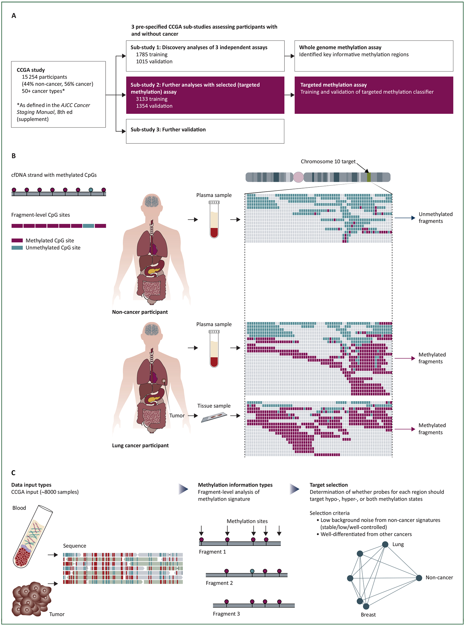
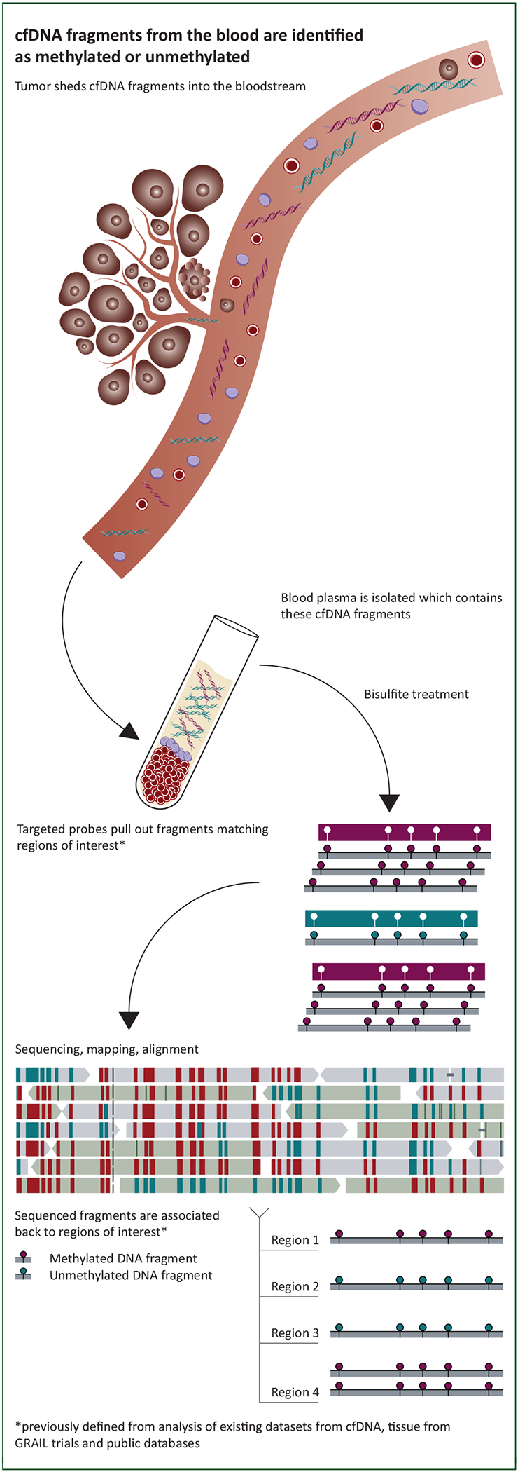
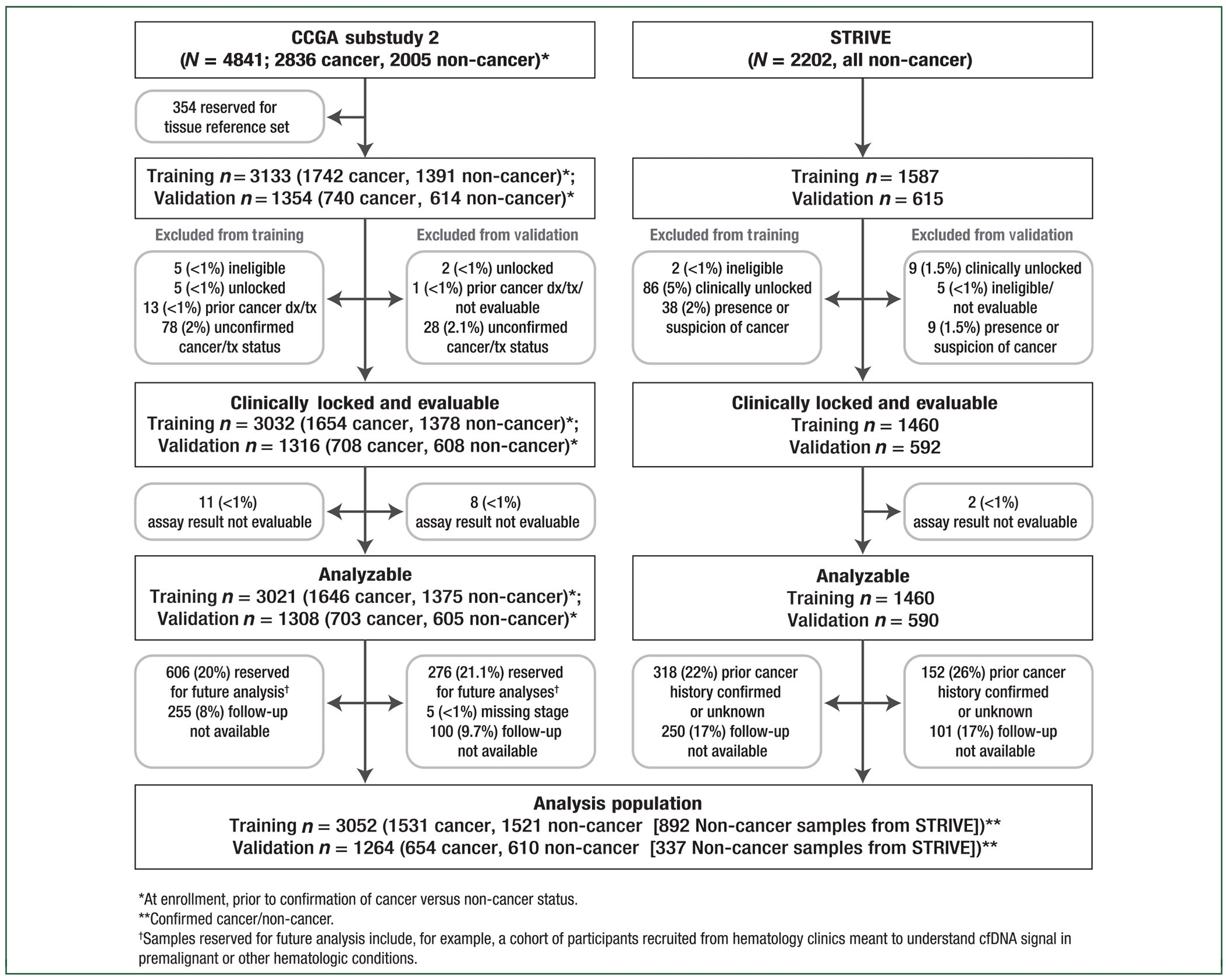
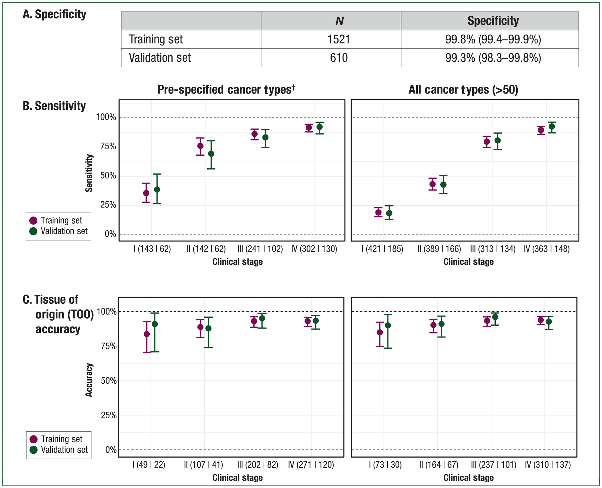
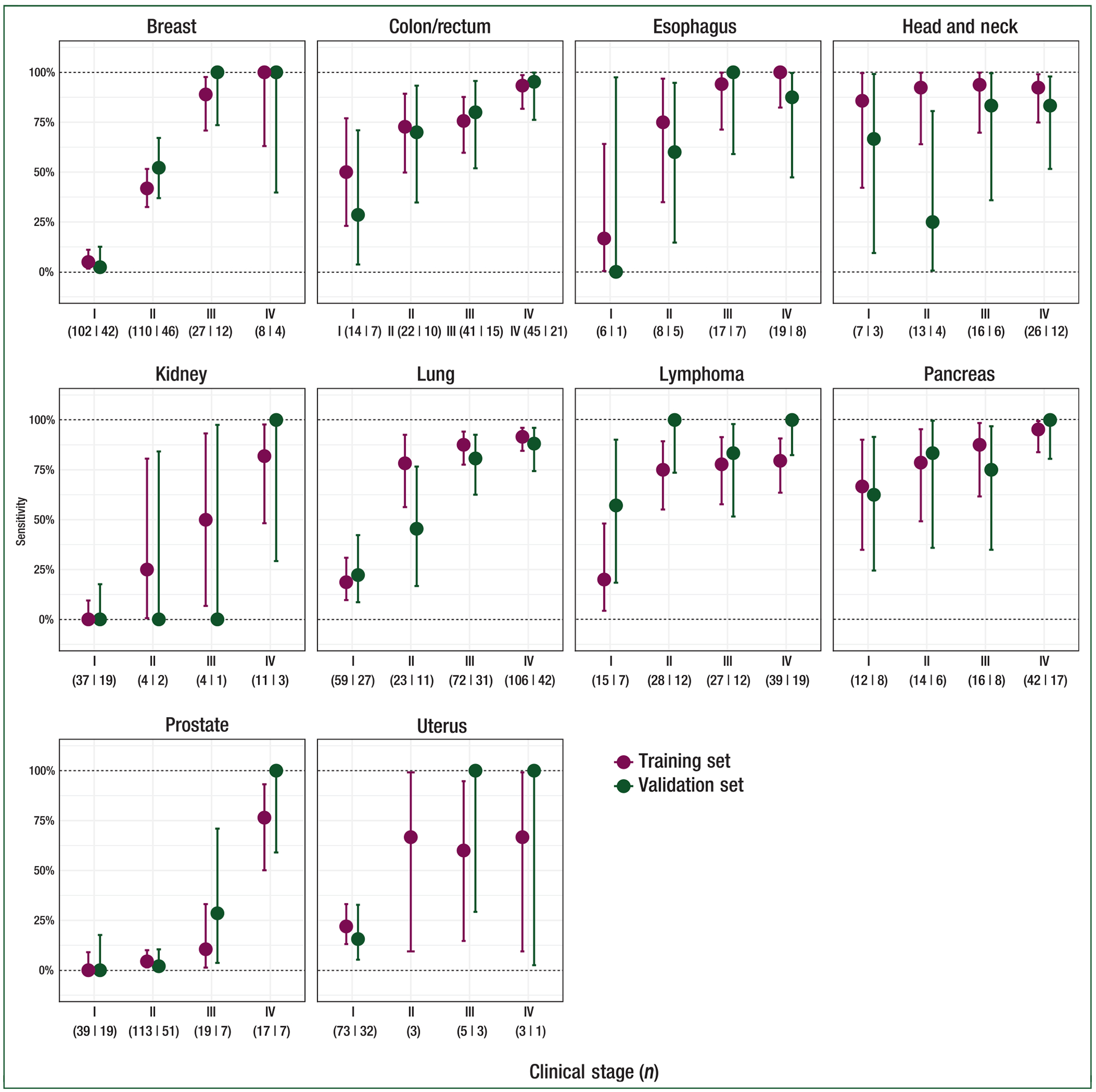
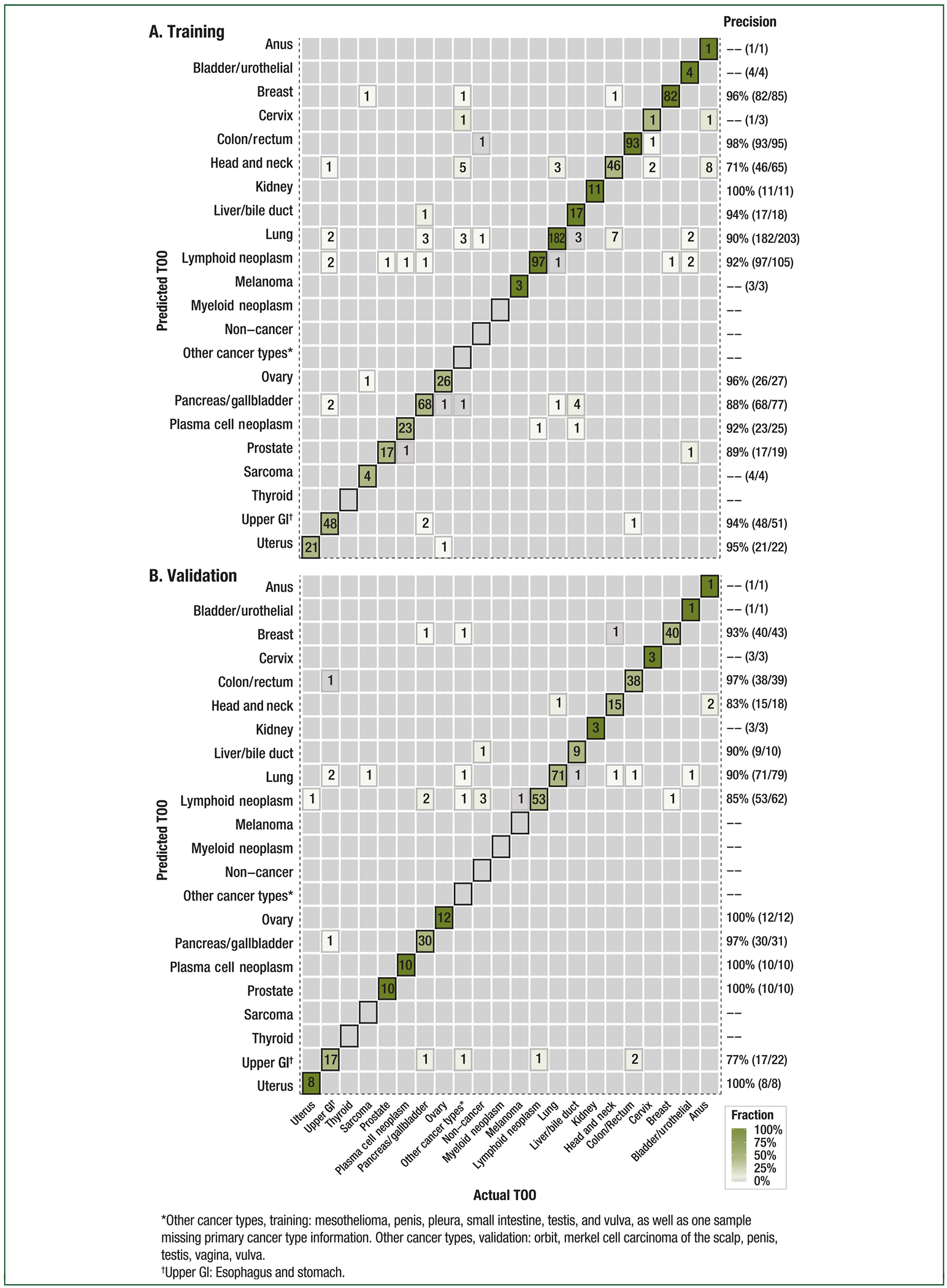
BACKGROUND: Early cancer detection could identify tumors at a time when outcomes are superior and treatment is less morbid. This prospective case-control sub-study (from NCT02889978 and NCT03085888) assessed the performance of targeted methylation analysis of circulating cell-free DNA (cfDNA) to detect and localize multiple cancer types across all stages at high specificity. PARTICIPANTS AND METHODS: The 6689 participants [2482 cancer (>50 cancer types), 4207 non-cancer] were divided into training and validation sets. Plasma cfDNA underwent bisulfite sequencing targeting a panel of >100 000 informative methylation regions. A classifier was developed and validated for cancer detection and tissue of origin (TOO) localization. RESULTS: Performance was consistent in training and validation sets. In validation, specificity was 99.3% [95% confidence interval (CI): 98.3% to 99.8%; 0.7% false-positive rate (FPR)]. Stage I-III sensitivity was 67.3% (CI: 60.7% to 73.3%) in a pre-specified set of 12 cancer types (anus, bladder, colon/rectum, esophagus, head and neck, liver/bile-duct, lung, lymphoma, ovary, pancreas, plasma cell neoplasm, stomach), which account for ∼63% of US cancer deaths annually, and was 43.9% (CI: 39.4% to 48.5%) in all cancer types. Detection increased with increasing stage: in the pre-specified cancer types sensitivity was 39% (CI: 27% to 52%) in stage I, 69% (CI: 56% to 80%) in stage II, 83% (CI: 75% to 90%) in stage III, and 92% (CI: 86% to 96%) in stage IV. In all cancer types sensitivity was 18% (CI: 13% to 25%) in stage I, 43% (CI: 35% to 51%) in stage II, 81% (CI: 73% to 87%) in stage III, and 93% (CI: 87% to 96%) in stage IV. TOO was predicted in 96% of samples with cancer-like signal; of those, the TOO localization was accurate in 93%. CONCLUSIONS: cfDNA sequencing leveraging informative methylation patterns detected more than 50 cancer types across stages. Considering the potential value of early detection in deadly malignancies, further evaluation of this test is justified in prospective population-level studies.
Overview
The claims and statistics of this paper haven’t been checked yet.
Run the check — free, no account needed. It reads the open-access text and checks the sources it cites.
Integrity signals
Registry
Cites
Statistics
Figures
Verification
Claims the paper makes
The claims and statistics of this paper haven’t been checked yet.
Run the check — free, no account needed. It reads the open-access text and checks the sources it cites.
Claims other papers make about this one
Each is checked against this paper’s own text: does it say what the citing paper says it does?
In the citing paper
“Perception-based approaches have been used to classify various disease conditions on the basis of different patterns in methylation of DNA sequencing13, volatile organic compounds using electronic noses14, small metabolites using mass spectrometry15, and image analysis of pathology, computerized tomography scans and magnetic resonance imaging data16,17.”
From Detection of ovarian cancer via the spectral fingerprinting of quantum-defect-modified carbon nanotubes in serum by machine learning (2022) · cites this paper as [13]
Self-check
Percentages, sums and reported differences re-computed from the paper’s own numbers.
The self-check needs the paper’s full text.
Citations
Cites
Initial sequencing and analysis of the human genome
International Human Genome Sequencing Consortium, Whitehead Institute for Biomedical Research, C. F. G. R., Lander, E. S., Linton, L. M., Birren, B., Nusbaum, C., Zody, M. C., Baldwin, J., Devon, K., Dewar, K., et al. (2001) · Nature · doi:10.1038/35057062 · Cited by 24,216
The Human Genome Browser at UCSC
Kent, W. J., Sugnet, C. W., Furey, T. S., Roskin, K. M., Pringle, T. H., Zahler, A. M., & Haussler, A. D. (2002) · Genome Research · doi:10.1101/gr.229102 · Cited by 10,400
Vogelstein, B., Papadopoulos, N., Velculescu, V. E., Zhou, S., Diaz, L. A., & Kinzler, K. W. (2013) · Science · doi:10.1126/science.1235122 · Cited by 7,863
Amin, M. B., Greene, F. L., Edge, S. B., Compton, C. C., Gershenwald, J. E., Brookland, R. K., Meyer, L., Gress, D. M., Byrd, D. R., & Winchester, D. P. (2017) · CA A Cancer Journal for Clinicians · doi:10.3322/caac.21388 · Cited by 6,531
Detection and localization of surgically resectable cancers with a multi-analyte blood test
Cohen, J. D., Li, L., Wang, Y., Thoburn, C., Afsari, B., Danilova, L., Douville, C., Javed, A. A., Wong, F., Mattox, A., et al. (2018) · Science · doi:10.1126/science.aar3247 · Cited by 2,783
Genome-wide cell-free DNA fragmentation in patients with cancer
Cristiano, S., Leal, A., Phallen, J., Fiksel, J., Adleff, V., Bruhm, D. C., Jensen, S. Ø., Medina, J. E., Hruban, C., White, J. R., et al. (2019) · Nature · doi:10.1038/s41586-019-1272-6 · Cited by 1,476
Results of Initial Low-Dose Computed Tomographic Screening for Lung Cancer
The National Lung Screening Trial Research Team (2013) · New England Journal of Medicine · doi:10.1056/nejmoa1209120 · Cited by 1,083
Sensitive tumour detection and classification using plasma cell-free DNA methylomes
Shen, S. Y., Singhania, R., Fehringer, G., Chakravarthy, A., Roehrl, M. H. A., Chadwick, D., Zuzarte, P. C., Borgida, A., Wang, T. T., Li, T., et al. (2018) · Nature · doi:10.1038/s41586-018-0703-0 · Cited by 983
Self-citationMerker, J. D., Oxnard, G. R., Compton, C., Diehn, M., Hurley, P., Lazar, A. J., Lindeman, N., Lockwood, C. M., Rai, A. J., Schilsky, R. L., et al. (2018) · Journal of Clinical Oncology · doi:10.1200/jco.2017.76.8671 · Cited by 959
Self-citationHigh-intensity sequencing reveals the sources of plasma circulating cell-free DNA variants
Razavi, P., Li, B. T., Brown, D. N., Jung, B., Hubbell, E. A., Shen, R., Abida, W. M., Juluru, K., de Bruijn, I., Hou, C., et al. (2019) · Nature Medicine · doi:10.1038/s41591-019-0652-7 · Cited by 808
Self-citation
Research works printed but not linked: 1
- Cohen, 2018, Detection and localization of surgically resectable cancers with a multi-analyte blood test, Science, 359, 926, 10.1126/science.aar3247 · DOI · Paper page
Cited by
Sorted by citations
Items 201–300 of 1,000
FinaleDB: a browser and database of cell-free DNA fragmentation patterns
Zheng, H., Zhu, M. S., & Liu, Y. (2020) · Bioinformatics · doi:10.1093/bioinformatics/btaa999 · Cited by 54
Circulating Tumor DNA: An Emerging Tool in Gastrointestinal Cancers
Alese, O. B., Cook, N., Franco, A. O., Ulanja, M. B., Tan, L., & Tie, J. (2022) · American Society of Clinical Oncology Educational Book · doi:10.1200/edbk_349143 · Cited by 54
Provencio, M., Calvo, V., Romero, A., Spicer, J. D., & Cruz‐Bermúdez, A. (2022) · American Society of Clinical Oncology Educational Book · doi:10.1200/edbk_358995 · Cited by 56
Manoochehri, M., Borhani, N., Gerhäuser, C., Assenov, Y., Schönung, M., Hielscher, T., Christensen, B. C., Lee, M. K., Gröne, H., Lipka, D. B., et al. (2022) · International Journal of Cancer · doi:10.1002/ijc.34337 · Cited by 54
Self-citationReese, K.-L., Pantel, K., & Smit, D. J. (2024) · Journal of Experimental & Clinical Cancer Research · doi:10.1186/s13046-024-03166-w · Cited by 56
Liquid biopsies across the cancer care continuum
Landon, B. V., Annapragada, A. V., Niknafs, N., Velculescu, V. E., & Anagnostou, V. (2025) · Nature Medicine · doi:10.1038/s41591-025-04093-9 · Cited by 55
Critical Factors in the Analytical Work Flow of Circulating Tumor DNA-Based Molecular Profiling
van der Leest, P., & Schuuring, E. M. D. (2024) · Clinical Chemistry · doi:10.1093/clinchem/hvad194 · Cited by 55
Li, P., Li, Z., Linghu, E., & Ji, J. (2024) · Chinese Medical Journal · doi:10.1097/cm9.0000000000003101 · Cited by 53
Gao, Y., Zhao, H., An, K., Liu, Z., Hai, L., Li, R., Zhou, Y., Zhao, W., Jia, Y., Wu, N., et al. (2022) · Clinical and Translational Medicine · doi:10.1002/ctm2.1014 · Cited by 52
Kwon, H., Shin, S. H., Kim, H. H., Min, N. Y., Lim, Y., Joo, T., Lee, K. J., Jeong, M., Kim, H., Yun, S.-Y., et al. (2023) · Scientific Reports · doi:10.1038/s41598-023-40611-w · Cited by 52
Self-citationPhage Cocktails Constrain the Growth of Enterococcus
Wandro, S., Ghatbale, P., Attai, H., Hendrickson, C. L., Samillano, C., Suh, J., Dunham, S. J. B., Pride, D. T., & Whiteson, K. (2022) · mSystems · doi:10.1128/msystems.00019-22 · Cited by 54
The Role of ctDNA in Gastric Cancer
Mencel, J., Slater, S., Cartwright, E., & Starling, N. (2022) · Cancers · doi:10.3390/cancers14205105 · Cited by 54
Liu, F., Beck, S., Yang, L., Luo, H., & Zhang, K. C. (2026) · Nature reviews. Cancer · doi:10.1038/s41568-026-00922-2 · Cited by 57
Pons-Belda, O. D., Fernandez-Uriarte, A., & Diamandis, E. P. (2021) · Diagnostics · doi:10.3390/diagnostics11122171 · Cited by 52
Making the Rounds: Exploring the Role of Circulating Tumor DNA (ctDNA) in Non-Small Cell Lung Cancer
Shields, M. D., Chen, K., Dutcher, G., Patel, I., & Pellini, B. (2022) · International Journal of Molecular Sciences · doi:10.3390/ijms23169006 · Cited by 51
Owens, L., Gulati, R., & Etzioni, R. (2022) · Cancer Epidemiology Biomarkers & Prevention · doi:10.1158/1055-9965.epi-22-0024 · Cited by 49
Crook, T., Leonard, R. C., Mokbel, K., Thompson, A. M., Michell, M. J., Page, R. L., Vaid, A. K., Mehrotra, R., Ranade, A. A., Limaye, S. A., et al. (2022) · Cancers · doi:10.3390/cancers14143341 · Cited by 49
Lapin, M., Edland, K. H., Tjensvoll, K., Oltedal, S., Austdal, M., Garresori, H., Rozenholc, Y., Gilje, B., & Nordgård, O. (2023) · Clinical Cancer Research · doi:10.1158/1078-0432.ccr-22-3526 · Cited by 49
Genetics of Hepatocellular Carcinoma: From Tumor to Circulating DNA
Campani, C., Zucman‐Rossi, J., & Nault, J. (2023) · Cancers · doi:10.3390/cancers15030817 · Cited by 52
Mittelstadt, S., Kelemen, O., Admard, J., Gschwind, A., Koch, A., Wörz, S. E., OBERLECHNER, E., Engler, T., Bonzheim, I., Staebler, A., et al. (2023) · British Journal of Cancer · doi:10.1038/s41416-023-02233-x · Cited by 49
Kim, S. Y., Jeong, S., Lee, W., Jeon, Y., Kim, Y.-J., Park, S., Lee, D., Go, D., Song, S., Lee, S., et al. (2023) · Experimental & Molecular Medicine · doi:10.1038/s12276-023-01119-5 · Cited by 52
Self-citationEarly detection of pancreatic cancer by a high-throughput protease-activated nanosensor assay
Mira, J. L. M., Quentel, A., Patel, R. K., Keith, D., Sousa, M., Minnier, J., Kingston, B. R., David, L. L., Esener, S. C., Sears, R. C., et al. (2025) · Science Translational Medicine · doi:10.1126/scitranslmed.adq3110 · Cited by 49
Artificial intelligence and machine learning in cell-free-DNA-based diagnosticsPreprint
Tsui, W., Ding, S. C., Jiang, P., & Lo, Y. M. D. (2025) · Genome Research · doi:10.1101/gr.278413.123 · Cited by 51
Detection of Cell Types Contributing to Cancer From Circulating, Cell-Free Methylated DNA
Barefoot, M. E., Loyfer, N., Kiliti, A. J., McDeed, A. P., Kaplan, T., & Wellstein, A. (2021) · Frontiers in Genetics · doi:10.3389/fgene.2021.671057 · Cited by 48
The Role of Cell-Free DNA in Cancer Treatment Decision Making
Telekes, A., & Horváth, A. (2022) · Cancers · doi:10.3390/cancers14246115 · Cited by 50
Valenza, C., Saldanha, E. F., Gong, Y., Placido, P. D., Gritsch, D., Ortiz, H., Trapani, D., Conforti, F., Cremolini, C., Peters, S., et al. (2025) · Annals of Oncology · doi:10.1016/j.annonc.2025.03.019 · Cited by 52
Detection of colorectal cancer in urine using DNA methylation analysis
Bach, S., Paulis, I., Sluiter, N. R., Tibbesma, M., Martin, I., van de Wiel, M. A., Tuynman, J. B., Bahce, I., Geert Kazemier, M., & Steenbergen, R. D. M. (2021) · Scientific Reports · doi:10.1038/s41598-021-81900-6 · Cited by 47
Liquid biopsies for residual disease and recurrence
Wan, J. C. M., Mughal, T. I., Razavi, P., Dawson, S., Moss, E. L., Govindan, R., Tan, I. B., Yap, Y. S., Robinson, W. A., Morris, C. D., et al. (2021) · Med · doi:10.1016/j.medj.2021.11.001 · Cited by 48
Philipson, T., Durie, T., Cong, Z., & Fendrick, A. M. (2023) · BMC Health Services Research · doi:10.1186/s12913-023-09738-4 · Cited by 46
Wang, J., Huang, J., Hu, Y., Guo, Q., Zhang, S., Tian, J., Niu, Y., Ji, L., Xu, Y., Tang, P., et al. (2024) · Nature Communications · doi:10.1038/s41467-023-44461-y · Cited by 50
Self-citationDNA methylation in human diseases
Younesian, S., Mohammadi, M. H., Younesian, O., Momeny, M., Ghaffari, S. H., & Bashash, D. (2024) · Heliyon · doi:10.1016/j.heliyon.2024.e32366 · Cited by 48
Circulating cell-free DNA-based methylation patterns for breast cancer diagnosis
Zhang, X., Zhao, D., Yin, Y., Yang, T., You, Z., Li, D., Chen, Y., Jiang, Y., Xu, S., Geng, J., et al. (2021) · npj Breast Cancer · doi:10.1038/s41523-021-00316-7 · Cited by 46
Non-invasive lung cancer diagnosis and prognosis based on multi-analyte liquid biopsy
Chen, K., Sun, J., Zhao, H., Jiang, R., Zheng, J., Li, Z., Peng, J., Shen, H., Zhang, K., Zhao, J., et al. (2021) · Molecular Cancer · doi:10.1186/s12943-021-01323-9 · Cited by 47
Self-citationLiquid Biopsies beyond Mutation Calling: Genomic and Epigenomic Features of Cell-Free DNA in Cancer
Angeles, A. K., Janke, F., Bauer, S., Christopoulos, P., Riediger, A. L., & Sültmann, H. (2021) · Cancers · doi:10.3390/cancers13225615 · Cited by 46
Tracing the Origin of Cell-Free DNA Molecules through Tissue-Specific Epigenetic Signatures
Oberhofer, A., Bronkhorst, A. J., Uhlig, C., Ungerer, V., & Holdenrieder, S. (2022) · Diagnostics · doi:10.3390/diagnostics12081834 · Cited by 46
Current and future applications of liquid biopsy in non-small-cell lung cancer—a narrative review
Tomasik, B., Skrzypski, M. T., Bieńkowski, M., Dziadziuszko, R., & Jassem, J. (2023) · Translational Lung Cancer Research · doi:10.21037/tlcr-22-742 · Cited by 48
Knudsen, A. B., Trentham‐Dietz, A., Kim, J. J., Mandelblatt, J. S., Meza, R., Zauber, A. G., Castle, P. E., & Feuer, E. J. (2023) · JAMA Network Open · doi:10.1001/jamanetworkopen.2023.44698 · Cited by 45
Guo, D., Huang, A., Wang, Y., Zhou, S., Wang, H., Xing, X., Zhang, S., Cheng, J., Xie, K., Yang, Q., et al. (2024) · Clinical and Translational Medicine · doi:10.1002/ctm2.1652 · Cited by 45
Self-citationMarinello, A., Tagliamento, M., Pagliaro, A., Conci, N., Cella, E., Vasseur, D., Remón, J., Lévy, A., Dall’Olio, F. G., & Besse, B. (2024) · Cancer Treatment Reviews · doi:10.1016/j.ctrv.2024.102791 · Cited by 46
Integrated multiomics signatures to optimize the accurate diagnosis of lung cancer
Zhao, M., Xue, G., He, B., Deng, J., Wang, T., Zhong, Y., Li, S., Wang, Y., He, Y., Chen, T., et al. (2025) · Nature Communications · doi:10.1038/s41467-024-55594-z · Cited by 48
Self-citationBoukouris, A. E., Michaelidou, K., Joosse, S. A., Charpidou, A. G., Mavroudis, D., Syrigos, K. N., & Agelaki, S. (2025) · npj Precision Oncology · doi:10.1038/s41698-025-00984-9 · Cited by 46
Chibuk, J., Flory, A., Kruglyak, K. M., Leibman, N. F., Nahama, A., Dharajiya, N. G., van den Boom, D., Jensen, T. J., Friedman, J. S., Shen, M. R., et al. (2021) · Frontiers in Veterinary Science · doi:10.3389/fvets.2021.664718 · Cited by 45
Cholangiocarcinoma: Recent Advances in Molecular Pathobiology and Therapeutic Approaches
Khosla, D., Misra, S., Chu, P. L., Guan, P., Nada, R., Gupta, R., Kaewnarin, K., Ko, T. K., Heng, H. L., Srinivasalu, V. K., et al. (2024) · Cancers · doi:10.3390/cancers16040801 · Cited by 44
“Additionally, the Circulating Cell-free Genome Atlas (CCGA) consortium published two studies that used NGS-based targeted methylation panels to study bisulfite-converted cfDNA in a variety of cancers, including CCA [71,72].”
BackgroundComputational challenges in detection of cancer using cell-free DNA methylation
Sharma, M., Verma, R. K., Kumar, S., & Kumar, V. (2021) · Computational and Structural Biotechnology Journal · doi:10.1016/j.csbj.2021.12.001 · Cited by 44
Metasurface Biosensors Enabling Single-Molecule Sensing of Cell-Free DNA
Iwanaga, M., Hironaka, T., Ikeda, N., Sugasawa, T., & Takekoshi, K. (2023) · Nano Letters · doi:10.1021/acs.nanolett.3c01527 · Cited by 47
Cost-Effectiveness of Liquid Biopsy for Colorectal Cancer Screening in Patients Who Are Unscreened
Aziz, Z., Wagner, S., Agyekum, A. A., Pumpalova, Y. S., Prest, M. T., Lim, F., Rustgi, S. D., Kastrinos, F., Grady, W. M., & Hur, C. (2023) · JAMA Network Open · doi:10.1001/jamanetworkopen.2023.43392 · Cited by 49
The biomolecule corona of lipid nanoparticles contains circulating cell-free DNA
Gardner, L., Warrington, J. I., Rogan, J., Rothwell, D. G., Brady, G., Dive, C., Kostarelos, K., & Hadjidemetriou, M. (2020) · Nanoscale Horizons · doi:10.1039/d0nh00333f · Cited by 44
Validation of a Transcriptome-Based Assay for Classifying Cancers of Unknown Primary Origin
Michuda, J., Breschi, A., Kapilivsky, J., Manghnani, K., McCarter, C., Hockenberry, A. J., Mineo, B., Igartua, C., Dudley, J. T., Stumpe, M. C., et al. (2023) · Molecular Diagnosis & Therapy · doi:10.1007/s40291-023-00650-5 · Cited by 43
Moss, J., Ben‐Ami, R., Shai, E., Gal-Rosenberg, O., Kalish, Y., Klochendler, A., Cann, G. M., Gläser, B., Arad, A., Shemer, R., et al. (2023) · Nature Communications · doi:10.1038/s41467-023-43310-2 · Cited by 43
Self-citationProtein biomarkers and alternatively methylated cell-free DNA detect early stage pancreatic cancer
Ben‐Ami, R., Wang, Q.-L., Zhang, J., Supplee, J., Fahrmann, J. F., Lehmann‐Werman, R., Brais, L. K., Nowak, J. A., Yuan, C., Loftus, M., et al. (2023) · Gut · doi:10.1136/gutjnl-2023-331074 · Cited by 43
Self-citationPhotonic Technologies for Liquid Biopsies: Recent Advances and Open Research Challenges
Dell’Olio, F., Su, J., Huser, T. R., Sottile, V., Cortés‐Hernández, L. E., & Alix‐Panabières, C. (2020) · Laser & Photonics Review · doi:10.1002/lpor.202000255 · Cited by 44
Non-Invasive Biomarkers for Earlier Detection of Pancreatic Cancer—A Comprehensive Review
Brezgyte, G., Shah, V. M., Jach, D., & Crnogorac‐Jurcevic, T. (2021) · Cancers · doi:10.3390/cancers13112722 · Cited by 46
Herbst, J., Pantel, K., Effenberger, K. E., & Wikman, H. (2022) · British Journal of Cancer · doi:10.1038/s41416-022-01868-6 · Cited by 43
Tafazzoli, A., Ramsey, S. D., Shaul, A., Chavan, A., Ye, W., Kansal, A. R., Ofman, J., & Fendrick, A. M. (2022) · PharmacoEconomics · doi:10.1007/s40273-022-01181-3 · Cited by 43
Wu, H., Guo, S., Liu, X., Li, Y., Su, Z., He, Q., Liu, X., Zhang, Z., Yu, L., Shi, X., et al. (2022) · BMC Medicine · doi:10.1186/s12916-022-02647-z · Cited by 43
Mutations in normal tissues—some diagnostic and clinical implications
Fiala, C., & Diamandis, E. P. (2020) · BMC Medicine · doi:10.1186/s12916-020-01763-y · Cited by 42
Ibrahim, J., de Beeck, K. O., Fransén, E., Peeters, M., & Camp, G. V. (2022) · Molecular Oncology · doi:10.1002/1878-0261.13176 · Cited by 42
Modelled mortality benefits of multi-cancer early detection screening in England
Sasieni, P. D., Smittenaar, R., Hubbell, E. A., Broggio, J., Neal, R. D., & Swanton, C. (2023) · British Journal of Cancer · doi:10.1038/s41416-023-02243-9 · Cited by 41
Self-citationExpanding the Reach and Grasp of Lung Cancer Screening
Osarogiagbon, R. U., Yang, P., & Sequist, L. V. D. (2023) · American Society of Clinical Oncology Educational Book · doi:10.1200/edbk_389958 · Cited by 41
Cerebrospinal fluid: A unique source of circulating tumor DNA with broad clinical applications
Hickman, R. A., Miller, A. M., & Arcila, M. E. (2023) · Translational Oncology · doi:10.1016/j.tranon.2023.101688 · Cited by 42
Aredo, J. V., Jamali, A., Zhu, J., Heater, N. K., Wakelee, H. A., Vaklavas, C., Anagnostou, V., & Lu, J. M. (2025) · American Society of Clinical Oncology Educational Book · doi:10.1200/edbk-25-481114 · Cited by 42
Sahoo, K., Lingasamy, P., Khatun, M., Sudhakaran, S. L., Salumets, A., Sundararajan, V., & Modhukur, V. (2025) · Epigenetics & Chromatin · doi:10.1186/s13072-025-00595-5 · Cited by 42
Pan-Cancer Early Detection: Hype or Hope?
Srivastava, S., & Hanash, S. (2020) · Cancer Cell · doi:10.1016/j.ccell.2020.05.021 · Cited by 41
Liquid biopsy versus tumor biopsy for clinical-trial recruitment
Corcoran, R. B. (2020) · Nature Medicine · doi:10.1038/s41591-020-01169-6 · Cited by 42
Vittone, J. L., Gill, D. M., Goldsmith, A., Klein, E. A., & Karlitz, J. J. (2024) · npj Precision Oncology · doi:10.1038/s41698-024-00568-z · Cited by 40
Self-citationState-of-the-Art Management of Colorectal Cancer: Treatment Advances and Innovation
Pathak, P., Chan, G. H. J., Deming, D. A., & Chee, C. E. (2024) · American Society of Clinical Oncology Educational Book · doi:10.1200/edbk_438466 · Cited by 43
Integration of multiomics features for blood-based early detection of colorectal cancer
Gao, Y., Cao, D., Li, M., Zhao, F., Wang, P., Mei, S., Song, Q., Wang, P., Nie, Y., Zhao, W., et al. (2024) · Molecular Cancer · doi:10.1186/s12943-024-01959-3 · Cited by 40
Self-citationLongitudinal ultrasensitive ctDNA monitoring for high-resolution lung cancer risk prediction
Black, J. R., Karasaki, T., Abbott, C. W., Li, B., Veeriah, S., Bakir, M. A., Liu, W. K., Huebner, A., Martínez‐Ruiz, C., Pawlik, P., et al. (2025) · Cell · doi:10.1016/j.cell.2025.10.020 · Cited by 42
Self-citationDNA methylation age as a biomarker for cancer
Lau, C. E., & Robinson, O. (2021) · International Journal of Cancer · doi:10.1002/ijc.33451 · Cited by 40
From APC to the genetics of hereditary and familial colon cancer syndromes
Olkinuora, A. P., Peltomäki, P., Aaltonen, L. A., & Rajamäki, K. (2021) · Human Molecular Genetics · doi:10.1093/hmg/ddab208 · Cited by 41
Combinatorial Power of cfDNA, CTCs and EVs in Oncology
Keup, C., Kimmig, R., & Kasimir‐Bauer, S. (2022) · Diagnostics · doi:10.3390/diagnostics12040870 · Cited by 40
Circulating Cell-Free Tumour DNA for Early Detection of Pancreatic Cancer
Jaworski, J. J., Morgan, R. D., & Sivakumar, S. (2020) · Cancers · doi:10.3390/cancers12123704 · Cited by 39
Somatic variation in normal tissues: friend or foe of cancer early detection?
Acha‐Sagredo, A., Ganguli, P., & Ciccarelli, F. D. (2022) · Annals of Oncology · doi:10.1016/j.annonc.2022.09.156 · Cited by 41
DNA methylation drives a new path in gastric cancer early detection: Current impact and prospects
Wang, X., Dong, Y., Zhang, H., Zhao, Y., Miao, T., Mohseni, G., Du, L., & Wang, C. (2023) · Genes & Diseases · doi:10.1016/j.gendis.2023.02.038 · Cited by 39
Self-citationLeeVan, E., & Pinsky, P. F. (2023) · Clinical Chemistry · doi:10.1093/clinchem/hvad134 · Cited by 39
Artificial intelligence in oncology: From bench to clinic
Elkhader, J. A., & Elemento, O. (2021) · Seminars in Cancer Biology · doi:10.1016/j.semcancer.2021.04.013 · Cited by 39
Meng, Z., Ren, Q., Zhong, G., Li, S., Chen, Y., Wu, W., Feng, Y., Mao, M., Zhang, F., & Long, G. (2021) · Journal of Molecular Diagnostics · doi:10.1016/j.jmoldx.2021.06.003 · Cited by 39
Xiao, Y., Ju, L., Qian, K., Jin, W., Wang, G., Zhao, Y., Jiang, W. I., Liu, N., Wu, K., Peng, M., et al. (2022) · Clinical and Translational Medicine · doi:10.1002/ctm2.1008 · Cited by 39
Self-citationPrediction of tissue-of-origin of early stage cancers using serum miRNomes
Matsuzaki, J., Kato, K., Oono, K., Tsuchiya, N., Sudo, K., Shimomura, A., Tamura, K., Shiino, S., Kinoshita, T., Daiko, H., et al. (2022) · JNCI Cancer Spectrum · doi:10.1093/jncics/pkac080 · Cited by 40
Heidrich, I., Abdalla, T. S. A., Reeh, M., & Pantel, K. (2021) · Cancers · doi:10.3390/cancers13184500 · Cited by 38
Using all our genomes: Blood‐based liquid biopsies for the early detection of cancer
Adams, E. W., Sepich‐Poore, G. D., Miller‐Montgomery, S., & Knight, R. D. (2022) · Open Research (University of Surrey) · doi:10.1002/viw.20200118 · Cited by 38
Patient Preferences for Multi-Cancer Early Detection (MCED) Screening Tests
Gelhorn, H. L., Ross, M., Kansal, A. R., Fung, E. T., Seiden, M. V., Krucien, N., & Chung, K. (2022) · Patient · doi:10.1007/s40271-022-00589-5 · Cited by 38
Self-citationProspects and Current Challenges of Extracellular Vesicle-Based Biomarkers in Cancer
Lawrence, S. R., & Shah, K. M. (2024) · Biology · doi:10.3390/biology13090694 · Cited by 39
Iden, C. R., Mustafa, S. M., Øgaard, N., Henriksen, T. V., Jensen, S. Ø., Ahlborn, L. B., Egebjerg, K., Bæksgaard, L., Garbyal, R. S., Nedergaard, M. K., et al. (2024) · Gastric Cancer · doi:10.1007/s10120-024-01556-9 · Cited by 38
From concept to clinic: a roadmap for DNA methylation biomarkers in liquid biopsies
Pharo, H. D., Vedeld, H. M., Sjurgard, I. V., Pinto, R., & Lind, G. E. (2025) · Oncogene · doi:10.1038/s41388-025-03624-5 · Cited by 40
Liquid Biopsy is Instrumental for 3PM Dimensional Solutions in Cancer Management
Líšková, A., Samec, M., Koklesová, L., Giordano, F. A., Kubatka, P., & Golubnitschaja, O. (2020) · Journal of Clinical Medicine · doi:10.3390/jcm9092749 · Cited by 38
Genome-wide study of the effect of blood collection tubes on the cell-free DNA methylome
Paemel, R. V., Koker, A. D., Caggiano, C., Morlion, A., Mestdagh, P., Wilde, B. D., Vandesompele, J., & Preter, K. D. (2020) · Epigenetics · doi:10.1080/15592294.2020.1827714 · Cited by 38
Guo, X. M., Miller, H., Matsuo, K., Roman, L. D., & Salhia, B. (2021) · Cancers · doi:10.3390/cancers13040838 · Cited by 38
Network approach in liquidomics landscape
Santini, D., Botticelli, A., Galvano, A., Iuliani, M., Incorvaia, L., Gristina, V., Taffon, C., Foderaro, S., Paccagnella, E., Simonetti, S., et al. (2023) · Journal of Experimental & Clinical Cancer Research · doi:10.1186/s13046-023-02743-9 · Cited by 37
Nguyễn, L. H. Đ., Nguyễn, T. H. H., Le, V. H., Quang, B. V., Nguyen, L. H., Phạm, N. H., Phan, T. H., Nguyen, H. T., Tran, V. S., Bui, C. V., et al. (2025) · BMC Medicine · doi:10.1186/s12916-025-03929-y · Cited by 37
Precision Oncology: Current Landscape, Emerging Trends, Challenges, and Future Perspectives
Qiao, D., Wang, R. C., & Wang, Z. (2025) · Cells · doi:10.3390/cells14221804 · Cited by 39
Clinical implementation and current advancement of blood liquid biopsy in cancer
Watanabe, K., Nakamura, Y., & Low, S. (2021) · Journal of Human Genetics · doi:10.1038/s10038-021-00939-5 · Cited by 37
Memarzia, A., Saadat, S., Asgharzadeh, F., Behrouz, S., Folkerts, G., & Boskabady, M. H. (2023) · Journal of Cellular and Molecular Medicine · doi:10.1111/jcmm.17936 · Cited by 37
Lokshin, A. E., Bast, R. C., & Rodland, K. (2021) · Cancers · doi:10.3390/cancers13040802 · Cited by 37
Prenatal BRCA1 epimutations contribute significantly to triple-negative breast cancer development
Nikolaienko, O., Eikesdal, H. P., Ognedal, E., Gilje, B., Lundgren, S., Blix, E. S., Espelid, H., Geisler, J., Geisler, S. B., Janssen, E. A. M., et al. (2023) · Genome Medicine · doi:10.1186/s13073-023-01262-8 · Cited by 36
A high-throughput test enables specific detection of hepatocellular carcinoma
Cheishvili, D., Wong, C., Karim, M. M., Kibria, M. G., Jahan, N., Das, P. C., Yousuf, M. A. K., Islam, M. A., Das, D. C., Noor-E-Alam, S. M., et al. (2023) · Nature Communications · doi:10.1038/s41467-023-39055-7 · Cited by 36
Detection of ovarian cancer using plasma cell-free DNA methylomes
Lu, H., Liu, Y., Wang, J., Fu, S., Wang, L., Huang, C., Li, J., Xie, L., Wang, D., Li, D., et al. (2022) · Clinical Epigenetics · doi:10.1186/s13148-022-01285-9 · Cited by 35
García‐Ortiz, M. V., Cano-Ramírez, P., Toledano‐Fonseca, M., Aranda, E., & Rodríguez‐Ariza, A. (2023) · Biomarker Research · doi:10.1186/s40364-023-00528-y · Cited by 35
Gao, M., Wu, X., Jiao, X., Hu, Y., Wang, Y., Zhuo, N., Dong, F., Wang, Y., Wang, F., Cao, Y., et al. (2024) · Journal for ImmunoTherapy of Cancer · doi:10.1136/jitc-2022-006616 · Cited by 35
Self-citationLiquid biopsy: An arsenal for tumour screening and early diagnosis
Zhang, Q., Zhang, X., Xie, P., & Zhang, W. (2024) · Cancer Treatment Reviews · doi:10.1016/j.ctrv.2024.102774 · Cited by 36
Metadata
- Authors
- Liu, M. C., Oxnard, G. R., Klein, E. A., Swanton, C., Seiden, M. V., Liu, M. C., Oxnard, G. R., Klein, E. A., Smith, D., Richards, D., Yeatman, T. J., Cohn, A. L., Lapham, R., Clement, J., Parker, A. S., Tummala, M. K., McIntyre, K., Sekeres, M. A., Bryce, A. H., Siegel, R., Wang, X., Cosgrove, D. P., Abu-Rustum, N. R., Trent, J., Thiel, D. D., Becerra, C., Agrawal, M., Garbo, L. E., Giguere, J. K., Michels, R. M., Harris, R. P., Richey, S. L., McCarthy, T. A., Waterhouse, D. M., Couch, F. J., Wilks, S. T., Krie, A. K., Balaraman, R., Restrepo, A., Meshad, M. W., Rieger-Christ, K., Sullivan, T., Lee, C. M., Greenwald, D. R., Oh, W., Tsao, C.-K., Fleshner, N., Kennecke, H. F., Khalil, M. F., Spigel, D. R., Manhas, A. P., Ulrich, B. K., Kovoor, P. A., Stokoe, C., Courtright, J. G., Yimer, H. A., Larson, T. G., Swanton, C., Seiden, M. V., Cummings, S. R., Absalan, F., Alexander, G., Allen, B., Amini, H., Aravanis, A. M., Bagaria, S., Bazargan, L., Beausang, J. F., Berman, J., Betts, C., Blocker, A., Bredno, J., Calef, R., Cann, G., Carter, J., Chang, C., Chawla, H., Chen, X., Chien, T. C., Civello, D., Davydov, K., Demas, V., Desai, M., Dong, Z., Fayzullina, S., Fields, A. P., Filippova, D., Freese, P., Fung, E. T., Gnerre, S., Gross, S., Halks-Miller, M., Hall, M. P., Hartman, A.-R., Hou, C., Hubbell, E., Hunkapiller, N., Jagadeesh, K., Jamshidi, A., Jiang, R., Jung, B., Kim, T., Klausner, R. D., Kurtzman, K. N., Lee, M., Lin, W., Lipson, J., Liu, H., Liu, Q., Lopatin, M., Maddala, T., Maher, M. C., Melton, C., Mich, A., Nautiyal, S., Newman, J., Newman, J., Nicula, V., Nicolaou, C., Nikolic, O., Pan, W., Patel, S., Prins, S. A., Rava, R., Ronaghi, N., Sakarya, O., Satya, R. V., Schellenberger, J., Scott, E., Sehnert, A. J., Shaknovich, R., Shanmugam, A., Shashidhar, K. C., Shen, L., Shenoy, A., Shojaee, S., Singh, P., Steffen, K. K., Tang, S., Toung, J. M., Valouev, A., Venn, O., Williams, R. T., Wu, T., Xu, H. H., Yakym, C., Yang, X., Yecies, J., Yip, A. S., Youngren, J., Yue, J., Zhang, J., Zhang, L., Zhang, L. (., Zhang, N., Curtis, C., & Berry, D. A.
- Affiliations
- Compass Oncology (US)
- Texas Oncology (US)
- Rocky Mountain Cancer Centers (US)
- Spartanburg Regional Healthcare System (US)
- Hartford Hospital (US)
- Mayo Clinic in Florida (US)
- Texas Health Dallas (US)
- Cleveland Clinic (US)
- Mayo Clinic (US)
- University of Miami Health System (US)
- Maryland Oncology Hematology (US)
- New York Oncology Hematology (US)
- Prisma Health (US)
- Virginia Cancer Specialists (US)
- Oncology Hematology Care (US)
- Avera Health (US)
- Lahey Hospital and Medical Center (US)
- Sansum Medical Clinic (US)
- Mount Sinai Hospital (US)
- University of Toronto (CA)
- Benaroya Research Institute (US)
- Lehigh Valley Health Network (US)
- Sarah Cannon (US)
- Minnesota Oncology (US)
- Sutter Health (US)
- Grail (United States) (US)
- Stanford University (US)
- The University of Texas MD Anderson Cancer Center (US)
- ORCID
- Smith, David
- Yeatman, Timothy J.
- Clement, Jessica
- Parker, Alexander S.
- McIntyre, Kristi
- Sekeres, Mikkael A.
- Bryce, Alan H.
- Siegel, Robert
- Wang, Xuezhong
- Cosgrove, David P.
- Trent, Jonathan
- Thiel, David D.
- Becerra, Carlos
- Agrawal, Manish
- Michels, Ross M.
- Richey, Stephen L.
- Waterhouse, David M.
- Couch, Fergus J.
- Wilks, Sharon T.
- Krie, Amy K.
- Sullivan, Travis
- Lee, Christine M.
- Oh, William
- Tsao, Che-Kai
- Fleshner, Neil
- Kennecke, Hagen F.
- Spigel, David R.
- Larson, Timothy G.
- Cummings, Steven R.
- Alexander, Gregory
- Amini, Hamed
- Aravanis, Alexander M.
- Bagaria, Siddhartha
- Blocker, Alexander
- Bredno, Joerg
- Chang, Christopher
- Chen, Xiaoji
- Fields, Alexander P.
- Fung, Eric T.
- Hall, Megan P.
- Hartman, Anne-Renee
- Hubbell, Earl
- Jagadeesh, Karthik
- Jamshidi, Arash
- Jung, Byoungsok
- Kim, TaeHyung
- Kurtzman, Kathryn N.
- Lipson, Jafi
- Lopatin, Margarita
- Maher, M. Cyrus
- Nautiyal, Shivani
- Newman, Jonathan
- Nicolaou, Cosmos
- Pan, Wenying
- Scott, Eric
- Sehnert, Amy J.
- Shaknovich, Rita
- Shen, Ling
- Williams, Richard T.
- Yang, Xiao
- Youngren, Jack
- Zhang, Lily
- Zhang, Nan
- Curtis, Christina
- Berry, Donald A.
- Venue
- Annals of Oncology
- Publisher
- Elsevier BV
- Published
- 2020-06
- Volume
- 31
- Issue
- 6
- Pages
- 745-759
- ISSN
- 0923-7534 (ISSN-L)
- PMC
- PMC8274402
- OpenAlex
- W3013924915
- Cited by
- 1,495
- References
- 28
- Citations by year
- 2026: 202 · 2025: 245 · 2024: 253 · 2023: 267 · 2022: 248 · 2021: 205 · 2020: 74
- FWCI
- 70.07
- Type
- journal article
- Language
- en
- Open access
- hybrid
- doi.org
- Licence
- Locations
- Annals of Oncology (publishedVersion, cc-by-nc-nd)
- PubMed (publishedVersion)
- PubMed Central (acceptedVersion, cc-by-nc-nd)
- UCL Discovery (University College London) (submittedVersion)
- Figshare (submittedVersion, cc-by-nc-nd)
- Funders
- GRAIL, Inc
- Princess Margaret Cancer Centre’s McCain GU BioBank
- Francis Crick Institute
- Cancer Research UK (FC001169, FC001202)
- UK Medical Research Council (FC001169, FC001202)
- Wellcome Trust (FC001169, FC001202)
- CRUK Lung Cancer Centre of Excellence
- Rosetrees Trust
- Breast Cancer Research Foundation
- Topics
- Life Sciences › Biochemistry, Genetics and Molecular Biology › Cancer Research › Cancer Genomics and Diagnostics
- Life Sciences › Biochemistry, Genetics and Molecular Biology › Molecular Biology › Epigenetics and DNA Methylation
- Life Sciences › Biochemistry, Genetics and Molecular Biology › Molecular Biology › Advanced biosensing and bioanalysis techniques
- Keywords
- multi-cancer early detection, cell-free DNA, DNA methylation signature, liquid biopsy, tumor stage, bisulfite sequencing, false positive rate, early-stage cancer detection, cancer mortality reduction
- MeSH terms
- Cell-Free Nucleic Acids, DNA, Neoplasm, Female, Humans, Neoplasms, Prospective Studies, Biomarkers, Tumor, DNA Methylation
About these checks
Registry, retraction, Cites and statistics results are computed, not judged, with one exception: a printed reference that no index lists is read once by a model before it counts as a citation error. Claims are extracted by a model and kept only when their quote appears verbatim in the text; each cited source is then checked, with a model’s help, for whether it says what the claim relies on. Those verdicts can be wrong — read the claim’s quote and the cited source before relying on one.
Statistics and arithmetic are recomputed, not judged.
Confidence bands are shown only for fields calibrated on our annotated test set. None is calibrated yet, so no band is shown.
What counts as a claim ›How we measure confidence ›How we check this ›
Are you an author, or do you see a problem with this page? Request a review
