

The DOI matches its Crossref record; 6 of the 6 references it cites were found in a registry or an index.
Click chemistry extracellular vesicle/peptide/chemokine nanocarriers for treating central nervous system injuries
Overview
The claims and statistics of this paper haven’t been checked yet.
Run the check — free, no account needed. It reads the open-access text and checks the sources it cites.
Integrity signals
Registry
Cites
Statistics
Figures
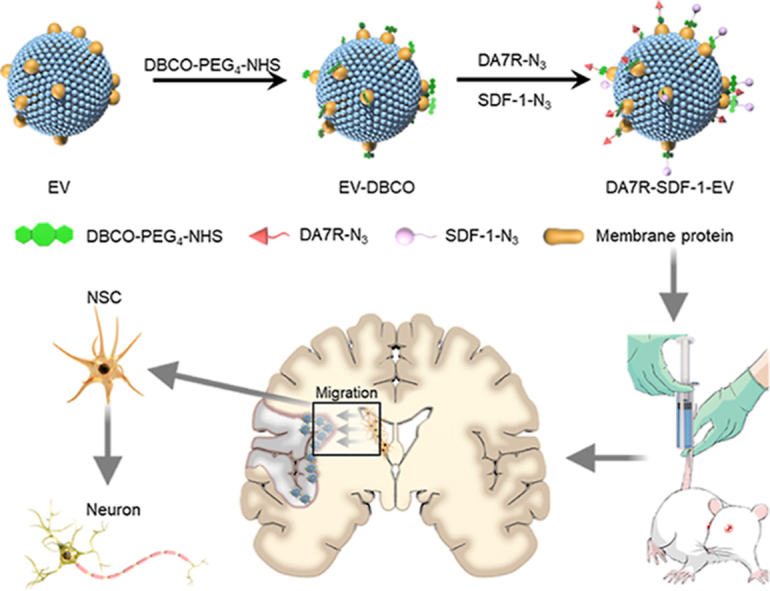
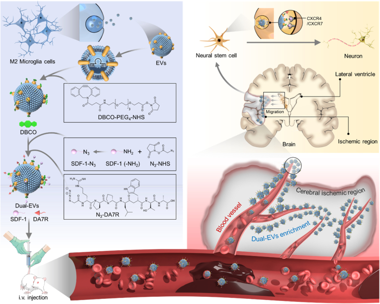
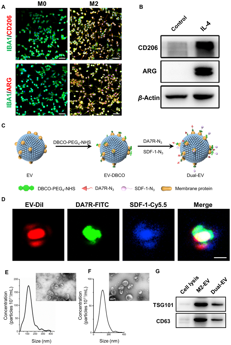
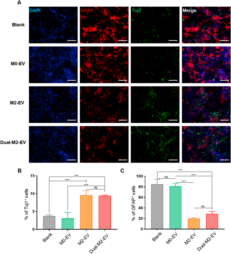
Verification
Claims the paper makes
The claims and statistics of this paper haven’t been checked yet.
Run the check — free, no account needed. It reads the open-access text and checks the sources it cites.
Claims other papers make about this one
Each is checked against this paper’s own text: does it say what the citing paper says it does?
In the citing paper
“IL-4- or IL-13-stimulated microglia can communicate with NSC, promoting neurogenesis and facilitating brain tissue repair [13, 14].”
From M2 microglia-derived small extracellular vesicles modulate NSC fate after ischemic stroke via miR-25-3p/miR-93-5p-TGFBR/PTEN/FOXO3 axis (2025) · cites this paper as [14]
Self-check
Percentages, sums and reported differences re-computed from the paper’s own numbers.
The self-check needs the paper’s full text.
Citations
Cites
Neuroinflammation and microglial activation in Alzheimer disease: where do we go from here?
Leng, F., & Edison, P. (2021) · Nature Reviews Neurology · doi:10.1038/s41582-020-00435-y · Cited by 3,179
RNA delivery by extracellular vesicles in mammalian cells and its applications
O’Brien, K., Breyne, K., Ughetto, S., Laurent, L. C., & Breakefield, X. O. (2020) · Nature Reviews Molecular Cell Biology · doi:10.1038/s41580-020-0251-y · Cited by 1,811
Zhou, B. O., Yue, R., Murphy, M. M., Peyer, J. G., & Morrison, S. J. (2014) · Cell Stem Cell · doi:10.1016/j.stem.2014.06.008 · Cited by 1,381 · cited 1×
“Acta Pharmaceutica Sinica B 2023;13(5):2202e2218”
Adult haematopoietic stem cell niches
Crane, G. M., Jeffery, E., & Morrison, S. J. (2017) · Nature Reviews Immunology · doi:10.1038/nri.2017.53 · Cited by 712
Song, Y., Li, Z., He, T., Qu, M., Jiang, L., Li, W., Shi, X., Pan, J., Zhang, L., Wang, Y., et al. (2019) · Theranostics · doi:10.7150/thno.30879 · Cited by 487
Self-citationNeural stem cell therapy for subacute and chronic ischemic stroke
Boese, A. C., Le, Q.-S. E., Pham, D., Hamblin, M. H., & Lee, J.-P. (2018) · Stem Cell Research & Therapy · doi:10.1186/s13287-018-0913-2 · Cited by 205
Research works printed but not linked: 94
- Xia, 2018, Tissue repair and regeneration with endogenous stem cells, Nat Rev Mater, 3, 174, 10.1038/s41578-018-0027-6 · DOI
- Bernardo, 2013, Mesenchymal stromal cells: sensors and switchers of inflammation, Cell Stem Cell, 13, 392, 10.1016/j.stem.2013.09.006 · DOI
- Xia, 2021, Stem cell recruitment based on scaffold features for bone tissue engineering, Biomater Sci, 9, 1189, 10.1039/D0BM01591A · DOI
- Blanpain, 2010, Stem cells: skin regeneration and repair, Nature, 464, 686, 10.1038/464686a · DOI
- Grégoire, 2015, Endogenous neural stem cell responses to stroke and spinal cord injury, Glia, 63, 1469, 10.1002/glia.22851 · DOI
- Yiu, 2006, Glial inhibition of CNS axon regeneration, Nat Rev Neurosci, 7, 617, 10.1038/nrn1956 · DOI
- Bernstock, 2017, Neural stem cell transplantation in ischemic stroke: a role for preconditioning and cellular engineering, J Cereb Blood Flow Metab, 37, 2314, 10.1177/0271678X17700432 · DOI
- Li, 2018, A broad-spectrum ROS-eliminating material for prevention of inflammation and drug-induced organ toxicity, Adv Sci (Weinh), 5
- Luo, 2019, Transplantation of NSCs promotes the recovery of cognitive functions by regulating neurotransmitters in rats with traumatic brain injury, Neurochem Res, 44, 2765, 10.1007/s11064-019-02897-z · DOI
- Uezono, 2018, Prior treatment with anti-high mobility group Box-1 antibody boosts human neural stem cell transplantation-mediated functional recovery after spinal cord injury, Stem Cell, 36, 737, 10.1002/stem.2802 · DOI
- Deng, 2019, EMSCs build an all-in-one niche via cell‒cell lipid raft assembly for promoted neuronal but suppressed astroglial differentiation of neural stem cells, Adv Mater, 31
- Xin, 2017, MicroRNA cluster miR-17-92 cluster in exosomes enhance neuroplasticity and functional recovery after stroke in rats, Stroke, 48, 747, 10.1161/STROKEAHA.116.015204 · DOI
- Williams, 2019, Mesenchymal stem cell-derived exosomes provide neuroprotection and improve long-term neurologic outcomes in a swine model of traumatic brain injury and hemorrhagic shock, J Neurotrauma, 36, 54, 10.1089/neu.2018.5711 · DOI
- Zhang, 2019, Exosomes-beyond stem cells for restorative therapy in stroke and neurological injury, Nat Rev Neurol, 15, 193, 10.1038/s41582-018-0126-4 · DOI · Paper page
- Webb, 2018, Human neural stem cell extracellular vesicles improve recovery in a porcine model of ischemic stroke, Stroke, 49, 1248, 10.1161/STROKEAHA.117.020353 · DOI
- Webb, 2018, Human neural stem cell extracellular vesicles improve tissue and functional recovery in the murine thromboembolic stroke model, Transl Stroke Res, 9, 530, 10.1007/s12975-017-0599-2 · DOI
- Vay, 2018, The plasticity of primary microglia and their multifaceted effects on endogenous neural stem cells in vitro and in vivo, J Neuroinflammation, 15, 226, 10.1186/s12974-018-1261-y · DOI
- Kabba, 2018, Microglia: housekeeper of the central nervous system, Cell Mol Neurobiol, 38, 53, 10.1007/s10571-017-0504-2 · DOI
- Wang, 2020, Peripheral nerve resident macrophages share tissue-specific programming and features of activated microglia, Nat Commun, 11, 2552, 10.1038/s41467-020-16355-w · DOI
- Armstrong, 2017, Re-engineering extracellular vesicles as smart nanoscale therapeutics, ACS Nano, 11, 69, 10.1021/acsnano.6b07607 · DOI
- Kooijmans, 2016, PEGylated and targeted extracellular vesicles display enhanced cell specificity and circulation time, J Control Release, 224, 77, 10.1016/j.jconrel.2016.01.009 · DOI
- Chowdhury, 2019, Dendronized fluorosurfactant for highly stable water-in-fluorinated oil emulsions with minimal inter-droplet transfer of small molecules, Nat Commun, 10, 4546, 10.1038/s41467-019-12462-5 · DOI
- Ying, 2018, Myristic acid-modified DA7R peptide for whole-process glioma-targeted drug delivery, ACS Appl Mater Interfaces, 10, 19473, 10.1021/acsami.8b05235 · DOI
- Qin, 2020, Solvent-controlled topological evolution from nanospheres to superhelices, Small, 16, 10.1002/smll.202004756 · DOI
- Ruan, 2021, Stapled RAP12 peptide ligand of LRP1 for micelles-based multifunctional glioma-targeted drug delivery, Chem Eng J, 403, 10.1016/j.cej.2020.126296 · DOI
- Ruan, 2017, Stapled RGD peptide enables glioma-targeted drug delivery by overcoming multiple barriers, ACS Appl Mater Interfaces, 9, 17745, 10.1021/acsami.7b03682 · DOI
- Shi, 2021, Stroke subtype-dependent synapse elimination by reactive gliosis in mice, Nat Commun, 12, 6943, 10.1038/s41467-021-27248-x · DOI
- Pan, 2020, MicroRNA-126-3p/-5p overexpression attenuates blood‒brain barrier disruption in a mouse model of middle cerebral artery occlusion, Stroke, 51, 619, 10.1161/STROKEAHA.119.027531 · DOI
- Ma, 2020, Endothelial progenitor cell transplantation alleviated ischemic brain injury via inhibiting C3/C3aR pathway in mice, J Cereb Blood Flow Metab, 40, 2374, 10.1177/0271678X19892777 · DOI
- Luo, 2019, L-Glutamine protects mouse brain from ischemic injury via up-regulating heat shock protein 70, CNS Neurosci Ther, 25, 1030, 10.1111/cns.13184 · DOI
- Gertz, 2012, Essential role of interleukin-6 in post-stroke angiogenesis, Brain, 135, 1964, 10.1093/brain/aws075 · DOI
- Zhang, 2018, Tetramethylpyrazine nitrone activates the BDNF/Akt/CREB pathway to promote post-ischaemic neuroregeneration and recovery of neurological functions in rats, Br J Pharmacol, 175, 517, 10.1111/bph.14102 · DOI
- Brown, 2021, Neural stem cells derived from primitive mesenchymal stem cells reversed disease symptoms and promoted neurogenesis in an experimental autoimmune encephalomyelitis mouse model of multiple sclerosis, Stem Cell Res Ther, 12, 499, 10.1186/s13287-021-02563-8 · DOI
- Xiong, 2020, DPYSL2 is a novel regulator for neural stem cell differentiation in rats: revealed by Panax notoginseng saponin administration, Stem Cell Res Ther, 11, 155, 10.1186/s13287-020-01652-4 · DOI
- Imamura, 2020, Donepezil-induced oligodendrocyte differentiation is mediated through estrogen receptors, J Neurochem, 155, 494, 10.1111/jnc.14927 · DOI
- Jiang, 2016, miR-124 promotes the neuronal differentiation of mouse inner ear neural stem cells, Int J Mol Med, 38, 1367, 10.3892/ijmm.2016.2751 · DOI
- Reekmans, 2011, Clinical potential of intravenous neural stem cell delivery for treatment of neuroinflammatory disease in mice?., Cell Transplant, 20, 851, 10.3727/096368910X543411 · DOI
- Zhou, 2012, microRNA-222 targeting PTEN promotes neurite outgrowth from adult dorsal root ganglion neurons following sciatic nerve transection, PLoS One, 7
- Soleimani, 2017, Dissection of regulatory elements during direct conversion of somatic cells into neurons, J Cell Biochem, 118, 3158, 10.1002/jcb.25944 · DOI
- Nasirishargh, 2021, Exosomal microRNAs from mesenchymal stem/stromal cells: biology and applications in neuroprotection, World J Stem Cell, 13, 776, 10.4252/wjsc.v13.i7.776 · DOI
- Jiang, 2018, Exosomes from MiR-30d-5p-ADSCs reverse acute ischemic stroke-induced, autophagy-mediated brain injury by promoting M2 microglial/macrophage polarization, Cell Physiol Biochem, 47, 864, 10.1159/000490078 · DOI
- Wang, 2021, Knockdown of XIST attenuates cerebral ischemia/reperfusion injury through regulation of miR-362/ROCK2 axis, Neurochem Res, 46, 2167, 10.1007/s11064-021-03354-6 · DOI
- Wen, 2020, Overexpression of microRNA-221 promotes the differentiation of stem cells from human exfoliated deciduous teeth to neurons through activation of Wnt/beta-catenin pathway via inhibition of CHD8, Cell Cycle, 19, 3231, 10.1080/15384101.2020.1816308 · DOI
- Hamada, 2012, MicroRNA expression profiling of NGF-treated PC12 cells revealed a critical role for miR-221 in neuronal differentiation, Neurochem Int, 60, 743, 10.1016/j.neuint.2012.03.010 · DOI
- Liu, 2021, miR-221/222 suppression induced by activation of the cAMP/PKA/CREB1 pathway is required for cAMP-induced bidirectional differentiation of glioma cells, FEBS Lett, 595, 2829, 10.1002/1873-3468.14208 · DOI
- Chen, 2020, Overexpression of miR-221 stimulates proliferation of rat neural stem cell with activating Phosphatase and tensin homolog/protein kinase B signaling pathway, Neuroreport, 31, 1015, 10.1097/WNR.0000000000001513 · DOI
- Wang, 2020, Study on the ameliorating effect of miR-221-3p on the nerve cells injury induced by sevoflurane, Int J Neurosci, 1
- Ai, 2021, Bone marrow mesenchymal stem cells-derived extracellular vesicles carrying microRNA-221-3p protect against ischemic stroke via ATF3, Brain Res Bull, 172, 220, 10.1016/j.brainresbull.2021.04.022 · DOI
- Wu, 2019, MicroRNA-129 modulates neuronal migration by targeting Fmr1 in the developing mouse cortex, Cell Death Dis, 10, 287, 10.1038/s41419-019-1517-1 · DOI
- He, 2018, MicroRNA expression profiles of neural stem cells following valproate inducement, J Cell Biochem, 119, 6204, 10.1002/jcb.26831 · DOI
- Wang, 2019, miR-155-5p promotes dorsal root ganglion neuron axonal growth in an inhibitory microenvironment via the cAMP/PKA pathway, Int J Biol Sci, 15, 1557, 10.7150/ijbs.31904 · DOI
- Ruan, 2018, A novel peptide ligand RAP12 of LRP1 for glioma targeted drug delivery, J Control Release, 279, 306, 10.1016/j.jconrel.2018.04.035 · DOI
- Lv, 2018, Bioengineered boronic ester modified dextran polymer nanoparticles as reactive oxygen species responsive nanocarrier for ischemic stroke treatment, ACS Nano, 12, 5417, 10.1021/acsnano.8b00477 · DOI
- Shi, 2020, Bioinspired nanosponge for salvaging ischemic stroke via free radical scavenging and self-adapted oxygen regulating, Nano Lett, 20, 780, 10.1021/acs.nanolett.9b04974 · DOI
- Wei, 2015, A d-Peptide ligand of nicotine acetylcholine receptors for brain-targeted drug delivery, Angew Chem Int Ed Engl, 54, 3023, 10.1002/anie.201411226 · DOI
- Ying, 2016, Stabilized heptapeptide A7R for enhanced multifunctional liposome-based tumor-targeted drug delivery, ACS Appl Mater Interfaces, 8, 13232, 10.1021/acsami.6b01300 · DOI
- Xiao, 2020, Sodium butyrate inhibits neovascularization partially via TNXIP/VEGFR2 pathway, Oxid Med Cell Longev, 2020, 10.1155/2020/6415671 · DOI
- Xia, 2020, Small extracellular vesicles secreted by human iPSC-derived MSC enhance angiogenesis through inhibiting STAT3-dependent autophagy in ischemic stroke, Stem Cell Res Ther, 11, 313, 10.1186/s13287-020-01834-0 · DOI
- Peng, 2020, MicroRNA-221 participates in cerebral ischemic stroke by modulating endothelial cell function by regulating the PTEN/PI3K/AKT pathway, Exp Ther Med, 19, 443
- Zhang, 2019, CP-25 exerts anti-angiogenic effects on a rat model of adjuvant-induced arthritis by promoting GRK2-induced downregulation of CXCR4-ERK1/2 signaling in endothelial cells, Mol Med Rep, 20, 4831
- Cunha, 2019, Electrical stimulation directs migration, enhances and orients cell division and upregulates the chemokine receptors CXCR4 and CXCR2 in endothelial cells, J Vasc Res, 56, 39, 10.1159/000495311 · DOI
- Zhang, 2017, CXCL12 enhances angiogenesis through CXCR7 activation in human umbilical vein endothelial cells, Sci Rep, 7, 8289, 10.1038/s41598-017-08840-y · DOI
- Cheng, 2017, The role of SDF-1/CXCR4/CXCR7 in neuronal regeneration after cerebral ischemia, Front Neurosci, 11, 590, 10.3389/fnins.2017.00590 · DOI
- Ma, 2012, Effects of vascular endothelial growth factor in ischemic stroke, J Neurosci Res, 90, 1873, 10.1002/jnr.23088 · DOI
- Ruan, 2015, Coupling of neurogenesis and angiogenesis after ischemic stroke, Brain Res, 1623, 166, 10.1016/j.brainres.2015.02.042 · DOI
- Wang, 2021, The effect of aging on VEGF/VEGFR2 signal pathway genes expression in rat liver sinusoidal endothelial cell, Mol Cell Biochem, 476, 269, 10.1007/s11010-020-03903-7 · DOI
- Heusermann, 2016, Exosomes surf on filopodia to enter cells at endocytic hot spots, traffic within endosomes, and are targeted to the ER, J Cell Biol, 213, 173, 10.1083/jcb.201506084 · DOI · Paper page
- van Niel, 2018, Shedding light on the cell biology of extracellular vesicles, Nat Rev Mol Cell Biol, 19, 213, 10.1038/nrm.2017.125 · DOI · Paper page
- Tian, 2018, Surface functionalized exosomes as targeted drug delivery vehicles for cerebral ischemia therapy, Biomaterials, 150, 137, 10.1016/j.biomaterials.2017.10.012 · DOI · Paper page
- Mathieu, 2019, Specificities of secretion and uptake of exosomes and other extracellular vesicles for cell-to-cell communication, Nat Cell Biol, 21, 9, 10.1038/s41556-018-0250-9 · DOI
- Li, 2021, M2 microglial small extracellular vesicles reduce glial scar formation via the miR-124/STAT3 pathway after ischemic stroke in mice, Theranostics, 11, 1232, 10.7150/thno.48761 · DOI · Paper page
- Kooijmans, 2016, PEGylated and targeted extracellular vesicles display enhanced cell specificity and circulation time, J Control Release, 224, 77, 10.1016/j.jconrel.2016.01.009 · DOI
- Lathwal, 2021, Engineering exosome polymer hybrids by atom transfer radical polymerization, Proc Natl Acad Sci U S A, 118
- Yang, 2021, Biologically modified nanoparticles as theranostic bionanomaterials, Prog Mater Sci, 118, 10.1016/j.pmatsci.2020.100768 · DOI
- Zhang, 2022, Microalgae-based oral microcarriers for gut microbiota homeostasis and intestinal protection in cancer radiotherapy, Nat Commun, 13, 1413, 10.1038/s41467-022-28744-4 · DOI
- Zhong, 2021, Orally deliverable strategy based on microalgal biomass for intestinal disease treatment, Sci Adv, 7, 10.1126/sciadv.abi9265 · DOI
- Liu, 2021, Arsenene nanodot with selective killing effects and their low-dose combination with ß-elemene for cancer therapy, Adv Mater, 33, 10.1002/adma.202170292 · DOI
- Gao, 2021, Targeting hypoxic tumors with hybrid nanobullets for oxygen-independent synergistic photothermal-thermodynamic therapy, Nanomicro Lett, 13, 99
- Li, 2021, Nature-derived bionanomaterials for sustained release of 5-fluorouracil to inhibit subconjunctival fibrosis, Mater Today Adv, 11
- Jin, 2022, An NIR photothermal-responsive hybrid hydrogel for enhanced wound healing, Bioact Mater, 16, 162
- Ouyang, 2020, In situ sprayed NIR-responsive, analgesic black phosphorus-based gel for diabetic ulcer treatment, Proc Natl Acad Sci U S A, 117, 28667, 10.1073/pnas.2016268117 · DOI
- Yang, 2020, Fighting immune cold and reprogramming immunosuppressive tumor microenvironment with red blood cell membrane-camouflaged nanobullets, ACS Nano, 14, 17442, 10.1021/acsnano.0c07721 · DOI
- Zhou, 2018, pH-sensitive poly (β-amino ester)s nanocarriers facilitate the inhibition of drug resistance in breast cancer cells, Nanomaterials, 8, 952, 10.3390/nano8110952 · DOI
- Lei, 2020, Bio-inspired ionic skin for theranostics hydrogel, Adv Funct Mater, 31
- Wang, 2021, Immunogenic-cell-killing and immunosuppression-inhibiting nanomedicine, Bioact Mater, 6, 1513
- Parekh, 2018, Nano-carriers for targeted delivery and biomedical imaging enhancement, Ther Deliv, 9, 451, 10.4155/tde-2018-0013 · DOI
- Jangili, 2022, DNA-damage-response-targeting mitochondria-activated multifunctional prodrug strategy for self-defensive tumor therapy, Angew Chem, 134
- Zhang, 2015, Tea and cancer prevention, J Cancer Res Updates, 4, 65
- Gao, 2021, NIR/MRI-guided oxygen-independent carrier-free anti-tumor nano-theranostics, Small
- Zhu, 2022, Emerging porous organic polymers for biomedical applications, Chem Soc Rev, 51, 1377, 10.1039/D1CS00871D · DOI
- Ji, 2021, Capturing functional two-dimensional nanosheets from sandwich-structure vermiculite for cancer theranostics, Nat Commun, 12, 1124, 10.1038/s41467-021-21436-5 · DOI
- Kong, 2021, Arsenene-mediated multiple independently targeted reactive oxygen species burst for cancer therapy, Nat Commun, 12, 4777, 10.1038/s41467-021-24961-5 · DOI
- Yang, 2021, A non-printed integrated-circuit textile for wireless theranostics, Nat Commun, 12, 4876, 10.1038/s41467-021-25075-8 · DOI
- Tang, 2020, A materials-science perspective on tackling COVID-19, Nat Rev Mater, 5, 847, 10.1038/s41578-020-00247-y · DOI
Cited by
Sorted by citations
Mitophagy and cGAS–STING crosstalk in neuroinflammation
Zhou, X., Wang, J., Yu, L., Qiao, G., Qin, D., Yuen-Kwan Law, B., Ren, F., Wu, J., & Wu, A. (2024) · Acta Pharmaceutica Sinica B · doi:10.1016/j.apsb.2024.05.012 · Cited by 131
Zhang, Q., Yi, Y., Chen, T., Ai, Y., Chen, Z., Liu, G., Tang, Z., Chen, J., Xu, T., Chen, X., et al. (2025) · Journal of Nanobiotechnology · doi:10.1186/s12951-025-03390-2 · Cited by 27
Metadata
- Authors
- Ruan, H., Li, Y., Wang, C., Jiang, Y., Han, Y., Li, Y., Zheng, D., Ye, J., Chen, G., Yang, G.-Y., Deng, L., Guo, M., Zhang, X., Tang, Y., & Cui, W.
- Venue
- Acta Pharmaceutica Sinica B
- Publisher
- Elsevier BV
- Published
- 2023-05
- Volume
- 13
- Issue
- 5
- Pages
- 2202-2218
- ISSN
- 2211-3835
- PMC
- PMC10213615
- Cited by
- 95
- References
- 6
- Language
- en
- Licence
- Funders
- National Key Research and Development Program of China (2019YFA0112000)
- China Postdoctoral Science Foundation (2019M661546)
- Shanghai Municipal Education Commission
- Natural Science Foundation of Zhejiang Province (LQ21H300009)
- Shanghai Jiao Tong University (ZH2018ZDA04)
- National Postdoctoral Program for Innovative Talents (BX20200212)
- National Natural Science Foundation of China (81801170, 81930051, 82003658)
About these checks
Registry, retraction, Cites and statistics results are computed, not judged, with one exception: a printed reference that no index lists is read once by a model before it counts as a citation error. Claims are extracted by a model and kept only when their quote appears verbatim in the text; each cited source is then checked, with a model’s help, for whether it says what the claim relies on. Those verdicts can be wrong — read the claim’s quote and the cited source before relying on one.
Statistics and arithmetic are recomputed, not judged.
Confidence bands are shown only for fields calibrated on our annotated test set. None is calibrated yet, so no band is shown.
What counts as a claim ›How we measure confidence ›How we check this ›
Are you an author, or do you see a problem with this page? Request a review
