

The DOI matches its Crossref record; 21 of the 21 references it cites were found in a registry or an index.
Deep Profiling of Mouse Splenic Architecture with CODEX Multiplexed Imaging
Open accessPublished version of a preprint
Abstract
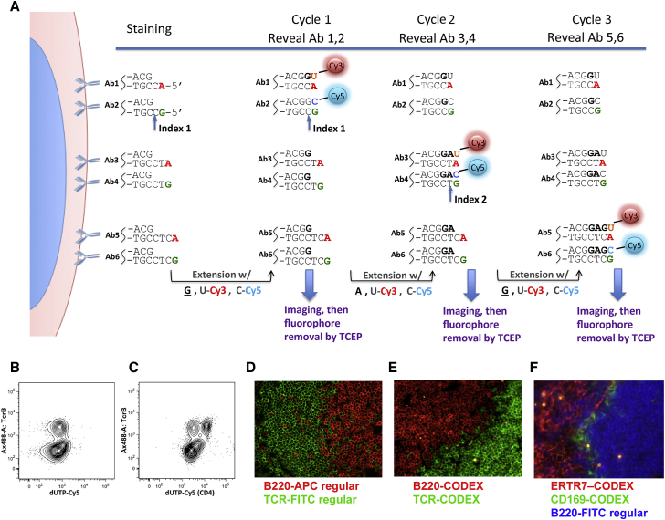
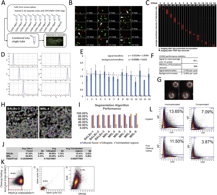
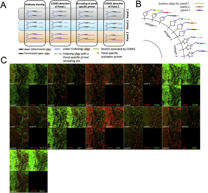
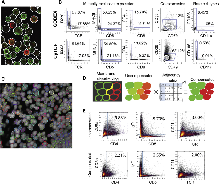
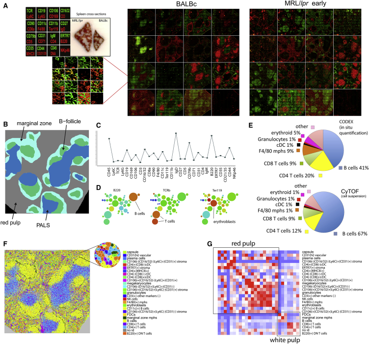
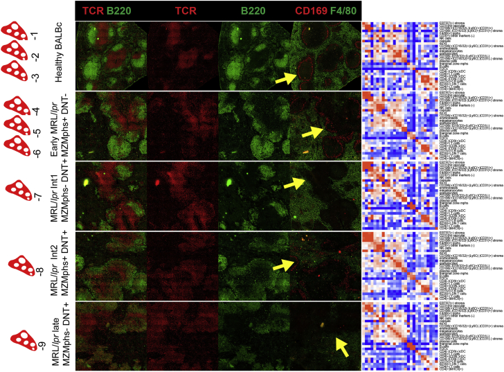
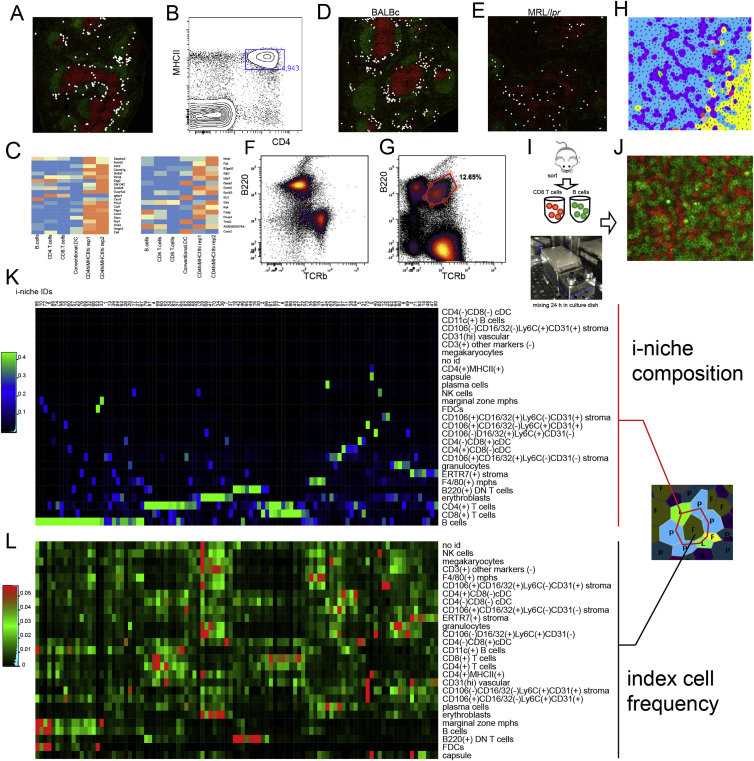
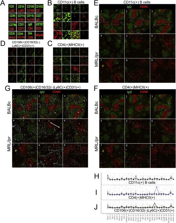
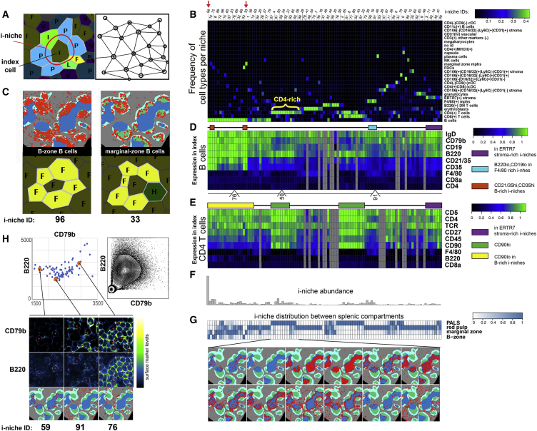
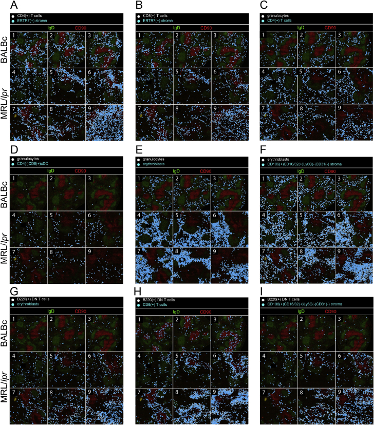
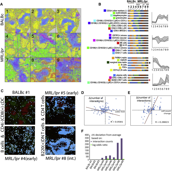
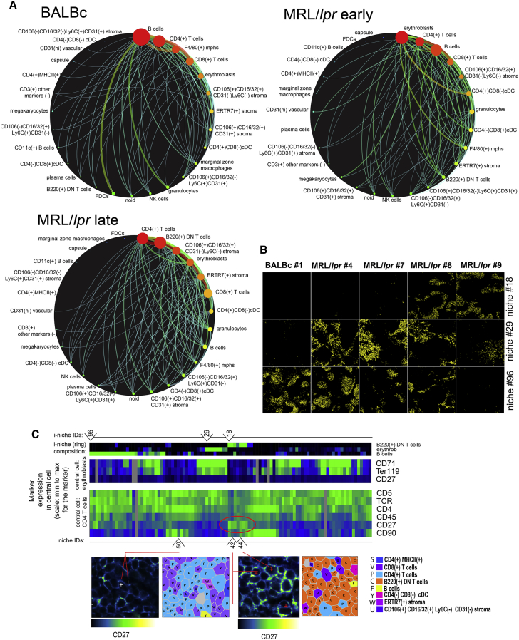
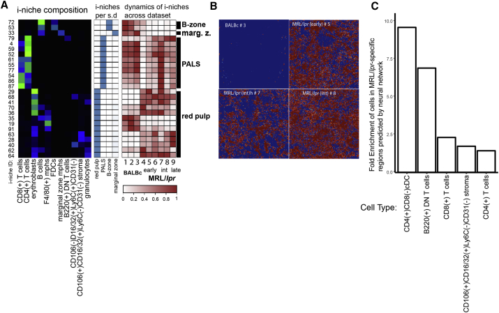
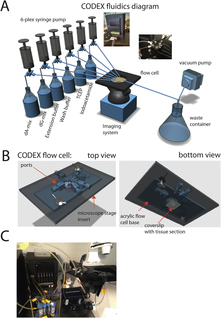
A highly multiplexed cytometric imaging approach, termed co-detection by indexing (CODEX), is used here to create multiplexed datasets of normal and lupus (MRL/lpr) murine spleens. CODEX iteratively visualizes antibody binding events using DNA barcodes, fluorescent dNTP analogs, and an in situ polymerization-based indexing procedure. An algorithmic pipeline for single-cell antigen quantification in tightly packed tissues was developed and used to overlay well-known morphological features with de novo characterization of lymphoid tissue architecture at a single-cell and cellular neighborhood levels. We observed an unexpected, profound impact of the cellular neighborhood on the expression of protein receptors on immune cells. By comparing normal murine spleen to spleens from animals with systemic autoimmune disease (MRL/lpr), extensive and previously uncharacterized splenic cell-interaction dynamics in the healthy versus diseased state was observed. The fidelity of multiplexed spatial cytometry demonstrated here allows for quantitative systemic characterization of tissue architecture in normal and clinically aberrant samples.
Overview
The claims and statistics of this paper haven’t been checked yet.
Run the check — free, no account needed. It reads the open-access text and checks the sources it cites.
Integrity signals
Registry
Cites
Statistics
Figures
Verification
Claims the paper makes
The claims and statistics of this paper haven’t been checked yet.
Run the check — free, no account needed. It reads the open-access text and checks the sources it cites.
Claims other papers make about this one
Each is checked against this paper’s own text: does it say what the citing paper says it does?
In the citing paper
“For example, multiplexed antibody-based imaging (mABI) technologies such as imaging mass cytometry (IMC) [31] and codetection by indexing (CODEX) [32] can detect 20–50 selected genes simultaneously in each cell at the protein level, which is a useful complement to transcript-based detection – especially given the potential for poor correlation of transcriptomic and proteomic data sets implicated”
From The heterogeneity of cellular senescence: insights at the single-cell level (2023) · cites this paper as [32]
Self-check
Percentages, sums and reported differences re-computed from the paper’s own numbers.
The self-check needs the paper’s full text.
Citations
Cites
Globally optimal stitching of tiled 3D microscopic image acquisitions
Preibisch, S., Saalfeld, S., & Tomancak, P. (2009) · Bioinformatics · doi:10.1093/bioinformatics/btp184 · Cited by 2,452
Bendall, S. C., Simonds, E. F., Qiu, P., Amir, E.-A. D., Krutzik, P. O., Finck, R., Bruggner, R. V., Melamed, R., Trejo, A., Ornatsky, O. I., et al. (2011) · Science · doi:10.1126/science.1198704 · Cited by 2,279
Self-citationSystemic Immunity Is Required for Effective Cancer Immunotherapy
Spitzer, M. H., Carmi, Y., Reticker-Flynn, N. E., Kwek, S. S., Madhireddy, D., Martins, M. M., Gherardini, P. F., Prestwood, T. R., Chabon, J., Bendall, S. C., et al. (2017) · Cell · doi:10.1016/j.cell.2016.12.022 · Cited by 963
A New Statistical Approach to Geographic Variation Analysis
Gabriel, K. R., & Sokal, R. R. (1969) · Systematic Zoology · doi:10.2307/2412323 · Cited by 854 · cited 1×
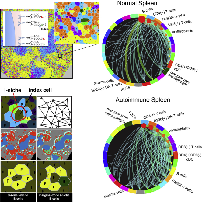
“Pairwise and Combinatorial (i-niches) Statistics of Cellto-Cell Contacts in Mouse Spleen To provide a high-level view of the cell-type interaction landscape, the total counts of contacts between every pair of cell types in the Delaunay neighborhood graph (Gabriel and Sokal, 1969) (Figure 4A and associated Mendeley dat…”
Rubtsov, A. V., Rubtsova, K., Fischer, A., Meehan, R. T., Gillis, J. Z., Kappler, J. W., & Marrack, P. (2011) · Blood · doi:10.1182/blood-2011-01-331462 · Cited by 808
Highly multiplexed single-cell analysis of formalin-fixed, paraffin-embedded cancer tissue
Gerdes, M. J., Sevinsky, C. J., Sood, A., Adak, S., Bello, M. O., Bordwell, A., Can, A. B., Corwin, A. D., Dinn, S. R., Filkins, R. J., et al. (2013) · Proceedings of the National Academy of Sciences · doi:10.1073/pnas.1300136110 · Cited by 716
Robinette, M. L., Fuchs, A. G., Cortez, V. S., Lee, J. S., Wang, Y., Durum, S. K., Gilfillan, S., & Colonna, M. (2015) · Nature Immunology · doi:10.1038/ni.3094 · Cited by 662
Erythroblastic islands: niches for erythropoiesis
Chasis, J. A., & Mohandas, N. (2008) · Blood · doi:10.1182/blood-2008-03-077883 · Cited by 506
Analyzing proteome topology and function by automated multidimensional fluorescence microscopy
SCHUBERT, W. J., Bonnekoh, B., Pommer, A. J., Philipsen, L., Böckelmann, R., Malykh, Y. N., Gollnick, H. P. M., Friedenberger, M., Bode, M., & Dress, A. (2006) · Nature Biotechnology · doi:10.1038/nbt1250 · Cited by 476 · cited 1×
“Approaches have been developed to overcome such limitations (Schubert et al., 2006; Gerdes et al., 2013), but these protocols have required multiple stain/ strip/wash cycles of the antibodies that can be time consuming or lead to sample degradation over the iterations.”
Gerner, M. Y., Kastenmüller, W., Ifrim, I., Kabát, J., & Germain, R. N. (2012) · Immunity · doi:10.1016/j.immuni.2012.07.011 · Cited by 446
Cited by
Sorted by citations
Items 901–1,000 of 1,002
From modality-specific to compositional foundation models for cell biology
Bahrami, M., Richter, T., Schmacke, N. A., Lavandera, A. E., & Theis, F. J. (2026) · Cell Systems · doi:10.1016/j.cels.2026.101534 · Cited by 7
Toward next-generation machine learning and deep learning for spatial omics
Zirem, Y., Fournier, I., & Salzet, M. (2026) · Briefings in Bioinformatics · doi:10.1093/bib/bbag131 · Cited by 5
“Imaging-based platforms such as MERFISH [3], seqFISH [4], CODEX [5], imaging mass cytometry, and MALDI-based imaging [6, 7] expanded this concept to proteins, lipids, and metabolites, enabling multi-layered molecular profiling at single-cell or subcellular resolution.”
Multiplex Protein Imaging through PACIFIC: Photoactive Immunofluorescence with Iterative Cleavage
Ji, F., Hur, M., Hur, S., Wang, S., Sarkar, P., Shao, S., Aispuro, D., Xu, C., Hu, Y., Li, Z., et al. (2023) · ACS Bio & Med Chem Au · doi:10.1021/acsbiomedchemau.3c00018 · Cited by 5
McMahon, N. P., Solanki, A., Wang, L. G., Montaño, A. R., Jones, J. A., Samkoe, K. S., Tichauer, K. M., & Gibbs, S. L. (2024) · Theranostics · doi:10.7150/thno.93256 · Cited by 6
“…constraints; however, an evolution of highly multiplexed immunostaining techniques using two distinct methods has emerged: (1) conventional antibody staining (i.e., IF or IHC) in a cyclic fashion [59, 60] or (2) mass spectrometry imaging (MSI) using rare earth metal labeled antibodies [23, 61-71].”
MethodInvestigating Spatial Dynamics in Spatial Omics Data with StarTrail
Chen, J., Xiong, C., Sun, Q., Song, Y., Wang, G. W., Gupta, G. P., Halder, A., Li, D., & Li, D. (2026) · Journal of the American Statistical Association · doi:10.1080/01621459.2026.2654225 · Cited by 4
Towards deciphering the bone marrow microenvironment with spatial multi-omics
Yip, R. K. H., Hawkins, E. D., Bowden, R. J., & Rogers, K. L. (2025) · Seminars in Cell and Developmental Biology · doi:10.1016/j.semcdb.2025.01.001 · Cited by 5
Statistical modeling and analysis of cell counts from multiplexed imaging data
Bost, P., Casanova, R., Mor, U., Haberecker, M., Pauli, C., Dettwiler, S., & Bodenmiller, B. (2025) · Cell Systems · doi:10.1016/j.cels.2025.101296 · Cited by 6
Peritumoral macrophages recruit eosinophils to promote antitumor immune responses in breast cancer
Saglimbeni, J. A., Esteva, E., Canales, J., Perez, O. A., Eichinger, A., Huntley, W., Khanna, K. M., Dolgalev, I., Klar, N. J., Adams, S., et al. (2025) · Proceedings of the National Academy of Sciences · doi:10.1073/pnas.2504645122 · Cited by 6
Kuczkiewicz‐Siemion, O., Sokół, K., Puton, B., Borkowska, A. M., & Szumera‐Ciećkiewicz, A. (2022) · Cancers · doi:10.3390/cancers14153833 · Cited by 5
Multiplex Immunofluorescence Image Quality Checking Using DAPI Channel–referenced Evaluation
Jiang, J., Moore, R. M., Jordan, C. E., Guo, R., Maus, R. L. G., Liu, H., Goode, E. L., Markovic, S. N., & Wang, C. (2023) · Journal of Histochemistry & Cytochemistry · doi:10.1369/00221554231161693 · Cited by 5
Rodriguez, S., Sun, B., McAllen, S., Jiang, M., & Parra, E. R. (2023) · Journal of Visualized Experiments · doi:10.3791/64758 · Cited by 5
Organelle landscape analysis using a multiparametric particle-based method
Kurikawa, Y., Koyama‐Honda, I., Igarashi, K., Tamura, N., Koike, S., & Mizushima, N. (2024) · PLoS Biology · doi:10.1371/journal.pbio.3002777 · Cited by 4
McMahon, N. P., Jones, J., Eng, J. R., Kwon, S., Chang, Y. H., Thibault, G., Chin, K., Nederlof, M. A., Gray, J. W., & Gibbs, S. L. (2019) · Imaging, Manipulation, and Analysis of Biomolecules, Cells, and Tissues · doi:10.1117/12.2510573 · Cited by 5
Oguchi, Y., Shintaku, H., & Uemura, S. (2020) · Communications Biology · doi:10.1038/s42003-020-01499-8 · Cited by 5
Single-Cell Quantitative Phenotyping via the Aptamer-Mounted Nest-PCR (Apt-nPCR)
Gao, T., Zhao, S., Sun, J., Huang, Q., Long, S., Lv, M., Ma, J., Guo, Z., & Li, G. (2022) · Analytical Chemistry · doi:10.1021/acs.analchem.1c03865 · Cited by 5
Causes and functional intricacies of inter- and intratumor heterogeneity of primary liver cancers
Khatib, S. A., & Wang, X. W. (2022) · Advances in cancer research · doi:10.1016/bs.acr.2022.01.006 · Cited by 6
mxnorm: An R Package to Normalize Multiplexed Imaging Data
Harris, C. R., Wrobel, J., & Vandekar, S. (2022) · The Journal of Open Source Software · doi:10.21105/joss.04180 · Cited by 5
“These imaging technologies, which include co-detection by indexing (CODEX), multiplexed ion beam imaging (MIBI), and multiplexed immunofluorescence imaging (MxIF), provide detailed information about spatial interactions between cells (Angelo et al., 2014; Gerdes et al., 2013; Goltsev et al., 2018).”
Cathcart, A. M., Smith, H. L., Labrie, M., & Mills, G. B. (2022) · Expert Review of Proteomics · doi:10.1080/14789450.2022.2070065 · Cited by 6
“CODEX also uses iterative antibody binding but employs DNA barcodes and fluorescent dNTP analogs to visualize binding events [25].”
Real-time spatial registration for 3D human atlas
Chen, L., Teng, D., Zhu, T., Kong, J., Herr, B. W., Bueckle, A., Börner, K., & Wang, F. (2022) · International Workshop on Analytics for Big Geospatial Data · doi:10.1145/3557917.3567618 · Cited by 5
“With the significant development of tissue imaging technologies[2] [3] [4] [5], enormous amounts of information about our human body are now produced at both cellular and subcellar levels.”
BackgroundToward the clinical development of synthetic immunity to cancer
Garcia, J. M., Burnett, C. E., & Roybal, K. T. (2023) · Immunological Reviews · doi:10.1111/imr.13245 · Cited by 9
“…orientation of the tumor immune compartment with multiplexed immunohistochemistry, 75 Multiplexed Ion Beam Imaging (MIBI), 76,77 CODEX imaging, 78 or RNAscope 79 provide new modalities and insight into understanding the TME and requirements for tool building in difficult to treat human tumors.”
BackgroundRealization of cellomics to dive into the whole-body or whole-organ cell cloud
Mitani, T. T., Susaki, E. A., Matsumoto, K., & Ueda, H. R. (2024) · Nature Methods · doi:10.1038/s41592-024-02307-5 · Cited by 5
Mapping cell phenotypes in breast cancer
Keren, L., & Angelo, M. (2020) · Nature Cancer · doi:10.1038/s43018-020-0031-9 · Cited by 5
Compressed sensing for imaging transcriptomicsPreprint
Cleary, B., Simonton, B., Bezney, J., Murray, E. M., Alam, S., Sinha, A., Habibi, E., Marshall, J. L., Lander, E. S., Chen, F., et al. (2019) · bioRxiv (Cold Spring Harbor Laboratory) · doi:10.1101/743039 · Cited by 6
“We use simulations to test parameters for (1) the total number of measurements m, (2) the maximum number of measurements in which each gene was included, (3) the individual genes for each measurement, and (4) the size d of the module dictionary and sparsity k of the linear combinations.”
MethodFluorescent in situ sequencing of DNA barcoded antibodiesPreprint
Kohman, R. E., & Church, G. M. (2020) · bioRxiv (Cold Spring Harbor Laboratory) · doi:10.1101/2020.04.27.060624 · Cited by 5
Multiscale profiling of enzyme activity in cancerPreprint
Amini, A. P., Kirkpatrick, J. D., Wang, C. S., Jaeger, A. M., Su, S., Naranjo, S., Zhong, Q., Cabana, C. M., Jacks, T., & Bhatia, S. N. (2021) · bioRxiv (Cold Spring Harbor Laboratory) · doi:10.1101/2021.11.11.468288 · Cited by 6
“Although single-cell 51 transcriptomics has enabled characterization of intratumoral heterogeneity [2, 3, 4], and techniques 52 to localize proteins [5, 6] and nucleic acid sequences [7, 8, 9] in situ are starting to enable study 53 of tumors in a spatial context [10], analogous techniques for single-cell and spatial…”
MethodLearning predictive models of tissue cellular neighborhoods from cell phenotypes with graph poolingPreprint
Hu, Y., Rong, J., Xie, R., Xu, Y., Peng, J. J., Gao, L., & Tan, K. (2022) · bioRxiv (Cold Spring Harbor Laboratory) · doi:10.1101/2022.11.06.515344 · Cited by 5
“The ground-truth labels of cells among four known splenic compartments, red pulp, marginal zone, B-cell zone and periarteriolar lymphoid sheath (PALS), were obtained from the authors of the original study 14 .”
BackgroundMethodWu, X., Xu, W., Deng, L., Li, Y., Wang, Z., Sun, L., Gao, A., Wang, H., Yang, X., Wu, C., et al. (2023) · bioRxiv (Cold Spring Harbor Laboratory) · doi:10.1101/2023.01.07.523058 · Cited by 5
Eng, J. R., Bucher, E., Hu, Z., Sanders, M. E., Chakravarthy, A. B., González, P., Pietenpol, J. A., Gibbs, S. L., Sears, R. C., & Chin, K. (2023) · bioRxiv (Cold Spring Harbor Laboratory) · doi:10.1101/2023.01.31.525753 · Cited by 5
SEG: Segmentation Evaluation in absence of Ground truth labelsPreprint
Sims, Z., Strgar, L., Thirumalaisamy, D., Heussner, R. T., Thibault, G., & Chang, Y. H. (2023) · bioRxiv (Cold Spring Harbor Laboratory) · doi:10.1101/2023.02.23.529809 · Cited by 8
Addressing persistent challenges in digital image analysis of cancerous tissuesPreprint
Prabhakaran, S., Yapp, C., Baker, G. J., Beyer, J., Chang, Y. H., Creason, A., Krueger, R., Muhlich, J. L., Patterson, N. H., Sidak, K., et al. (2023) · bioRxiv (Cold Spring Harbor Laboratory) · doi:10.1101/2023.07.21.548450 · Cited by 5
Spatial Metabolomics and the Vulnerable Atherosclerotic Plaque
Luo, L., & Leeper, N. J. (2023) · Arteriosclerosis Thrombosis and Vascular Biology · doi:10.1161/atvbaha.123.319739 · Cited by 5
Escalating High-dimensional Imaging using Combinatorial Channel Multiplexing and Deep LearningPreprint
Ben-Uri, R., Shabat, L. B., Shainshein, D., Bar-Tal, O., Bussi, Y., Maimon, N., Haran, T. K., Milo, I., Goliand, I., Addadi, Y., et al. (2023) · bioRxiv (Cold Spring Harbor Laboratory) · doi:10.1101/2023.09.09.556962 · Cited by 5
Deep Learning Model Imputes Missing Stains in Multiplex ImagesPreprint
Shaban, M., Lassoued, W., Canubas, K., Bailey, S., Liu, Y., Allen, C. T., Strauss, J., Gulley, J. L., Jiang, S., Mahmood, F., et al. (2023) · bioRxiv (Cold Spring Harbor Laboratory) · doi:10.1101/2023.11.21.568088 · Cited by 6
Pati, P., Karkampouna, S., Bonollo, F., Compérat, É., Radić, M., Spahn, M., Martinelli, A. L., Wartenberg, M., Julio, M. K., & Rapsomaniki, M. (2023) · bioRxiv (Cold Spring Harbor Laboratory) · doi:10.1101/2023.11.29.568996 · Cited by 5
“Recently, multiplexed imaging technologies ( e . g ., imaging mass cytometry - IMC [4], co-detection by indexing - CODEX [5], multiplexed ion beam imaging - MIBI [6]) have enabled the simultaneous quantification of dozens of markers on the same tissue, revolutionizing spatial biology [7].”
BackgroundKim, E. N., Seok, H., Koh, J., Yang, W., Lee, G. H., Choi, W. H., Lim, J. S., Ok, Y. J., Choi, J. S., Kim, C. J., et al. (2024) · bioRxiv (Cold Spring Harbor Laboratory) · doi:10.1101/2024.02.22.581315 · Cited by 5
High-Throughput Multiomics Profiling of Model Systems Using the AVITI24 PlatformPreprint
Lopez, T., Honigfort, D. J., Mah, A., Thompson, C., Dien, V. T., Dawood, M., Frankel, E., Fuller, D., Jamison, C., Kelley, R., et al. (2025) · bioRxiv (Cold Spring Harbor Laboratory) · doi:10.1101/2025.05.03.651997 · Cited by 5
OmicsNavigator : An auditable scientific partner for scalable hypothesis validation in spatial omicsPreprint
Yiyao, L., Vakharia, N., Liang, W., Mayer, A. T., Luo, R., Trevino, A. E., & Wu, Z. (2025) · bioRxiv (Cold Spring Harbor Laboratory) · doi:10.1101/2025.07.21.665821 · Cited by 5
Thousandfold Expansion MicroscopyPreprint
Hu, H., Krah, D., Ntolkeras, A., Chanda, S., Heimbrodt, A., Mondal, M., Altendorf, J., Jing, B., Berger, B., Shaib, A., et al. (2026) · bioRxiv (Cold Spring Harbor Laboratory) · doi:10.64898/2026.05.31.729018 · Cited by 5
Highly Sensitive Spatial Proteomics with Multicolor Cleavable Fluorescent Tyramide
Chen, Y., Wang, Y., LaBaer, J., & Guo, J. (2025) · Chemical & Biomedical Imaging · doi:10.1021/cbmi.4c00099 · Cited by 4
Using spatial proteomics to enhance cell type assignments in histology images
Dayao, M. T., Mayer, A. T., Trevino, A. E., & Bar‐Joseph, Z. (2025) · Cell Reports Methods · doi:10.1016/j.crmeth.2025.101204 · Cited by 4
Dette, A., Moers, F., Mayr, T., von Stillfried, S. F., Bernhardt, M., Förster, S., Werlein, C., Ackermann, M., Muders, M. H., Kristiansen, G., et al. (2025) · Journal of Virology · doi:10.1128/jvi.01384-25 · Cited by 5
Recent advances and applications of single-cell sequencing in insects
Nayyar, N., Arya, S. K., Harrison, D. A., & Palli, S. R. (2025) · Current Opinion in Insect Science · doi:10.1016/j.cois.2025.101455 · Cited by 5
Qiu, S., Wang, Z., Guo, S., Xie, D., Cai, Y., Wang, X., Yang, Q., Zhao, Q., Guan, Y., Lin, C., et al. (2026) · Journal of Future Foods · doi:10.1016/j.jfutfo.2026.01.017 · Cited by 4
Spatiotemporal decoding of skin biology: development, aging, disease, and regeneration
Xi, D., Huang, R. S., Li, Y., Xia, Z., & Ji, S. (2026) · Burns & Trauma · doi:10.1093/burnst/tkag019 · Cited by 5
“Because of these features, imaging-based spatial omics techniques (e.g. MERFISH/MERSCOPE [ 26], HCR-FISH [ 27], and CODEX/CyCIF [ 28, 29]) can be used to construct high-resolution 3D molecular maps in situ .”
Hoebel, K., Lindsay, J. R., Altreuter, J., Alessi, J. V., Weirather, J. L., Dryg, I. D., Giobbie‐Hurder, A., Li, Z., Yu, K., Awad, M. M., et al. (2026) · npj Precision Oncology · doi:10.1038/s41698-026-01314-3 · Cited by 6
Jurnečka, D., Hrovatin, M., Bokalj, B., & Turk, B. (2026) · FEBS Journal · doi:10.1111/febs.70514 · Cited by 5
AI proteomics: from protein identification to virtual cells
Sun, Y., Jun, A., Liu, Z., Sun, R., Qian, L., Payne, S., Bittremieux, W., Ralser, M., Li, C., Chen, Y., et al. (2026) · Nature Methods · doi:10.1038/s41592-026-03085-y · Cited by 7
“Imaging-based methods, such as co-detection by indexing (CODEX) 44 and multiplexed ion beam imaging (MIBI) 45 , provide high spatial resolution and wide tissue coverage, yet they typically offer limited proteomic depth due to antibody multiplexing constraints.”
Programmable DNA Reactions for Advanced Fluorescence Microscopy in Bioimaging
Fan, H. B. (2024) · Small Methods · doi:10.1002/smtd.202401279 · Cited by 3
Sakai, S. A., Nomura, R., Nagasawa, S., Chi, S., Suzuki, A., Suzuki, Y., Imai, M., Nakamura, Y., Yoshino, T., Ishikawa, S., et al. (2025) · PLoS Computational Biology · doi:10.1371/journal.pcbi.1012854 · Cited by 4
“Single-cell spatial omics platforms, such as Xenium, CosMx [1], and PhenoCycler [2], offer opportunities for the investigation of hundreds or thousands of genes in various organs and tissue types.”
mNSF: multi-sample non-negative spatial factorization
Wang, Y., Woyshner, K., Sriworarat, C., Stein-O’Brien, G., Goff, L. A., & Hansen, K. D. (2025) · Genome biology · doi:10.1186/s13059-025-03601-x · Cited by 5
Integrated Cells and Collagen Fibers Spatial Image Analysis
Vasiukov, G., Novitskaya, T., Senosain, M., Camai, A., Menshikh, A., Massion, P., Zijlstra, A., & Novitskiy, S. (2021) · Frontiers in Bioinformatics · doi:10.3389/fbinf.2021.758775 · Cited by 5
“The most popular open source tools that can be used: ImageJ (Rueden et al., 2017), FIJI, HistoCAT (Schapiro et al., 2017), CODEX toolkit (Goltsev et al., 2018), KNIME (Dietz and Berthold, 2016), Ilastik (Berg et al., 2019), CellPro fi ler (Carpenter et al., 2006) and Cytokit (Czech et al., 2019).”
MethodAlignment, segmentation and neighborhood analysis in cyclic immunohistochemistry data using CASSATT
Brockman, A. A., Khurana, R., Bartkowiak, T., Thomas, P. L., Sivagnanam, S., Betts, C. B., Coussens, L. M., Lovly, C. M., Irish, J. M., & Ihrie, R. A. (2023) · Cytometry Part B Clinical Cytometry · doi:10.1002/cyto.b.22114 · Cited by 4
“Thus, a cell's voronoi neighbors are defined as the collection of cells that are the nearest seed point in the voronoi diagram in every direction from the index cell (Figure 5a,b; Goltsev et al., 2018).”
BackgroundMethodInfluentialSingle-Cell Spatial MIST for Versatile, Scalable Detection of Protein Markers
Meah, A., Vedarethinam, V., Bronstein, R. D., Gujarati, N. A., Jain, T., Mallipattu, S. K., Li, Y., & Wang, J. (2023) · Biosensors · doi:10.3390/bios13090852 · Cited by 5
Optical microscopic imaging, manipulation, and analysis methods for morphogenesis research
Katoh, T. A., Fukai, Y. T., & Ishibashi, T. (2023) · Microscopy · doi:10.1093/jmicro/dfad059 · Cited by 5
“Additionally, highly multiplexed immunostaining can now be achieved through multi-round acquisition [184].”
BackgroundRodríguez-Bejarano, O. H., Roa, L., Vargas-Hernández, G., Botero-Espinosa, L., Parra-López, C., & Patarroyo, M. A. (2023) · Biochimica et Biophysica Acta (BBA) - Reviews on Cancer · doi:10.1016/j.bbcan.2023.189064 · Cited by 4
Exploring the intricate cross-talk between clonal expansion and the bone marrow niche
Ngo, S. D., Papazoglou, D., Encabo, H. H., & Bonnet, D. A. (2024) · Frontiers in Hematology · doi:10.3389/frhem.2024.1334807 · Cited by 4
Ram, S., Mojtahedzadeh, S., Aguilar, J.-K., Coskran, T., Powell, E. L., & O’Neil, S. P. (2024) · Scientific Reports · doi:10.1038/s41598-024-58372-5 · Cited by 5
Metabolic nanoscopy enhanced by experimental and computational approaches
Jang, H., Wu, S., Li, Y., Li, Z., & Shi, L. (2024) · npj Imaging · doi:10.1038/s44303-024-00062-y · Cited by 5
Stromal KITL/SCF Maintains Pancreas Tissue Homeostasis and Restrains Tumor Progression
Oñate, M. K., Oon, C. H., Bhattacharyya, S., Low, V., Chen, C., Zhao, X., Arnold, F., Yan, Z., Pramod, S., Hang, Y., et al. (2025) · Cancer Discovery · doi:10.1158/2159-8290.cd-24-1079 · Cited by 4
“Based on our transcriptional profiling results, we compared tissue microenvironments in Rosa26 mTmG/ + ;Kitl flox/flox ;Fabp4-Cre mice compared with Rosa26 mTmG/ + ;Fabp4-Cre controls using co-detection by indexing (CODEX), a barcode-based, multiplexed imaging approach (34).”
Chou, M.-Y., & Yang, M. (2025) · Translational Oncology · doi:10.1016/j.tranon.2025.102353 · Cited by 4
“Spatial proteomics, achieved through techniques like imaging mass cytometry [166], multiplexed ion beam imaging [167], and CODEX [168], offers unprecedented resolution of TIME heterogeneity.”
CellTune: an integrative software for accurate cell classification in spatial proteomics
Bussi, Y., Shainshein, D., Eli, O., Posner, S., Azulay, N., Maimon, N., Haran, T. K., Ben-Uri, R., Brown, C., Schuldiner, N., et al. (2026) · Nature Methods · doi:10.1038/s41592-026-03162-2 · Cited by 5
“For example, cycling cells will be positive for Ki67 and apoptotic cells will express cleaved caspase 3.”
Casella, G., Torcasso, M. S., Ai, J., Cao, T., Hara, S., Andrade, M. S., Ghosh, D., Shao, D., Chang, A., Ko, K., et al. (2025) · Journal of Clinical Investigation · doi:10.1172/jci192669 · Cited by 4
“Briefly, the mean pixel intensities for 26 cardinal markers were organized into decision trees (DTs) for cell class annotation (47, 55) (Supplemental Figure 3).”
Highly Multiplexed, Quantitative Tissue Imaging at Cellular Resolution
McCarthy, M. E., & Birtwistle, M. R. (2019) · Current Pathobiology Reports · doi:10.1007/s40139-019-00203-8 · Cited by 4
Pugh, M., Akarka, A. U., Hunter, K. J., Dojcinov, S. D., & Marafioti, T. (2020) · Diagnostic histopathology · doi:10.1016/j.mpdhp.2020.07.002 · Cited by 4
Draper, M., Willems, M., Malahe, R. S. R. K., Hamilton, A., & Tarasov, A. I. (2021) · Journal of Imaging · doi:10.3390/jimaging7010009 · Cited by 4
“Deep phenotypical profiling of living cells, arranged into an intact tissue, is done via high-content microscopy [13,14], which also characterizes specific proteins expressed on the plasma membrane.”
BackgroundCell–cell communication networks in tissue: Toward quantitatively linking structure with function
Luthria, G., Lauffenburger, D. A., & Miller, M. A. (2021) · Current Opinion in Systems Biology · doi:10.1016/j.coisb.2021.05.002 · Cited by 4
Microscopic image-based covariation network analysis for actin scaffold-modified insulin signaling
Noguchi, Y., Kano, F., Maiya, N., Iwamoto, C., Yamasaki, S., Otsubo, Y., Nakatsu, D., Kunishige, R., & Murata, M. (2021) · iScience · doi:10.1016/j.isci.2021.102724 · Cited by 4
“, 2015, 2018), and modified DNA barcoding approaches (Goltsev et al., 2018; Schürch et al., 2020), are applicable for the data production process in our PLOMCON analysis system, illustrating both the flexibility of the system and the broad scope of its potential use.”
MethodIn situ functional cell phenotyping reveals microdomain networks in colorectal cancer recurrence
Furman, S. A., Stern, A. M., Uttam, S., Taylor, D. L., Pullara, F., & Chennubhotla, S. C. (2021) · Cell Reports Methods · doi:10.1016/j.crmeth.2021.100072 · Cited by 6
“, 2017) or performed in a batch before imaging (Goltsev et al., 2018).”
InfluentialThree-dimensional tumour microenvironment reconstruction and tumour-immune interactions' analysis
Barmpoutis, P., Kayhanian, H., Waddingham, W., Alexander, D. C., & Jansen, M. (2021) · International Conference on Digital Image Computing: Techniques and Applications (DICTA) · doi:10.1109/dicta52665.2021.9647283 · Cited by 4
Zhu, W., Cai, E., Li, H., Wang, P., Shen, A., Popp, J., & Hu, J. (2021) · Angewandte Chemie · doi:10.1002/ange.202106136 · Cited by 4
Reddy, R., Yang, L., Liu, J., Liu, Z., & Wang, J. (2022) · Analytical Chemistry · doi:10.1021/acs.analchem.1c04970 · Cited by 4
Su, T., Wang, S., Huang, S., Cai, H., McKinley, E. T., Beeghly-Fadiel, A. C., Zheng, W., Shu, X., & Cai, Q. (2022) · Cancer Biomarkers · doi:10.3233/cbm-220071 · Cited by 4
A flexible open-source processing workflow for multiplexed fluorescence imaging based on cycles
Potier, G., Doméné, A., & Paul‐Gilloteaux, P. (2022) · F1000Research · doi:10.12688/f1000research.124990.1 · Cited by 4
Immunotyping and Quantification of Melanoma Tumor–Infiltrating Lymphocytes
Meneveau, M. O., Sahli, Z. T., Lynch, K., Mauldin, I. S., & Slingluff, C. L. (2021) · Methods in molecular biology · doi:10.1007/978-1-0716-1205-7_36 · Cited by 4
Cereceda, K., Jorquera, R., & Villarroel‐Espíndola, F. (2021) · Advances in Laboratory Medicine / Avances en Medicina de Laboratorio · doi:10.1515/almed-2021-0075 · Cited by 4
Spatial and temporal tools for building a human cell atlas
Cool, J., Conroy, R., Hanlon, S. E., Hughes, S. K., & Roy, A. L. (2019) · Molecular Biology of the Cell · doi:10.1091/mbc.e18-10-0667 · Cited by 4
Ion beam subcellular tomographyPreprint
Coskun, A. F., Han, G., Chen, S.-Y., Clavé, X. R., Jiang, S., Schürch, C. M., Bai, Y., Hitzman, C., & Nolan, G. P. (2019) · bioRxiv (Cold Spring Harbor Laboratory) · doi:10.1101/557728 · Cited by 4
“3,4 Multiplexed ion beam imaging (MIBI) can be employed to spatially visualize multiple (6-50) protein parameters in histology sections,5,6.”
BackgroundMultiplex tissue imaging: An introduction to the scope and challenges
Bellamy, C. O. C., & Prost, S. (2019) · American Journal of Transplantation · doi:10.1111/ajt.15767 · Cited by 4
Novel Human Kidney Cell Subsets Identified by Mux-SeqPreprint
Schroeder, A., Sur, S., Rashmi, P., Damm, I., Zarinsefat, A., Kretzler, M., Hodgin, J., Hartoularos, G. C., Sigdel, T. K., Ye, J. C., et al. (2020) · bioRxiv (Cold Spring Harbor Laboratory) · doi:10.1101/2020.03.02.973925 · Cited by 4
Divergent cancer etiologies drive distinct B cell signatures and tertiary lymphoid structuresPreprint
Ruffin, A. T., Cillo, A. R., Tabib, T., Liu, A., Onkar, S., Kunning, S. R., Lampenfeld, C., Abécassis, I., Qi, Z., Soose, R. J., et al. (2020) · bioRxiv (Cold Spring Harbor Laboratory) · doi:10.1101/2020.05.29.123265 · Cited by 4
Virus-Dependent Immune Conditioning of Tissue MicroenvironmentsPreprint
Jiang, S., Chan, C. N., Clavé, X. R., Chen, H. M., Bai, Y., Zhu, B., McCaffrey, E., Greenwald, N. F., Liu, C. C., Barlow, G. L., et al. (2021) · SSRN Electronic Journal · doi:10.2139/ssrn.3860320 · Cited by 4
Dhainaut, M., Rose, S. A., Aktürk, G., Wroblewska, A., Park, E. S., Nielsen, S. R., Buckup, M., Roudko, V., Pia, L., Bérichel, J. L., et al. (2021) · bioRxiv (Cold Spring Harbor Laboratory) · doi:10.1101/2021.07.13.451021 · Cited by 4
“As there are multiplex imaging techniques and instruments that can detect as many as 60 proteins on a single tissue section, much more extensive detection of cell types and cell states is possible (Goltsev et al., 2018; Jackson et al., 2020; Lin et al., 2018).”
BackgroundZhu, B., Bai, Y., Yeo, Y. Y., Lu, X., Clavé, X. R., Chen, H. M., Yeung, J., Gerber, G. K., Angelo, M., Shalek, A. K., et al. (2024) · bioRxiv (Cold Spring Harbor Laboratory) · doi:10.1101/2024.03.04.583400 · Cited by 4
Scalable Condition-relevant Cell Niche Analysis of Spatial Omics Data with TaichiPreprint
Cui, Y., & Yuan, Z. (2024) · bioRxiv (Cold Spring Harbor Laboratory) · doi:10.1101/2024.05.30.596656 · Cited by 4
Spatial proteomics with 100+ markers with Highly Multiplexed MALDI-IHCPreprint
Zhang, M., Abbey, J. A., Bost, P., Usui, G., Haefliger, S., Muenst, S. E., Lim, M. J., Yagnik, G. B., de Souza, N., Rothschild, K. J., et al. (2025) · bioRxiv (Cold Spring Harbor Laboratory) · doi:10.1101/2025.04.18.649415 · Cited by 4
(2025) · doi:10.1016/j.ymeth.2025.07.007 · Cited by 6
“Compared to other multiplexed imaging modalities like CODEX [34], which typically have three times higher resolution, this relatively low resolution along the Z-axis leads to more considerable shifts in cell shape between 2D cells belonging to the same 3D cell in adjacent slices (and thus requires…”
Single-cell spatial proteomics.
Liyanage, S. H., & Guo, J. (2025) · PubMed · doi:10.14670/hh-18-861 · Cited by 3
Spatial Single‐Cell Technologies for Exploring Gastrointestinal Tissue Transcriptome
Kang, H. M., & Lee, J. H. (2023) · Comprehensive physiology · doi:10.1002/cphy.c210053 · Cited by 5
“…and in situ imaging technique is specifically amenable for the multiplexed detection of proteins, which has been recently implemented through the Co-Detection by Indexing (CODEX) platform (33), iterative indirect immunofluorescence imaging (4i) platform (34, 83), or Nanostring CosMx platform (37).”
Olivas, A. D., Ngai, P., Schahrer, E., Xing, J., Ghadiri, M., Olortegui, K. S., Cursio, J., Akiyama, S., Chang, E. B., Shen, L., et al. (2025) · Cellular and Molecular Gastroenterology and Hepatology · doi:10.1016/j.jcmgh.2025.101563 · Cited by 3
Kim, E. N., Seok, H., Koh, J., Bae, J. M., Yang, W., Lee, G. H., Choi, W. H., Lim, J. S., Ok, Y. J., Choi, J. S., et al. (2025) · Translational research · doi:10.1016/j.trsl.2025.05.008 · Cited by 3
Roadmap for spatial transcriptomics of HIV in tissues
Guan, Q., Eshetu, A., & Ho, Y. (2025) · Current Opinion in HIV and AIDS · doi:10.1097/coh.0000000000000961 · Cited by 5
Bao, H., Zhang, S., Yu, Z., & Xu, H. (2025) · Nature Communications · doi:10.1038/s41467-025-61907-7 · Cited by 4
Dynamic remodeling of the pancreas immune landscape in obesity
Koshkin, A., Tanagala, K. K. K., Eichinger, A., Chait, M., Young, A., Shakil, S., Yoshikawa, J., Sakamoto, Y., Wells, S. B., Fazlollahi, L., et al. (2026) · Nature Communications · doi:10.1038/s41467-026-75090-w · Cited by 3
Spatial multi-omics technologies and applications in cancer
Ji, L. (2025) · Biosystems · doi:10.1016/j.biosystems.2025.105576 · Cited by 3
Liu, W., Yang, Y.-Y., & Shi, Z. (2025) · World Journal of Stem Cells · doi:10.4252/wjsc.v17.i11.112702 · Cited by 3
“These approaches, particularly when combined with complementary antibody-based platforms such as mass cytometry or CODEX imaging, may allow for more comprehensive profiling of PTM heterogeneity at the single-cell level[121].”
Zhao, F., Xiao, J., He, H., & Li, K. (2026) · Analytical Chemistry · doi:10.1021/acs.analchem.5c07181 · Cited by 3
Opportunities and Challenges in Precision Neurotherapeutics
Mujica-Parodi, L. R., Öngür, D., & Richardson, R. M. (2026) · Annual Review of Biomedical Engineering · doi:10.1146/annurev-bioeng-110824-031709 · Cited by 4
Multimodal DNA Nanostructure Barcodes for Single-Cell Protein Profiling and Tumor Subtyping
Cui, M., Hu, C., Dong, J., Cheng, Y., Sun, B., Fan, C., Wang, L., & Chao, J. (2026) · ACS Nano · doi:10.1021/acsnano.5c18968 · Cited by 3
Unraveling the tapestry: Lessons from multi-omics and spatial biology in hepatocellular cancer
Massoudi, D., Srivastava, A., & Dhanasekaran, R. (2026) · Hepatology · doi:10.1097/hep.0000000000001716 · Cited by 3
Multiplexed optical barcoding and sequencing for spatial omics
Venkatramani, A. V., Ciftci, D., Pham, K., Cohen, L., Sepúlveda, L. A., Li, C., & Zhuang, X. (2026) · Scientific Reports · doi:10.1038/s41598-026-41186-y · Cited by 3
Metadata
- Authors
- Goltsev, Y., Samusik, N., Kennedy-Darling, J., Bhate, S., Hale, M., Vazquez, G., Black, S., & Nolan, G. P.
- Affiliations
- Stanford Medicine (US)
- Corresponding author
- Nolan, Garry P.
- Venue
- Cell
- Publisher
- Elsevier BV
- Published
- 2018-08
- Volume
- 174
- Issue
- 4
- Pages
- 968-981.e15
- ISSN
- 0092-8674 (ISSN-L)
- PMC
- PMC6086938
- OpenAlex
- W2949238013
- Cited by
- 1,501
- References
- 20
- Citations by year
- 2026: 231 · 2025: 244 · 2024: 232 · 2023: 198 · 2022: 194 · 2021: 194 · 2020: 125 · 2019: 72 · 2018: 9
- FWCI
- 35.39
- Type
- journal article
- Language
- en
- Open access
- hybrid
- doi.org
- Licence
- Locations
- Cell (publishedVersion, cc-by)
- bioRxiv (Cold Spring Harbor Laboratory) (acceptedVersion)
- Europe PMC (PubMed Central) (publishedVersion, cc-by)
- PubMed Central (publishedVersion, cc-by)
- PubMed (publishedVersion)
- Funders
- US NIH (U19AI057229, 1U19AI100627, R01CA184968, 1R33CA183654-01, R33CA183692, 1R01GM10983601, 1R21CA183660, 1R01NS08953301, 5UH2AR067676, 1R01CA19665701, R01HL120724, R01HL128173)
- Bill and Melinda Gates foundation (OPP1113682)
- Department of Defense (W81XWH-14-1-0180)
- Northrop-Grumman Corporation (7500108142)
- NIH (U54-CA209971)
- Rachford & Carlotta A. Harris Endowed Chair
- Topics
- Life Sciences › Biochemistry, Genetics and Molecular Biology › Molecular Biology › Single-cell and spatial transcriptomics
- Life Sciences › Biochemistry, Genetics and Molecular Biology › Biophysics › Cell Image Analysis Techniques
- Life Sciences › Biochemistry, Genetics and Molecular Biology › Biophysics › Advanced Fluorescence Microscopy Techniques
- Keywords
- codices, splenic architecture, MRL/lpr mice, systemic lupus erythematosus, cell-cell interactions, autoimmune disease
- MeSH terms
- Animals, Antibodies, Disease Models, Animal, Female, Image Processing, Computer-Assisted, Lupus Erythematosus, Systemic, Male, Mass Spectrometry, Spleen, Oligonucleotide Probes, Mice, Inbred MRL lpr, Mice
About these checks
Registry, retraction, Cites and statistics results are computed, not judged, with one exception: a printed reference that no index lists is read once by a model before it counts as a citation error. Claims are extracted by a model and kept only when their quote appears verbatim in the text; each cited source is then checked, with a model’s help, for whether it says what the claim relies on. Those verdicts can be wrong — read the claim’s quote and the cited source before relying on one.
Statistics and arithmetic are recomputed, not judged.
Confidence bands are shown only for fields calibrated on our annotated test set. None is calibrated yet, so no band is shown.
What counts as a claim ›How we measure confidence ›How we check this ›
Are you an author, or do you see a problem with this page? Request a review
