

The DOI matches its Crossref record; 21 of the 21 references it cites were found in a registry or an index.
Deep Profiling of Mouse Splenic Architecture with CODEX Multiplexed Imaging
Open accessPublished version of a preprint
Abstract
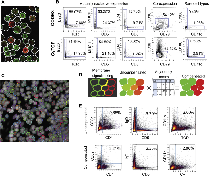
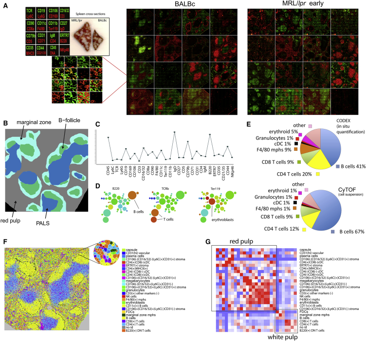
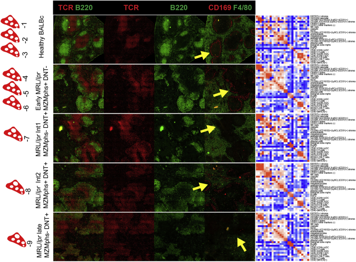
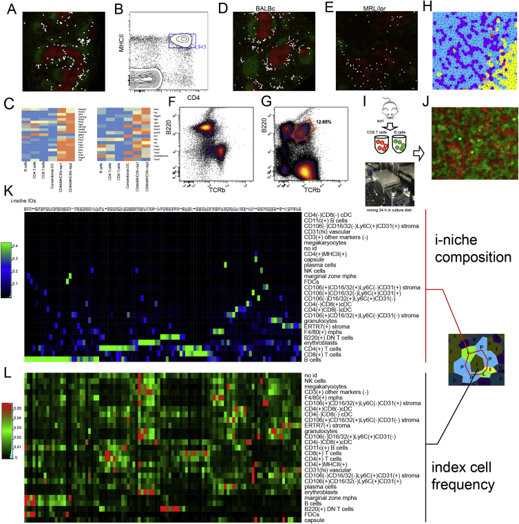
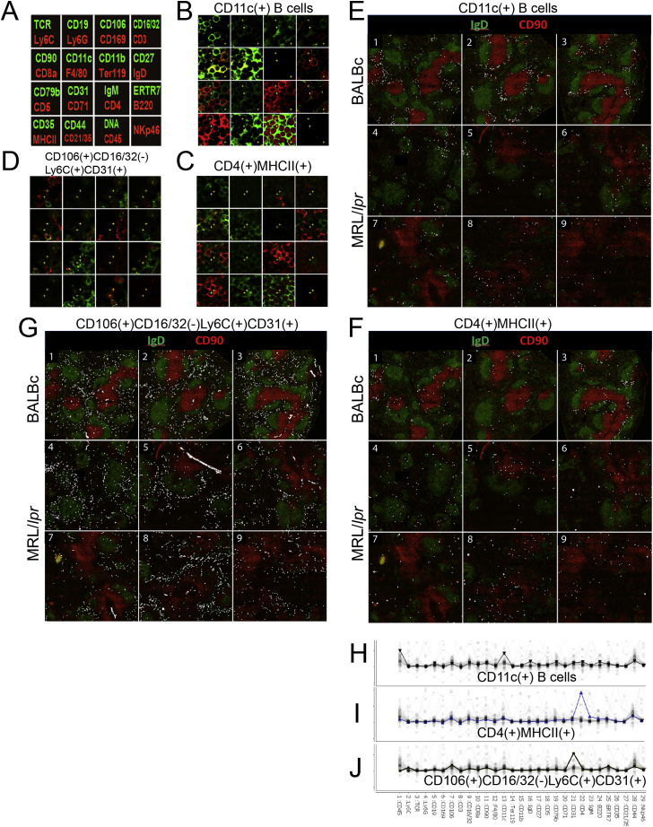
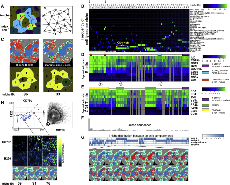
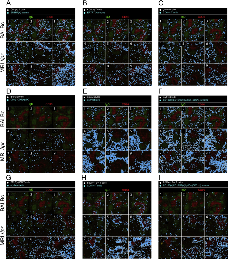
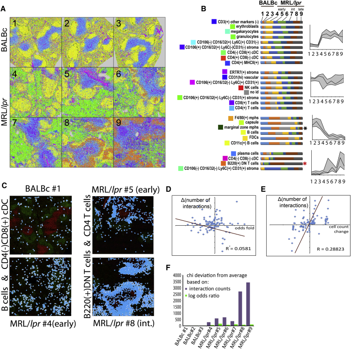
A highly multiplexed cytometric imaging approach, termed co-detection by indexing (CODEX), is used here to create multiplexed datasets of normal and lupus (MRL/lpr) murine spleens. CODEX iteratively visualizes antibody binding events using DNA barcodes, fluorescent dNTP analogs, and an in situ polymerization-based indexing procedure. An algorithmic pipeline for single-cell antigen quantification in tightly packed tissues was developed and used to overlay well-known morphological features with de novo characterization of lymphoid tissue architecture at a single-cell and cellular neighborhood levels. We observed an unexpected, profound impact of the cellular neighborhood on the expression of protein receptors on immune cells. By comparing normal murine spleen to spleens from animals with systemic autoimmune disease (MRL/lpr), extensive and previously uncharacterized splenic cell-interaction dynamics in the healthy versus diseased state was observed. The fidelity of multiplexed spatial cytometry demonstrated here allows for quantitative systemic characterization of tissue architecture in normal and clinically aberrant samples.
Overview
The claims and statistics of this paper haven’t been checked yet.
Run the check — free, no account needed. It reads the open-access text and checks the sources it cites.
Integrity signals
Registry
Cites
Statistics
Figures
Verification
Claims the paper makes
The claims and statistics of this paper haven’t been checked yet.
Run the check — free, no account needed. It reads the open-access text and checks the sources it cites.
Claims other papers make about this one
Each is checked against this paper’s own text: does it say what the citing paper says it does?
In the citing paper
“For example, multiplexed antibody-based imaging (mABI) technologies such as imaging mass cytometry (IMC) [31] and codetection by indexing (CODEX) [32] can detect 20–50 selected genes simultaneously in each cell at the protein level, which is a useful complement to transcript-based detection – especially given the potential for poor correlation of transcriptomic and proteomic data sets implicated”
From The heterogeneity of cellular senescence: insights at the single-cell level (2023) · cites this paper as [32]
Self-check
Percentages, sums and reported differences re-computed from the paper’s own numbers.
The self-check needs the paper’s full text.
Citations
Cites
Globally optimal stitching of tiled 3D microscopic image acquisitions
Preibisch, S., Saalfeld, S., & Tomancak, P. (2009) · Bioinformatics · doi:10.1093/bioinformatics/btp184 · Cited by 2,452
Bendall, S. C., Simonds, E. F., Qiu, P., Amir, E.-A. D., Krutzik, P. O., Finck, R., Bruggner, R. V., Melamed, R., Trejo, A., Ornatsky, O. I., et al. (2011) · Science · doi:10.1126/science.1198704 · Cited by 2,279
Self-citationSystemic Immunity Is Required for Effective Cancer Immunotherapy
Spitzer, M. H., Carmi, Y., Reticker-Flynn, N. E., Kwek, S. S., Madhireddy, D., Martins, M. M., Gherardini, P. F., Prestwood, T. R., Chabon, J., Bendall, S. C., et al. (2017) · Cell · doi:10.1016/j.cell.2016.12.022 · Cited by 963
A New Statistical Approach to Geographic Variation Analysis
Gabriel, K. R., & Sokal, R. R. (1969) · Systematic Zoology · doi:10.2307/2412323 · Cited by 854 · cited 1×
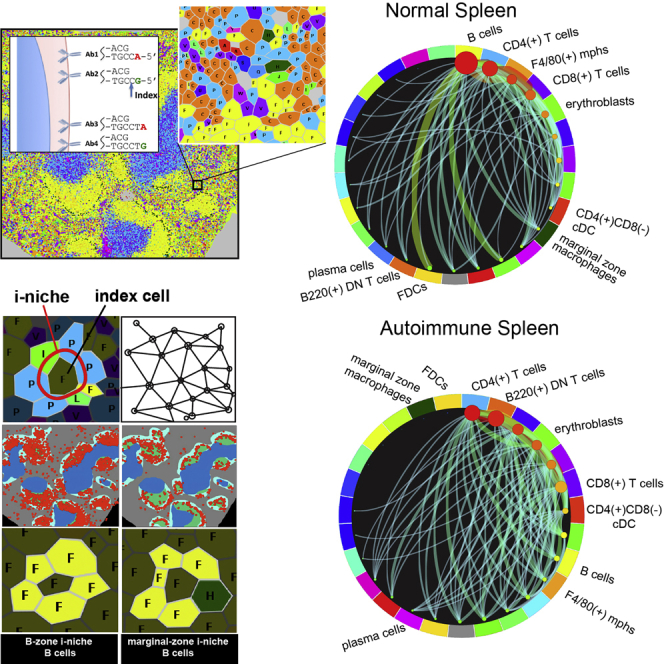
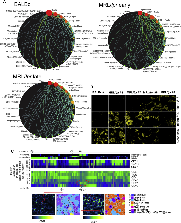
“Pairwise and Combinatorial (i-niches) Statistics of Cellto-Cell Contacts in Mouse Spleen To provide a high-level view of the cell-type interaction landscape, the total counts of contacts between every pair of cell types in the Delaunay neighborhood graph (Gabriel and Sokal, 1969) (Figure 4A and associated Mendeley dat…”
Rubtsov, A. V., Rubtsova, K., Fischer, A., Meehan, R. T., Gillis, J. Z., Kappler, J. W., & Marrack, P. (2011) · Blood · doi:10.1182/blood-2011-01-331462 · Cited by 808
Highly multiplexed single-cell analysis of formalin-fixed, paraffin-embedded cancer tissue
Gerdes, M. J., Sevinsky, C. J., Sood, A., Adak, S., Bello, M. O., Bordwell, A., Can, A. B., Corwin, A. D., Dinn, S. R., Filkins, R. J., et al. (2013) · Proceedings of the National Academy of Sciences · doi:10.1073/pnas.1300136110 · Cited by 716
Robinette, M. L., Fuchs, A. G., Cortez, V. S., Lee, J. S., Wang, Y., Durum, S. K., Gilfillan, S., & Colonna, M. (2015) · Nature Immunology · doi:10.1038/ni.3094 · Cited by 662
Erythroblastic islands: niches for erythropoiesis
Chasis, J. A., & Mohandas, N. (2008) · Blood · doi:10.1182/blood-2008-03-077883 · Cited by 506
Analyzing proteome topology and function by automated multidimensional fluorescence microscopy
SCHUBERT, W. J., Bonnekoh, B., Pommer, A. J., Philipsen, L., Böckelmann, R., Malykh, Y. N., Gollnick, H. P. M., Friedenberger, M., Bode, M., & Dress, A. (2006) · Nature Biotechnology · doi:10.1038/nbt1250 · Cited by 476 · cited 1×
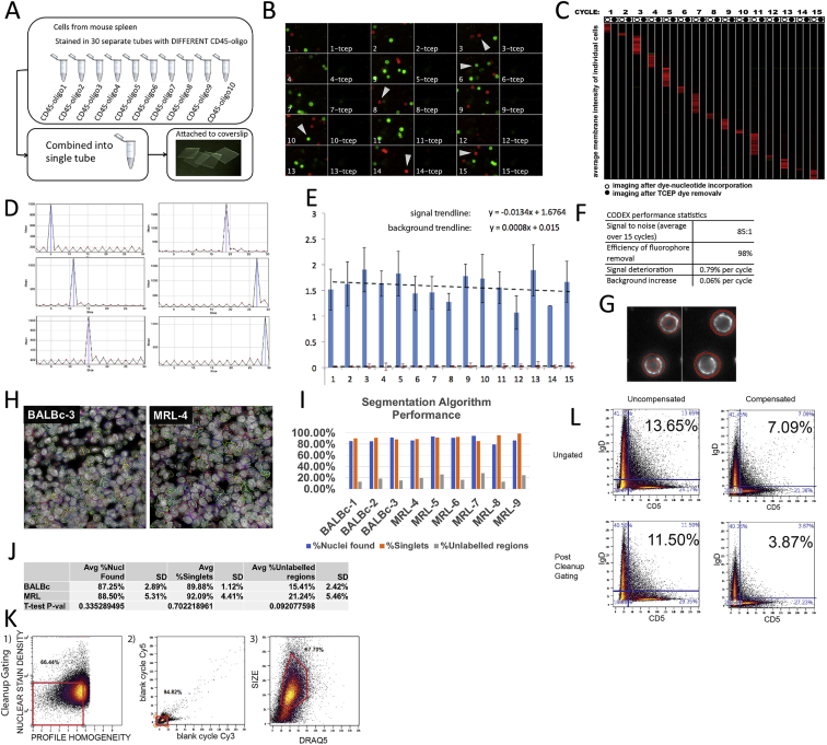
“Approaches have been developed to overcome such limitations (Schubert et al., 2006; Gerdes et al., 2013), but these protocols have required multiple stain/ strip/wash cycles of the antibodies that can be time consuming or lead to sample degradation over the iterations.”
Gerner, M. Y., Kastenmüller, W., Ifrim, I., Kabát, J., & Germain, R. N. (2012) · Immunity · doi:10.1016/j.immuni.2012.07.011 · Cited by 446
Cited by
Sorted by citations
Items 501–600 of 1,002
Reticker-Flynn, N. E., & Engleman, E. G. (2020) · eLife · doi:10.7554/elife.53839 · Cited by 23
“Additional cycles are performed enabling highly multiplexed imaging without requiring antibody staining between each cycle (Goltsev et al., 2018).”
BackgroundSeqStain is an efficient method for multiplexed, spatialomic profiling of human and murine tissues
Rajagopalan, A. R., Venkatesh, I., Aslam, R., Kirchenbüechler, D., Khanna, S., Cimbaluk, D. J., Kordower, J. H., & Gupta, V. (2021) · Cell Reports Methods · doi:10.1016/j.crmeth.2021.100006 · Cited by 20
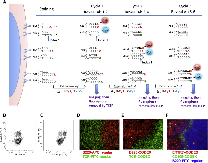
“The CODEX method combines DNA-tagged antibodies and in situ primer extension with fluorescently tagged nucleotides to achieve high-level multiplexing (Goltsev et al., 2018).”
InfluentialAlmekinders, M. M., Bismeijer, T., Kumar, T., Yang, F., Thijssen, B., van der Linden, R., van Rooijen, C. C. N., Vonk, S., Sun, B., Parra, E. R., et al. (2022) · British Journal of Cancer · doi:10.1038/s41416-022-01888-2 · Cited by 21
“Other promising avenues to gain deeper insight in the IME include multiplexed imaging such as CO-Detection by indexing (CODEX) [34], Digital Spatial Profiling technology [35] and MIBI-TOF (multiplexed ion beam imaging by time of flight) [36] allowing detection of a higher number of proteins.”
MethodHigh-multiplex tissue imaging in routine pathology—are we there yet?
Einhaus, J. F., Rochwarger, A., Mattern, S., Gaudilliere, B. L., & Schürch, C. M. (2023) · Archiv für Pathologische Anatomie und Physiologie und für Klinische Medicin · doi:10.1007/s00428-023-03509-6 · Cited by 23
“Technologies that use DNA-tagged antibodies, such as in CO-Detection by indEXing (CODEX) or Immunostaining with Signal Amplification By Exchange Reaction (ImmunoSABER) [13, 14], have more recently evolved as alternative HMTI platforms.”
DELVE: feature selection for preserving biological trajectories in single-cell data
Ranek, J. S., Stallaert, W., Milner, J. J., Redick, M. A., Wolff, S. C., Beltrán, A. S., Stanley, N., & Purvis, J. E. (2024) · Nature Communications · doi:10.1038/s41467-024-46773-z · Cited by 20
A new understanding of tissue biology from MS-based proteomics at single-cell resolution
Nordmann, T. M., Mund, A., & Mann, M. (2024) · Nature Methods · doi:10.1038/s41592-024-02541-x · Cited by 22
Liao, R., Mondal, M., Nazaroff, C. D., Mastroeni, D., Coleman, P. D., LaBaer, J., & Guo, J. (2021) · Frontiers in Cell and Developmental Biology · doi:10.3389/fcell.2020.614624 · Cited by 22
“, 2014) or high magnification objectives and long exposure time to detect the protein targets (Schubert et al., 2006; Duose et al., 2012; Schweller et al., 2012; Gerdes et al., 2013; Zrazhevskiy and Gao, 2013; Lin et al., 2015; Mondal et al., 2017; Goltsev et al., 2018; Gut et al., 2018; Liao et al., 2020).”
BackgroundSpatial-CITE-seq: spatially resolved high-plex protein and whole transcriptome co-mappingPreprint
Liu, Y., DiStasio, M., Su, G., Asashima, H., Enninful, A., Qin, X., Deng, Y., Bordignon, P., Cassano, M., Tomayko, M. M., et al. (2022) · bioRxiv (Cold Spring Harbor Laboratory) · doi:10.1101/2022.04.01.486788 · Cited by 21
Giotto Suite: a multi-scale and technology-agnostic spatial multi-omics analysis ecosystemPreprint
Chen, J. G., Chávez, J., O’Brien, M., Xu, J., Ruiz, E. C., Wang, W., Amin, I., Sarfraz, I., Guckhool, P., Sistig, A., et al. (2023) · bioRxiv (Cold Spring Harbor Laboratory) · doi:10.1101/2023.11.26.568752 · Cited by 21
“The package R.utils is required in the tutorials, but is not installed during GS installation.”
Highly multiplexed imaging in the omics era: understanding tissue structures in health and disease
Bodenmiller, B. (2024) · Nature Methods · doi:10.1038/s41592-024-02538-6 · Cited by 20
Entropy measures for quantifying complexity in digital pathology and spatial omics
Li, X., Ren, X., & Venugopal, R. (2025) · iScience · doi:10.1016/j.isci.2025.112765 · Cited by 25
McKenzie, M. E., Irac, S. E., Chen, Z., Moradi, A., Jenner, A. L., Nguyen, Q. H., & Rashidieh, B. (2026) · Seminars in Cancer Biology · doi:10.1016/j.semcancer.2026.01.002 · Cited by 24
Haist, M., Baertsch, M., Reticker-Flynn, N. E., Lu, G., Kempchen, T. N., Chu, P., Vazquez, G., Chen, H. M., Sunwoo, J. B., Zhang, W., et al. (2026) · Cancer Cell · doi:10.1016/j.ccell.2026.01.003 · Cited by 21
Toward mapping the human body at a cellular resolution
Roy, A. L., & Conroy, R. (2018) · Molecular Biology of the Cell · doi:10.1091/mbc.e18-04-0260 · Cited by 20
“A wide variety of sequencing approaches have Volume 29 August 1, 2018 Human atlases | 1781 also emerged over the past few years, each with its own strengths and weaknesses (Haque et al., 2017; Ziegenhain et al., 2017).”
BackgroundDNA‐Barcoded Fluorescence Microscopy for Spatial Omics
Schueder, F., Unterauer, E. M., Ganji, M., & Jungmann, R. (2020) · PROTEOMICS · doi:10.1002/pmic.201900368 · Cited by 21
Multimodal predictors for precision immunotherapy
Roelofsen, L. M., Kaptein, P., & Thommen, D. S. (2022) · Immuno-Oncology Technology · doi:10.1016/j.iotech.2022.100071 · Cited by 19
Membrane marker selection for segmenting single cell spatial proteomics data
Dayao, M. T., Brusko, M., Wasserfall, C. H., & Bar‐Joseph, Z. (2022) · Nature Communications · doi:10.1038/s41467-022-29667-w · Cited by 20
“Specifically, we compared the resulting segmentation to (1) the Cytokit segmentation that uses only the top-ranked protein marker for segmentation (Top 1) and (2) Cytokit’s default segmentation which is based on extending the nucleus segmentation by a specified radius (Nucl-ext).”
MethodInfluentialNeoadjuvant Immunotherapy for Hepatocellular Carcinoma
Marron, T. U., Schwartz, M., Corbett, V., & Mérad, M. (2022) · Journal of Hepatocellular Carcinoma · doi:10.2147/jhc.s340935 · Cited by 20
Tissue registration and exploration user interfaces in support of a human reference atlas
Börner, K., Bueckle, A., Herr, B. W., Cross, L. E., Quardokus, E. M., Record, E. G., Ju, Y., Silverstein, J. C., Browne, K., Jain, S. R., et al. (2022) · Communications Biology · doi:10.1038/s42003-022-03644-x · Cited by 22
Streamlining spatial omics data analysis with Pysodb
Lin, S., Zhao, F., Wu, Z., Yao, J., Zhao, Y. C., & Yuan, Z. (2023) · Nature Protocols · doi:10.1038/s41596-023-00925-5 · Cited by 20
Why tumour geography matters — and how to map it
Eisenstein, M. S. (2024) · Nature · doi:10.1038/d41586-024-03830-3 · Cited by 20
Encoding Multiple Virtual Signals in DNA Barcodes with Single-Molecule FRET
Kim, S. H., Kim, H., Jeong, H., & Yoon, T. (2021) · Nano Letters · doi:10.1021/acs.nanolett.0c04502 · Cited by 20
Targeting CD123 in hematologic malignancies: identifying suitable patients for targeted therapy
Patnaik, M. M., Mughal, T. I., Brooks, C. L., Lindsay, R. W. B., & Pemmaraju, N. (2021) · Leukemia & lymphoma/Leukemia and lymphoma · doi:10.1080/10428194.2021.1927021 · Cited by 20
“Progress in cyclic labeling and optical imaging methods has improved the spatial resolution and accuracy of surface protein detection in multiplex assays [57].”
MethodImmune cell topography predicts response to PD-1 blockade in cutaneous T cell lymphomaPreprint
Phillips, D. J., Matusiak, M., Rivero‐Gutiérrez, B., Bhate, S. S., Barlow, G. L., Jiang, S., Demeter, J., Smythe, K. S., Pierce, R. H., Fling, S. P., et al. (2020) · medRxiv · doi:10.1101/2020.12.06.20244913 · Cited by 20
Revealing gene function with statistical inference at single-cell resolution
Trapnell, C. (2024) · Nature Reviews Genetics · doi:10.1038/s41576-024-00750-w · Cited by 19
“…transcriptome profiling method that spatially labels nuclei using hashing oligos prior to recovering them from tissue Uses single-cell sequencing to profile randomly sampled nuclei from a tissue section CODEX 114 A multiplexed method for visualizing antibody binding events in tissue sections”
Isnard, P., & Humphreys, B. D. (2024) · American Journal Of Pathology · doi:10.1016/j.ajpath.2024.06.012 · Cited by 18
Xiong, X., Wang, X., Liu, C., Shao, Z.-M., & Yu, K. (2024) · Biomarker Research · doi:10.1186/s40364-024-00654-1 · Cited by 21
“…of sequencing technology, an increasing number of SCS and spatial sequencing technologies and commercial platforms are gradually being introduced, enabling researchers to select one or multiple sequencing technologies/ commercial platforms based on their research objectives [14, 41, 42, 52, 53, 73–”
BackgroundMi, H., Varadhan, R., Cimino‐Mathews, A., Emens, L. A., Santa-Maria, C. A., & Popel, A. S. (2024) · Modern Pathology · doi:10.1016/j.modpat.2024.100652 · Cited by 19
Profiling immune cell tissue niches in the spatial -omics era
Lee, C. Y. C., McCaffrey, J., McGovern, D., & Clatworthy, M. R. (2024) · Journal of Allergy and Clinical Immunology · doi:10.1016/j.jaci.2024.11.001 · Cited by 21
“8,32 These approaches enabled over 50 proteins to be measured per tissue section.”
Cancer therapy resistance from a spatial‐omics perspective
Zhang, Y., Yang, C., Chen, X., Wu, L. L., Yuan, Z., Zhang, F., & Qian, B. (2025) · Clinical and Translational Medicine · doi:10.1002/ctm2.70396 · Cited by 19
Decoding wound healing: cellular insights and technological advances
Berthiaume, K. A., Galvin, E. R., Kness-Knezinskis, E., Hostler, A. C., Chen, K., & Gurtner, G. C. (2026) · npj Biomedical Innovations. · doi:10.1038/s44385-025-00046-6 · Cited by 19
“Asthesemethodsbecomemorewidely available,theywillofferadditionalopportunitiestostudywoundhealingin aspatiotemporalmanner 38,39 .”
Germain, R. N., Radtke, A. J., Thakur, N., Schrom, E. C., Hor, J. L., Ichise, H., Arroyo-Mejías, A., Chu, C. J., & Grant, S. M. (2021) · Immunological Reviews · doi:10.1111/imr.13052 · Cited by 19
Single-Cell Epigenomics Reveals Mechanisms of Cancer Progression
LaFave, L. M., Savage, R. E., & Buenrostro, J. D. (2022) · Annual Review of Cancer Biology · doi:10.1146/annurev-cancerbio-070620-094453 · Cited by 20
Melanoma central nervous system metastases: An update to approaches, challenges, and opportunities
Karz, A., Dimitrova, M., Kleffman, K., Alvarez-Breckenridge, C. A., Atkins, M. B., Boire, A. A., Bosenberg, M., Brastianos, P. K., Cahill, D. P., Chen, Q., et al. (2022) · Pigment Cell & Melanoma Research · doi:10.1111/pcmr.13059 · Cited by 17
“These include scRNA-seq and CITE-seq, spatial transcriptomics, multiplex immunofluorescence imaging technologies (e.g., CODEX and GeoMx), and machine learning-based intercellular communication network inference (Armingol et al. 2021, Goltsev et al. 2018, Khan et al. 2021, Turei et al. 2021).”
MethodGentschev, I., Petrov, I. S., Ye, M., Kafuri, L., Toews, R., Cecil, A., Oelschaeger, T. A., & Szalay, A. A. (2022) · Cancers · doi:10.3390/cancers14246033 · Cited by 17
Semi-automated approaches for interrogating spatial heterogeneity of tissue samples
Navikas, V., Kowal, J., Rodriguez, D., Rivest, F., Brajkovic, S., Cassano, M., & Dupouy, D. (2024) · Scientific Reports · doi:10.1038/s41598-024-55387-w · Cited by 18
“…protein composition of tissue have recently been developed 40 including, but not limited to: cyclic immunofluorescence 41 , co-detection by indexing 42 , multiplex ion beam imaging 43 , seqIF 9 , in addition to manual immunofluorescence protocols as for example iterative indirect…”
Computational methods for alignment and integration of spatially resolved transcriptomics data
Liu, Y., & Yang, C. (2024) · Computational and Structural Biotechnology Journal · doi:10.1016/j.csbj.2024.03.002 · Cited by 19
Pont, F., Tosolini, M., Gao, Q., Perrier, M., Madrid-Mencía, M., Huang, T. S., Neuvial, P., Ayyoub, M., Nazor, K. L., & Fournié, J. (2020) · NAR Genomics and Bioinformatics · doi:10.1093/nargab/lqaa025 · Cited by 19
“Its versatility enables users to analyze likewise any kind of single cell data about chromatin accessibility (12,13), epigenomics (14), mutations (15), chromosome conformation (16), RNA modification (17), spatial transcriptomics (18,19) and spatial proteomics (20,21).”
BackgroundCurrent Status of Tissue Clearing and the Path Forward in Neuroscience
Zhao, J., Lai, H. M., Qi, Y., He, D., & Sun, H. (2020) · ACS Chemical Neuroscience · doi:10.1021/acschemneuro.0c00563 · Cited by 18
Wang, Y., Zeng, Y., Saka, S. K., Xie, W., Goldaracena, I., Kohman, R. E., Yin, P., & Church, G. M. (2018) · bioRxiv (Cold Spring Harbor Laboratory) · doi:10.1101/274456 · Cited by 19
A spatial human thymus cell atlas mapped to a continuous tissue axisPreprint
Yayon, N., Kedlian, V. R., Boehme, L., Suo, C., Wachter, B. T., Beuschel, R. T., Amsalem, O., Polański, K., Koplev, S., Tuck, E. J., et al. (2023) · bioRxiv (Cold Spring Harbor Laboratory) · doi:10.1101/2023.10.25.562925 · Cited by 19
Spatial omics shed light on the tumour organisation of glioblastoma
Whittle, J. R., Kriel, J., Fatunla, E. O., Lu, T., Moffet, J. J. D., Spiteri, M., Best, S. A., & Freytag, S. (2025) · Seminars in Cell and Developmental Biology · doi:10.1016/j.semcdb.2024.12.006 · Cited by 20
Wang, Z. J., Farooq, A., Chen, Y.-J., Bhargava, A., Xu, A. M., & Thomson, M. W. (2025) · Nature Biomedical Engineering · doi:10.1038/s41551-025-01357-0 · Cited by 19
“Recent advances in in situ molecular profiling techniques, including spatial transcriptomic 15,16 and proteomic 17,18 methods, simultaneously capture the spatial relationship of tens to thousands of molecular signals and T-cell localization where cells in the IMC images are pixelated and CD8 + T…”
Endothelial-driven TGFβ signaling supports lung interstitial macrophage development from monocytes
Peng, W., Vanneste, D., Bejarano, D. A., Abinet, J., Meunier, M., Radermecker, C., Perin, F., Cataldo, D. D., Bureau, F., Schlitzer, A., et al. (2025) · Science Immunology · doi:10.1126/sciimmunol.adr4977 · Cited by 17
Oh, C., Jung, H. S., Park, J., Awad, J., Jiang, D., Dowling, D. J., & Im, H. (2025) · ACS Nano · doi:10.1021/acsnano.5c04836 · Cited by 21
Automated cell annotation and classification on histopathology for spatial biomarker discovery
Li, Z., Mirjahanmardi, S. H., Sali, R., Eweje, F. R., Gopaulchan, M., Kloker, L. H., Zhang, X., Li, G., Jiang, Y., & Li, R. (2025) · Nature Communications · doi:10.1038/s41467-025-61349-1 · Cited by 21
“Recent development of highly multi-plexed imaging technology such as co-detection by indexing 44 or one-shot high-plex immuno fl uorescence 45 , may be used to identify additional cell types on the same tissue section as the H&E. Additionally, while traditional spatial transcriptomics only provide…”
Kim, D. Y., Lee, J., Choi, J., Shin, H., Lee, J. S., & Kim, E. J. (2025) · Seminars in Cancer Biology · doi:10.1016/j.semcancer.2025.12.009 · Cited by 20
Introduction to Multiparametric Flow Cytometry and Analysis of High-Dimensional Data
Sun, J. X., Kroeger, J. L., & Markowitz, J. (2020) · Methods in molecular biology · doi:10.1007/978-1-0716-0849-4_13 · Cited by 23
Endocrine Pancreas Development and Dysfunction Through the Lens of Single-Cell RNA-Sequencing
Szlachcic, W. J., Ziojla, N., Kizewska, D. K., Kempa, M., & Borowiak, M. (2021) · Frontiers in Cell and Developmental Biology · doi:10.3389/fcell.2021.629212 · Cited by 19
“Single-cell proteomics is also being extensively developed (Goltsev et al., 2018; Gut et al., 2018; Saka et al., 2019; Labib et al., 2020).”
BackgroundSpace in cancer biology: its role and implications
Khartchenko, A. F., Kashyap, A., Geiger, T., & Kaigala, G. V. (2022) · Trends in cancer · doi:10.1016/j.trecan.2022.07.008 · Cited by 19
Multiomics Empowers Predictive Pancreatic Cancer Immunotherapy
Montagne, J. M., Jaffee, E. M., & Fertig, E. J. (2023) · The Journal of Immunology · doi:10.4049/jimmunol.2200660 · Cited by 18
Deciphering the tumour immune microenvironment cell by cell
Nabhan, M., Egan, D., Kreileder, M., Zhernovkov, V., Timosenko, E., Slidel, T. W. F., Dovedi, S. J., Glennon, K., Brennan, D. J., & Kölch, W. (2023) · Immuno-Oncology Technology · doi:10.1016/j.iotech.2023.100383 · Cited by 17
CODEX multiplexed tissue imaging
Goltsev, Y., & Nolan, G. P. (2023) · Nature reviews. Immunology · doi:10.1038/s41577-023-00936-z · Cited by 18
Heller, G., Fuereder, T., Grandits, A. M., & Wieser, R. (2023) · Oncology Research Featuring Preclinical and Clinical Cancer Therapeutics · doi:10.32604/or.2023.044774 · Cited by 19
Thermal-plex: fluidic-free, rapid sequential multiplexed imaging with DNA-encoded thermal channels
Fan, H. B., Kishi, J. Y., Delgado, R. N., Jeong, J., Saka, S. K., Su, H., Cepko, C. L., & Yin, P. (2023) · Nature Methods · doi:10.1038/s41592-023-02115-3 · Cited by 18
“We expect thermal-plex to be versatile and applicable to other imaging targets beyond RNA, for example, chromosomes 19,30 and proteins 11,31 .”
BackgroundOne-Shot Single-Cell Proteome and Metabolome Analysis Strategy for the Same Single Cell
Wu, J., Xu, Q., Jiang, Y., Chen, J., Ying, W.-X., Fan, Q., Wang, H.-F., Wang, Y., Shi, S.-W., Pan, J., et al. (2024) · Analytical Chemistry · doi:10.1021/acs.analchem.3c05659 · Cited by 20
GraphCompass: spatial metrics for differential analyses of cell organization across conditions
Ali, M., Kuijs, M., Hediyeh-zadeh, S., Treis, T., Hrovatin, K., Palla, G., Schaar, A. C., & Theis, F. J. (2024) · Bioinformatics · doi:10.1093/bioinformatics/btae242 · Cited by 17
“…methods such as 10 × Genomics Visium (Ståhl et al. 2016) and Stereo-seq (Chen et al. 2022), as well as spatial proteomics methods such as CODEX (Goltsev et al. 2018) and multiplexed ion beam imaging by time of flight (MIBI-TOF) (Keren et al. 2019), can measure molecular profiles while…”
MethodPancreatlas: Applying an Adaptable Framework to Map the Human Pancreas in Health and Disease
Saunders, D. C., Messmer, J., Kusmartseva, I., Beery, M. L., Yang, M., Atkinson, M. A., Powers, A. C., Cartailler, J., & Briššová, M. (2020) · Patterns · doi:10.1016/j.patter.2020.100120 · Cited by 19
Next Generation Imaging Techniques to Define Immune Topographies in Solid Tumors
Pietrobon, V., Cesano, A., Marincola, F. M., & Kather, J. N. (2021) · Frontiers in Immunology · doi:10.3389/fimmu.2020.604967 · Cited by 18
Nanoscopic subcellular imaging enabled by ion beam tomography
Coskun, A. F., Han, G., Ganesh, S., Chen, S.-Y., Rovira‐Clavé, X., Harmsen, S., Jiang, S., Schürch, C. M., Bai, Y., Hitzman, C., et al. (2021) · Nature Communications · doi:10.1038/s41467-020-20753-5 · Cited by 22
Single-cell analysis of regions of interest (SCARI) using a photosensitive tag
van der Leun, A. M., Hoekstra, M. E., Reinalda, L., Scheele, C. L. G. J., Toebes, M., van de Graaff, M. J., Chen, L. Y. Y., Li, H., Bercovich, A., Lubling, Y., et al. (2021) · Nature Chemical Biology · doi:10.1038/s41589-021-00839-x · Cited by 19
SOAPy: a Python package to dissect spatial architecture, dynamics, and communication
Wang, H., Li, J., Jing, S., Lin, P., Qiu, Y., Yan, X., Yuan, J., Tang, Z., Li, Y., Zhang, H., et al. (2025) · Genome biology · doi:10.1186/s13059-025-03550-5 · Cited by 20
“Given an index cell, I-niche is defined as the proportion of cell types for its surrounding cells [55].”
Advances in Spatial Omics Technologies
Hui, T., Zhou, J., Yao, M., Xie, Y., & Zeng, H. (2025) · Small Methods · doi:10.1002/smtd.202401171 · Cited by 18
Advancements in Nanomaterials and Molecular Probes for Spatial Omics
Lin, Y., Hou, J., Li, B., Shu, W., & Wan, J. (2025) · ACS Nano · doi:10.1021/acsnano.4c18470 · Cited by 17
Single-cell spatiotemporal dissection of the human maternal–fetal interface
Wang, C., Zhou, Y., Wang, Y. J., Guha, T. K., Luo, Z., Mustafaraj, A., McIntyre, T. I., Schwab, M. E., Davidson, B. R., Reeder, G. C., et al. (2026) · Nature · doi:10.1038/s41586-026-10316-x · Cited by 17
Proteomic characterization of the human lens and Cataractogenesis
Cantrell, L. S., & Schey, K. L. (2021) · Expert Review of Proteomics · doi:10.1080/14789450.2021.1913062 · Cited by 17
“cron resolution in a semiquantitative manner [173].”
Unmixing for ultra-high-plex fluorescence imaging
Enninful, A., Baysoy, A., & Fan, R. (2022) · Nature Communications · doi:10.1038/s41467-022-31110-z · Cited by 15
Towards multiplexed immunofluorescence of 3D tissues
Cho, W., Kim, S., & Park, Y.-G. (2023) · Molecular Brain · doi:10.1186/s13041-023-01027-9 · Cited by 18
Spatial Proteomics for the Molecular Characterization of Breast Cancer
Brožová, K., Hantusch, B., Kenner, L., & Kratochwill, K. (2023) · Proteomes · doi:10.3390/proteomes11020017 · Cited by 19
Gosline, S. J. C., Veličković, M., Pino, J. C., Day, L. Z., Attah, I. K., Swensen, A., Danna, V., Posso, C., Rodland, K., Chen, J., et al. (2023) · Molecular & Cellular Proteomics · doi:10.1016/j.mcpro.2023.100592 · Cited by 19
Multiplex imaging in immuno-oncology
Zhao, C. N., & Germain, R. N. (2023) · Journal for ImmunoTherapy of Cancer · doi:10.1136/jitc-2023-006923 · Cited by 21
“…iterative bleaching extends multiplexity (IBEX), 31 tissue-based cyclic immunofluorescence (t-CyCIF), 38 multiepitope-ligand cartography, 39 and multiplexed immunofluorescence (MxIF, Cell DIVE, PhenoCycler (formerly CODEX)) 40 41 can detect more than 60 markers in a single tissue section (table 1).”
BackgroundApplication of spatial-omics to the classification of kidney biopsy samples in transplantation
Tasca, P., van den Berg, B. M., Rabelink, T. J., Wang, G., Heijs, B., van Kooten, C., de Vries, A. P. J., & Kers, J. (2024) · Nature Reviews Nephrology · doi:10.1038/s41581-024-00861-x · Cited by 17
“After the image acquisition, the reporters are removed with a solution that does not affect antibody bonds, and the cycle is repeated until all the antibodies of the panel are revealed 86 .”
Satou, A., Bennani, N. N., & Feldman, A. L. (2019) · Expert Review of Hematology · doi:10.1080/17474086.2019.1647777 · Cited by 17
Personal tumor antigens in blood malignancies: genomics-directed identification and targeting
Penter, L., & Wu, C. J. (2020) · Journal of Clinical Investigation · doi:10.1172/jci129209 · Cited by 17
“Likewise, the development of CRISPR/Cas9 gene editing (173), the discovery efforts with genome-wide screens (174), and developments in the area of spatial tissue-based characterization (175) will have important implications for the delivery of novel targets and subsequent engineering of immune responses.”
BackgroundBourriez, N., Bendidi, I., Cohen, E., Watkinson, G., Sanchez, M., Bollot, G., & Genovesio, A. (2024) · IEEE Computer Society Conference on Computer Vision and Pattern Recognition/Proceedings - IEEE Computer Society Conference on Computer Vision and Pattern Recognition/Proceedings · doi:10.1109/cvpr52733.2024.01098 · Cited by 16
“Unlike conventional pictures in RGB color format, microscopy images span a plethora of specialized types of image across various channels [21, 27, 44].”
BackgroundEmerging AI approaches for cancer spatial omics
Noorbakhsh, J., pour, A. F., & Chuang, J. H. (2025) · GigaScience · doi:10.1093/gigascience/giaf128 · Cited by 19
“Advances in multiplex techniques, such as CODEX [ 56 , 57 ], IMC [ 58 ], and Cell Dive [ 59 ], have allowed simultaneous measurement of many proteins at subcellular resolution.”
From Histology to High‐Resolution Mapping: The Rise of Spatial Omics in Immunology
Giovanni, M. D., Inverso, D., & Iannacone, M. (2025) · European Journal of Immunology · doi:10.1002/eji.70073 · Cited by 17
“CODEX (CO-Detection by Indexing, PhenoCycler platform from Akoya Biosciences) is a multiplexed imaging platform designed for high-resolution spatial proteomics, enabling the detection of up to 100 protein markers within intact tissue sections while preserving spatial context [30, 31].”
Integrative deep learning of spatial multi-omics with SWITCH
Li, Z., Qu, S., Liang, H., Tang, R., Zhang, X., Fan, L., Yang, J., Gan, Z., Gao, S., Zhang, Y., et al. (2025) · Nature Computational Science · doi:10.1038/s43588-025-00891-w · Cited by 20
“Spatial transcriptomics (ST) 1–4 was developed to overcome this constraint and has evolved to encompass spatial multi-omics, including spatial epigenomics 5–7 and proteomics 8,9 .”
Regulatory T cell therapies: from patient data to biological insights
Rodrigues, K. B., Eggenhuizen, P. J., Bacchetta, R., & Good, Z. (2025) · Frontiers in Immunology · doi:10.3389/fimmu.2025.1675114 · Cited by 18
“…panel of up to 6,000 genes and support custom probes for engineered proteins, such as CAR or TCR. Spatial proteomics technologies, including MIBI (87) and CODEX (64, 88), can be used instead of or in parallel with spatial transcriptomics methods to learn insights from the relevant tissue biopsies.”
He, G., He, S., Jing, X., Dai, Y., Guo, X. N., Gao, J., & Zhang, W. (2025) · Oncogene · doi:10.1038/s41388-025-03635-2 · Cited by 19
“Goltsev et al. demonstrated the feasibility of CODEX for high-dimensional proteomic imaging of spleen and tumor tissues [87].”
Single‐Cell and Spatial Multiomics: Applications for Diseases
Li, W., Chen, C., Zhu, X., & Zhang, C. (2025) · MedComm · doi:10.1002/mco2.70553 · Cited by 16
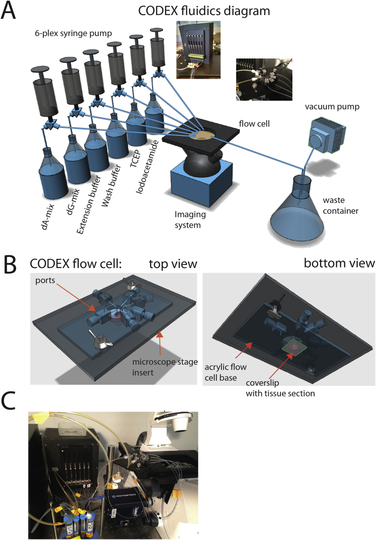
“CODEX (codetection by indexing), an alternative method to highly multiplexed spatial proteomics, uses a library of DNA-barcoded antibodies and successive cycles of hybridization, imaging, and dye inactivation to visualize dozens of proteins at once on a standard fluorescence microscope [ 50 ].”
Bridging scales: From cell biology to physiology using in situ single-cell technologies
Nagle, M. P., Tam, G. S., Maltz, E., Hemminger, Z., & Wollman, R. (2021) · Cell Systems · doi:10.1016/j.cels.2021.03.002 · Cited by 15
Lu, G., Baertsch, M., Hickey, J. W., Goltsev, Y., Rech, A. J., Mani, L., Forgó, E., Kong, C. S., Jiang, S., Nolan, G. P., et al. (2022) · Frontiers in Immunology · doi:10.3389/fimmu.2022.981825 · Cited by 16
“The CODEX dataset acquired on a Keyence microscope has the following parameters (1): tile: a single field of view (2); region: tissue region of interest composed of single tile or multiple tiles collected as rectangular grids (3); z: the number of z planes acquired in the axial direction that includes the best focus p…”
BackgroundMethodInfluentialImproving and evaluating deep learning models of cellular organization
Sun, H., Fu, X., Abraham, S., Shen, J., & Murphy, R. F. (2022) · Bioinformatics · doi:10.1093/bioinformatics/btac688 · Cited by 15
Spatial subcellular organelle networks in single cells
Venkatesan, M., Zhang, N. Y., Marteau, B. L., Yajima, Y., Garcia, N. O. D. Z., Fang, Z., Hu, T., Cai, S., Ford, A., Olszewski, H., et al. (2023) · Scientific Reports · doi:10.1038/s41598-023-32474-y · Cited by 17
Proteome-Scale Tissue Mapping Using Mass Spectrometry Based on Label-Free and Multiplexed Workflows
Kwon, Y. M., Woo, J. J., Yu, F., Williams, S., Markillie, L. M., Moore, R., Nakayasu, E. S., Chen, J., Campbell‐Thompson, M., Mathews, C. E., et al. (2024) · Molecular & Cellular Proteomics · doi:10.1016/j.mcpro.2024.100841 · Cited by 16
“While spatial transcriptomics provide valuable insights into cellular heterogeneity, they do not directly infer the expression levels of proteins, which are the actual functional molecules in signaling pathways 10, 11 .”
MuSpAn: a toolbox for multiscale spatial analysis
Bull, J. A., Moore, J. W., Corry, S. M., Lin, M., Belnoue-Davis, H. L., Mulholland-Illingworth, E. J., Leedham, S. J., & Byrne, H. M. (2026) · Nature Communications · doi:10.1038/s41467-026-75649-7 · Cited by 18
Baharlou, H., Canete, N. P., Bertram, K. M., Sandgren, K. J., Cunningham, A. L., Harman, A. N., & Patrick, E. (2020) · Bioinformatics · doi:10.1093/bioinformatics/btaa780 · Cited by 15
Visual cohort comparison for spatial single-cell omics-data
Somarakis, A., Ijsselsteijn, M. E., Luk, S. J., Kenkhuis, B., de Miranda, N. F. C. C., Lelieveldt, B. P. F., & Höllt, T. (2020) · IEEE Transactions on Visualization and Computer Graphics · doi:10.1109/tvcg.2020.3030336 · Cited by 19
Pfisterer, U., Bräunig, J., Brattås, P. L., Heidenblad, M., Karlsson, G., & Fioretos, T. (2021) · Genes Chromosomes and Cancer · doi:10.1002/gcc.22944 · Cited by 17
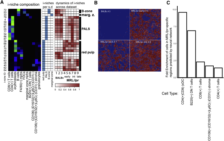
“pher differences in tissue composition in murine normal and diseased spleen at single-cell resolution.(159) Its applicability to clinical human samples, however, still needs to be shown in large-scale studies.”
BackgroundLü, M., Sha, Y., Silva, T. C., Colaprico, A., Sun, X., Ban, Y. J., Wang, L., Lehmann, B. D., & Chen, X. S. (2021) · Frontiers in Genetics · doi:10.3389/fgene.2021.708835 · Cited by 16
Multi-Omics Profiling of the Tumor Microenvironment
Oekelen, O. V., & Laganà, A. (2022) · Advances in experimental medicine and biology · doi:10.1007/978-3-030-91836-1_16 · Cited by 13
A multiplexed epitope barcoding strategy that enables dynamic cellular phenotypic screens
Kudo, T., Lane, K. M., & Covert, M. W. (2022) · Cell Systems · doi:10.1016/j.cels.2022.02.006 · Cited by 14
Cheng, Y., Burrack, R., & Li, Q. (2022) · Journal of Histochemistry & Cytochemistry · doi:10.1369/00221554221114174 · Cited by 16
“70(8) 571 –581 © The Author(s) 2022 Article reuse guidelines: sagepub.”
Winfree, S., McNutt, A. T., Khochare, S., Borgard, T. J., Barwinska, D., Sabo, A. R., Ferkowicz, M. J., Williams, J. C., Lingeman, J. E., Gulbronson, C. J., et al. (2023) · Laboratory Investigation · doi:10.1016/j.labinv.2023.100104 · Cited by 14
Hoeppli, R. E., & Pesenacker, A. M. (2019) · Frontiers in Immunology · doi:10.3389/fimmu.2019.00046 · Cited by 16
Metabolic regulation in adult and aging skeletal muscle stem cellsPreprint
Sartorelli, V., & Ciuffoli, V. (2024) · Genes & Development · doi:10.1101/gad.352277.124 · Cited by 19
Spatially Resolved Multiomics: Data Analysis from Monoomics to Multiomics
Huan, C., Li, J., Li, Y., Zhao, S., Yang, Q., Zhang, Z., Li, C., Li, S., Guo, Z., Yao, J., et al. (2024) · BME Frontiers · doi:10.34133/bmef.0084 · Cited by 17
“Positive interactions between macrophages and erythroid cells were observed in erythroblast islands in mouse spleens, whereas in the white pulp, homotypic cell associations played major roles [ 70 ].”
Multimodal spatial proteomic profiling in acute myeloid leukemia
Ly, C. P., Veletić, I., Pacheco, C. D., Dasdemir, E., Jelloul, F. Z., Borgogno, S. F., Basi, A., Gomez, J. A., Root, J. L., Reville, P. K., et al. (2025) · npj Precision Oncology · doi:10.1038/s41698-025-00897-7 · Cited by 15
Cellular neighborhoods in cancer
Ma, L., Xiong, B., Liu, M., & Tan, K. (2026) · Nature Cancer · doi:10.1038/s43018-025-01107-w · Cited by 16
Metadata
- Authors
- Goltsev, Y., Samusik, N., Kennedy-Darling, J., Bhate, S., Hale, M., Vazquez, G., Black, S., & Nolan, G. P.
- Affiliations
- Stanford Medicine (US)
- Corresponding author
- Nolan, Garry P.
- Venue
- Cell
- Publisher
- Elsevier BV
- Published
- 2018-08
- Volume
- 174
- Issue
- 4
- Pages
- 968-981.e15
- ISSN
- 0092-8674 (ISSN-L)
- PMC
- PMC6086938
- OpenAlex
- W2949238013
- Cited by
- 1,501
- References
- 20
- Citations by year
- 2026: 231 · 2025: 244 · 2024: 232 · 2023: 198 · 2022: 194 · 2021: 194 · 2020: 125 · 2019: 72 · 2018: 9
- FWCI
- 35.39
- Type
- journal article
- Language
- en
- Open access
- hybrid
- doi.org
- Licence
- Locations
- Cell (publishedVersion, cc-by)
- bioRxiv (Cold Spring Harbor Laboratory) (acceptedVersion)
- Europe PMC (PubMed Central) (publishedVersion, cc-by)
- PubMed Central (publishedVersion, cc-by)
- PubMed (publishedVersion)
- Funders
- US NIH (U19AI057229, 1U19AI100627, R01CA184968, 1R33CA183654-01, R33CA183692, 1R01GM10983601, 1R21CA183660, 1R01NS08953301, 5UH2AR067676, 1R01CA19665701, R01HL120724, R01HL128173)
- Bill and Melinda Gates foundation (OPP1113682)
- Department of Defense (W81XWH-14-1-0180)
- Northrop-Grumman Corporation (7500108142)
- NIH (U54-CA209971)
- Rachford & Carlotta A. Harris Endowed Chair
- Topics
- Life Sciences › Biochemistry, Genetics and Molecular Biology › Molecular Biology › Single-cell and spatial transcriptomics
- Life Sciences › Biochemistry, Genetics and Molecular Biology › Biophysics › Cell Image Analysis Techniques
- Life Sciences › Biochemistry, Genetics and Molecular Biology › Biophysics › Advanced Fluorescence Microscopy Techniques
- Keywords
- codices, splenic architecture, MRL/lpr mice, systemic lupus erythematosus, cell-cell interactions, autoimmune disease
- MeSH terms
- Animals, Antibodies, Disease Models, Animal, Female, Image Processing, Computer-Assisted, Lupus Erythematosus, Systemic, Male, Mass Spectrometry, Spleen, Oligonucleotide Probes, Mice, Inbred MRL lpr, Mice
About these checks
Registry, retraction, Cites and statistics results are computed, not judged, with one exception: a printed reference that no index lists is read once by a model before it counts as a citation error. Claims are extracted by a model and kept only when their quote appears verbatim in the text; each cited source is then checked, with a model’s help, for whether it says what the claim relies on. Those verdicts can be wrong — read the claim’s quote and the cited source before relying on one.
Statistics and arithmetic are recomputed, not judged.
Confidence bands are shown only for fields calibrated on our annotated test set. None is calibrated yet, so no band is shown.
What counts as a claim ›How we measure confidence ›How we check this ›
Are you an author, or do you see a problem with this page? Request a review
