

The DOI matches its Crossref record; 21 of the 21 references it cites were found in a registry or an index.
Deep Profiling of Mouse Splenic Architecture with CODEX Multiplexed Imaging
Open accessPublished version of a preprint
Abstract
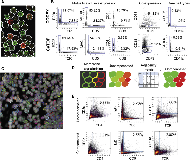
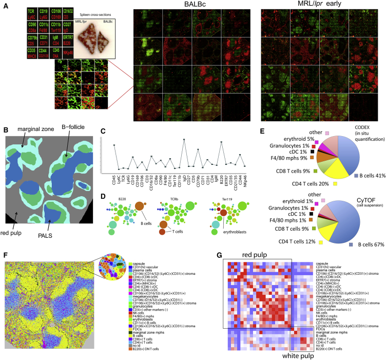
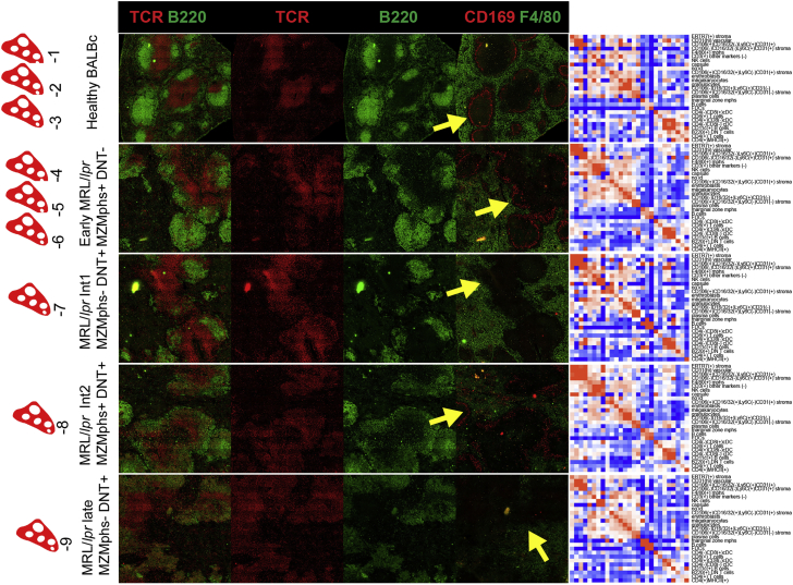
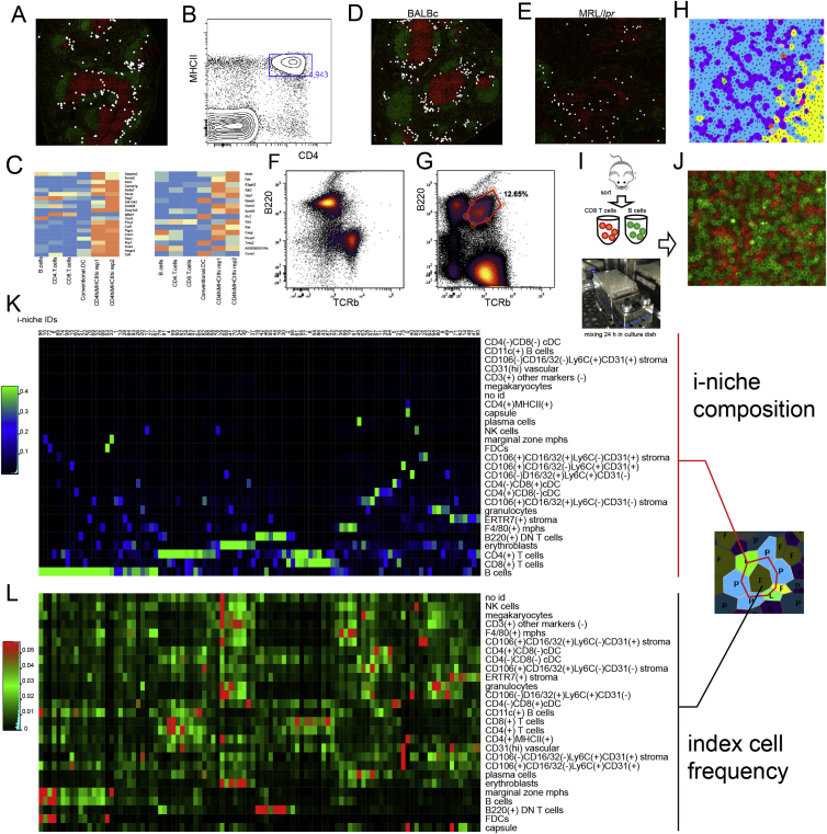
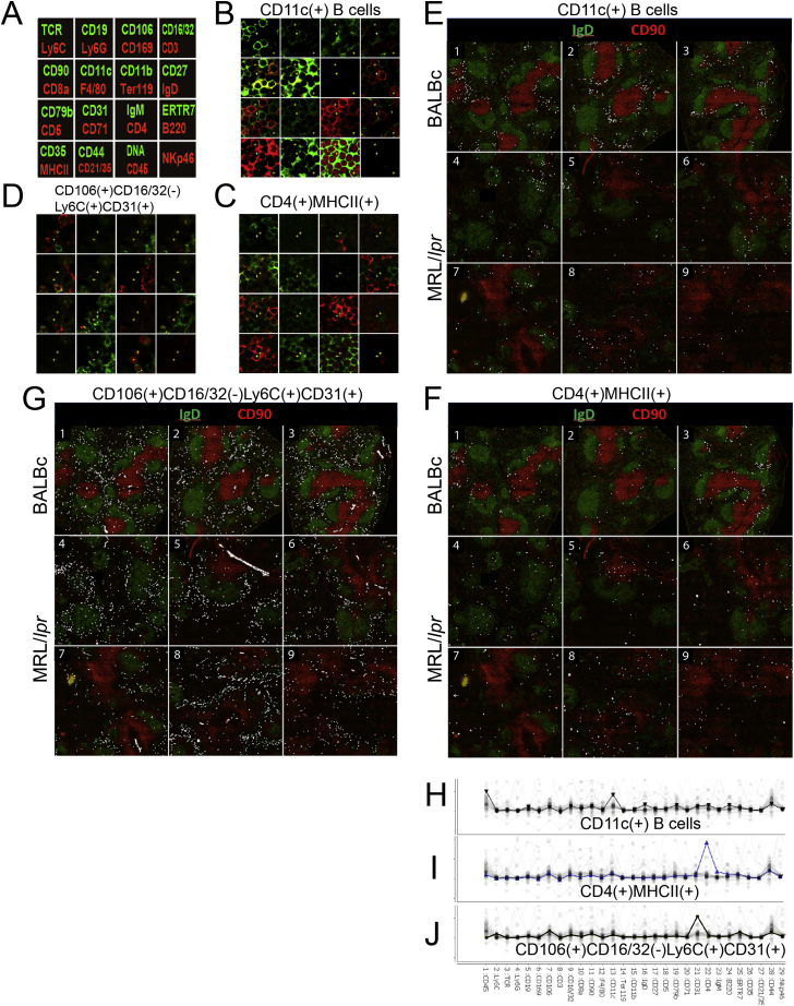
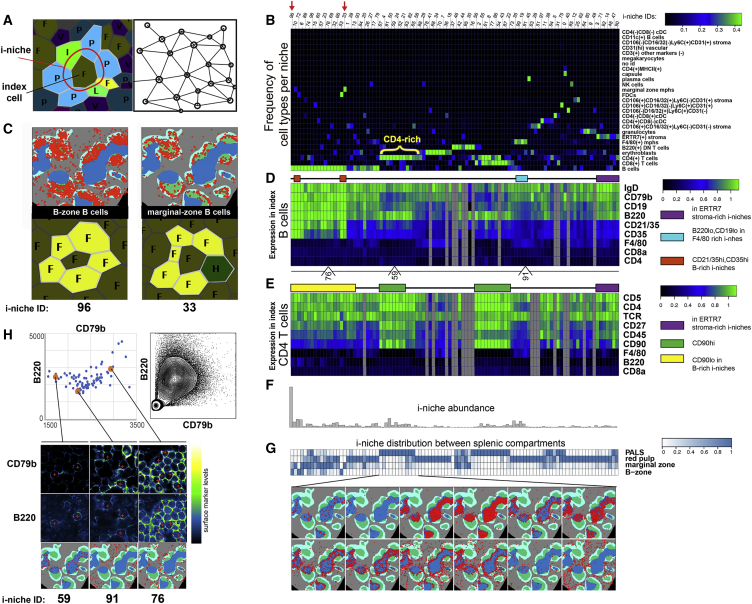
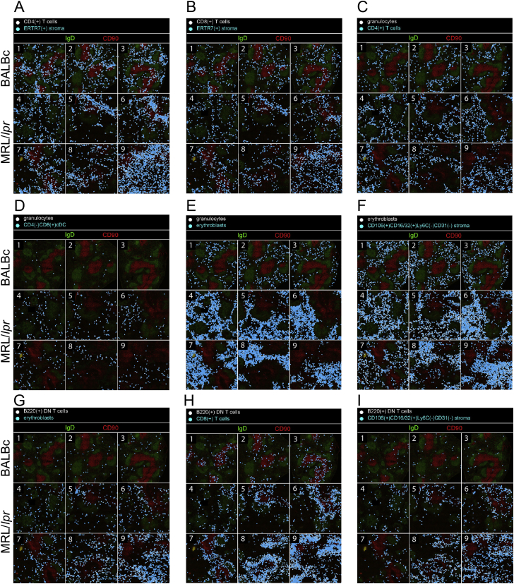
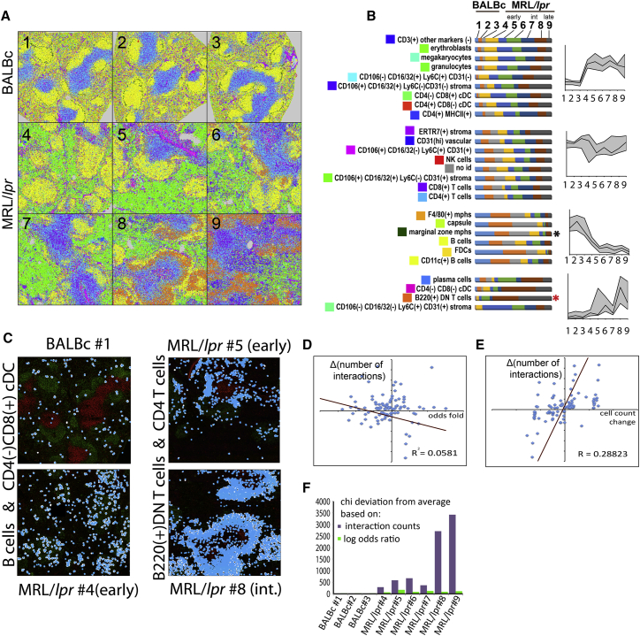
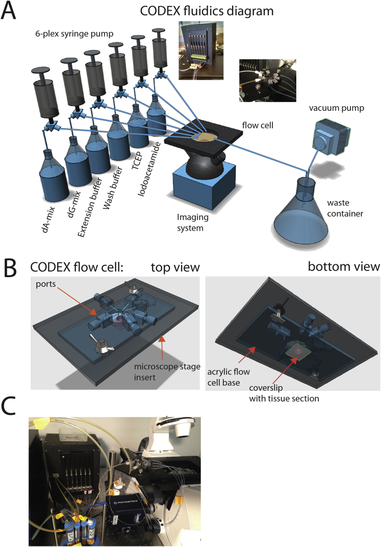
A highly multiplexed cytometric imaging approach, termed co-detection by indexing (CODEX), is used here to create multiplexed datasets of normal and lupus (MRL/lpr) murine spleens. CODEX iteratively visualizes antibody binding events using DNA barcodes, fluorescent dNTP analogs, and an in situ polymerization-based indexing procedure. An algorithmic pipeline for single-cell antigen quantification in tightly packed tissues was developed and used to overlay well-known morphological features with de novo characterization of lymphoid tissue architecture at a single-cell and cellular neighborhood levels. We observed an unexpected, profound impact of the cellular neighborhood on the expression of protein receptors on immune cells. By comparing normal murine spleen to spleens from animals with systemic autoimmune disease (MRL/lpr), extensive and previously uncharacterized splenic cell-interaction dynamics in the healthy versus diseased state was observed. The fidelity of multiplexed spatial cytometry demonstrated here allows for quantitative systemic characterization of tissue architecture in normal and clinically aberrant samples.
Overview
The claims and statistics of this paper haven’t been checked yet.
Run the check — free, no account needed. It reads the open-access text and checks the sources it cites.
Integrity signals
Registry
Cites
Statistics
Figures
Verification
Claims the paper makes
The claims and statistics of this paper haven’t been checked yet.
Run the check — free, no account needed. It reads the open-access text and checks the sources it cites.
Claims other papers make about this one
Each is checked against this paper’s own text: does it say what the citing paper says it does?
In the citing paper
“For example, multiplexed antibody-based imaging (mABI) technologies such as imaging mass cytometry (IMC) [31] and codetection by indexing (CODEX) [32] can detect 20–50 selected genes simultaneously in each cell at the protein level, which is a useful complement to transcript-based detection – especially given the potential for poor correlation of transcriptomic and proteomic data sets implicated”
From The heterogeneity of cellular senescence: insights at the single-cell level (2023) · cites this paper as [32]
Self-check
Percentages, sums and reported differences re-computed from the paper’s own numbers.
The self-check needs the paper’s full text.
Citations
Cites
Globally optimal stitching of tiled 3D microscopic image acquisitions
Preibisch, S., Saalfeld, S., & Tomancak, P. (2009) · Bioinformatics · doi:10.1093/bioinformatics/btp184 · Cited by 2,452
Bendall, S. C., Simonds, E. F., Qiu, P., Amir, E.-A. D., Krutzik, P. O., Finck, R., Bruggner, R. V., Melamed, R., Trejo, A., Ornatsky, O. I., et al. (2011) · Science · doi:10.1126/science.1198704 · Cited by 2,279
Self-citationSystemic Immunity Is Required for Effective Cancer Immunotherapy
Spitzer, M. H., Carmi, Y., Reticker-Flynn, N. E., Kwek, S. S., Madhireddy, D., Martins, M. M., Gherardini, P. F., Prestwood, T. R., Chabon, J., Bendall, S. C., et al. (2017) · Cell · doi:10.1016/j.cell.2016.12.022 · Cited by 963
A New Statistical Approach to Geographic Variation Analysis
Gabriel, K. R., & Sokal, R. R. (1969) · Systematic Zoology · doi:10.2307/2412323 · Cited by 854 · cited 1×
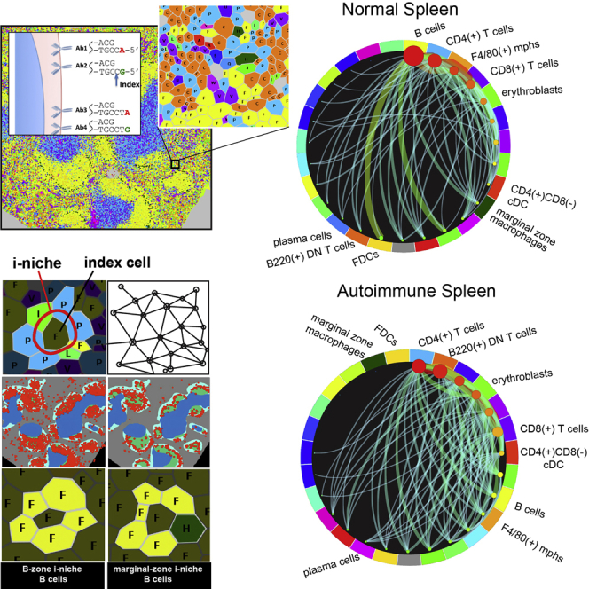
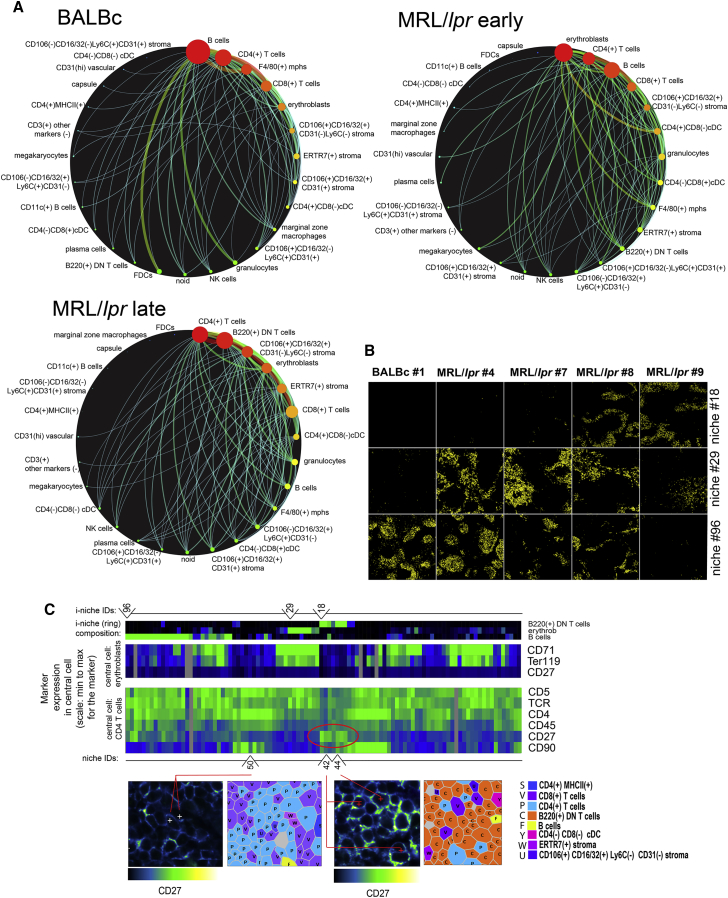
“Pairwise and Combinatorial (i-niches) Statistics of Cellto-Cell Contacts in Mouse Spleen To provide a high-level view of the cell-type interaction landscape, the total counts of contacts between every pair of cell types in the Delaunay neighborhood graph (Gabriel and Sokal, 1969) (Figure 4A and associated Mendeley dat…”
Rubtsov, A. V., Rubtsova, K., Fischer, A., Meehan, R. T., Gillis, J. Z., Kappler, J. W., & Marrack, P. (2011) · Blood · doi:10.1182/blood-2011-01-331462 · Cited by 808
Highly multiplexed single-cell analysis of formalin-fixed, paraffin-embedded cancer tissue
Gerdes, M. J., Sevinsky, C. J., Sood, A., Adak, S., Bello, M. O., Bordwell, A., Can, A. B., Corwin, A. D., Dinn, S. R., Filkins, R. J., et al. (2013) · Proceedings of the National Academy of Sciences · doi:10.1073/pnas.1300136110 · Cited by 716
Robinette, M. L., Fuchs, A. G., Cortez, V. S., Lee, J. S., Wang, Y., Durum, S. K., Gilfillan, S., & Colonna, M. (2015) · Nature Immunology · doi:10.1038/ni.3094 · Cited by 662
Erythroblastic islands: niches for erythropoiesis
Chasis, J. A., & Mohandas, N. (2008) · Blood · doi:10.1182/blood-2008-03-077883 · Cited by 506
Analyzing proteome topology and function by automated multidimensional fluorescence microscopy
SCHUBERT, W. J., Bonnekoh, B., Pommer, A. J., Philipsen, L., Böckelmann, R., Malykh, Y. N., Gollnick, H. P. M., Friedenberger, M., Bode, M., & Dress, A. (2006) · Nature Biotechnology · doi:10.1038/nbt1250 · Cited by 476 · cited 1×
“Approaches have been developed to overcome such limitations (Schubert et al., 2006; Gerdes et al., 2013), but these protocols have required multiple stain/ strip/wash cycles of the antibodies that can be time consuming or lead to sample degradation over the iterations.”
Gerner, M. Y., Kastenmüller, W., Ifrim, I., Kabát, J., & Germain, R. N. (2012) · Immunity · doi:10.1016/j.immuni.2012.07.011 · Cited by 446
Cited by
Sorted by citations
Items 301–400 of 1,002
Zhang, D., Kirby, L. A., Lin, Y., Song, M., Wang, L., Wang, L., Kanatani, S., Jimenez-Beristain, T., Dang, Y., Zhong, M., et al. (2024) · bioRxiv (Cold Spring Harbor Laboratory) · doi:10.1101/2024.07.28.605493 · Cited by 45
Dhanasekaran, R. (2021) · Seminars in Liver Disease · doi:10.1055/s-0040-1722261 · Cited by 47
Harnessing the Potential of Multiomics Studies for Precision Medicine in Infectious Disease
Ward, R., Aghaeepour, N., Bhattacharyya, R. P., Clish, C. B., Gaudilliere, B. L., Hacohen, N., Mansour, M. K., Mudd, P. A., Pasupneti, S., Presti, R. M., et al. (2021) · Open Forum Infectious Diseases · doi:10.1093/ofid/ofab483 · Cited by 45
“Additional complementary approaches such as multiplexed cytometric imaging (CODEX) and in situ hybridization (RNAscope) may be used to improve power of spatial transcriptomics [37, 51, 52].”
MethodMayer, A. T., Holman, D., Sood, A., Tandon, U., Bhate, S. S., Bodapati, S., Barlow, G. L., Chang, J. H., Black, S., Crenshaw, E. C., et al. (2023) · Science Advances · doi:10.1126/sciadv.add1166 · Cited by 44
“A multiplexed immunofluorescence-based imaging technology called CO-Detection by indEXing (CODEX) has allowed simultaneous visualization of up to 60 biomarkers on intact tissue with singlecell resolution (27, 28).”
MethodAquila: a spatial omics database and analysis platform
Zheng, Y., Chen, Y., Ding, X., Wong, K. H., & Cheung, E. C. W. (2022) · Nucleic Acids Research · doi:10.1093/nar/gkac874 · Cited by 44
Spatial omics advances for in situ RNA biology
Ren, J., Luo, S., Shi, H., & Wang, X. (2024) · Molecular Cell · doi:10.1016/j.molcel.2024.08.002 · Cited by 45
Spatial molecular profiling: platforms, applications and analysis tools
Zhang, M., Sheffield, T. Y., Zhan, X., Li, Q., Yang, D. M., Wang, Y., Wang, S., Xie, Y., Wang, T., & Xiao, G. (2020) · Briefings in Bioinformatics · doi:10.1093/bib/bbaa145 · Cited by 41
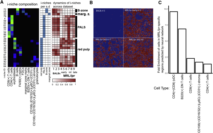
“Goltsev et al. [52] conducted a similar study and discovered that some surface-marker expressions in immune cells were highly correlated with the neighborhood cell type composition.”
BackgroundYounes, S. F., Rojansky, R. B., Menke, J. R., Gratzinger, D. A., & Natkunam, Y. (2021) · Cancers · doi:10.3390/cancers13123021 · Cited by 43
Proteomics approaches to characterize the immune responses in cancer
Urbiola-Salvador, V., Miroszewska, D., Jabłońska, A., Qureshi, T., & Chen, Z. J. (2022) · Biochimica et Biophysica Acta (BBA) - Molecular Cell Research · doi:10.1016/j.bbamcr.2022.119266 · Cited by 42
Multi‐omic and spatial dissection of immunotherapy response groups in non‐small cell lung cancer
Monkman, J. H., Kim, H., Mayer, A. T., Mehdi, A. M., Matigian, N., Cumberbatch, M., Bhagat, M., Ladwa, R., Mueller, S. N., Adams, M. N., et al. (2023) · Immunology · doi:10.1111/imm.13646 · Cited by 44
“The choice of 10 nearest neighbours and 10 clusters was chosen heuristically as parameters that worked well for a wide variety of datasets [21, 22].”
BackgroundAbeler‐Dörner, L., Laing, A. G., Lorenc, A., Ushakov, D. S., Clare, S., Speak, A. O., Correa, M. A. D., White, J. K., Ramírez‐Solis, R., Saran, N., et al. (2019) · Nature Immunology · doi:10.1038/s41590-019-0549-0 · Cited by 43
Deconstructing tumor heterogeneity: the stromal perspective
Vickman, R. E., Faget, D. V., Beachy, P. A., Beebe, D. J., Bhowmick, N. A., Cukierman, E., Deng, W.-M., Granneman, J. G., Hildesheim, J., Kalluri, R., et al. (2020) · Oncotarget · doi:10.18632/oncotarget.27736 · Cited by 43
“Despite recent technological advances, such as imaging mass cytometry and co-detection by indexing (CODEX) [52], histological analyses remain limited due to reliance on pre-existing antibodies.”
BackgroundReguant, A. P., Köhler, R., Mothes, R., Bauherr, S., Hernández, D. C., Uecker, R., Holzwarth, K., Kotsch, K., Seidl, M., Philipsen, L., et al. (2021) · Nature Communications · doi:10.1038/s41467-021-21994-8 · Cited by 42
Compressed sensing for highly efficient imaging transcriptomics
Cleary, B., Simonton, B., Bezney, J., Murray, E. M., Alam, S., Sinha, A., Habibi, E., Marshall, J. L., Lander, E. S., Chen, F., et al. (2021) · Nature Biotechnology · doi:10.1038/s41587-021-00883-x · Cited by 45
Next-Generation Digital Histopathology of the Tumor Microenvironment
Mungenast, F., Fernando, A., Nica, R., Boghiu, B., Lungu, B., Batra, J., & Ecker, R. (2021) · Genes · doi:10.3390/genes12040538 · Cited by 41
“CODEX (IF) Antibodies conjugated to a CODEX barcode; visualized by the binding of highly specific corresponding dye-labeled CODEX reporter • No limitation by the number of different antibody species -> no secondary antibodies • Fast, each round of extension and bleaching (10 min) • Up to 35 rounds with 3 markers • Spe…”
BackgroundOr‐Geva, N., & Steinman, L. (2025) · Proceedings of the National Academy of Sciences · doi:10.1073/pnas.2425670122 · Cited by 41
“Log odds ratios, as described by Goltsev et al. ( 8 ), quantify the likelihood of interactions between specific cell types by comparing their observed co - occurrence in MS lesions to control samples.”
High-resolution spatial mapping of cell state and lineage dynamics in vivo with PEtracer
Koblan, L. W., Yost, K. E., Zheng, P., Colgan, W. N., Jones, M. G., Yang, D., Kumar, A., Sandhu, J., Schnell, A., Sun, D., et al. (2025) · Science · doi:10.1126/science.adx3800 · Cited by 53
Uttam, S., Stern, A. M., Sevinsky, C. J., Furman, S. A., Pullara, F., Spagnolo, D. M., Nguyen, L., Gough, A., Ginty, F., Taylor, D. L., et al. (2020) · Nature Communications · doi:10.1038/s41467-020-17083-x · Cited by 39
Jiang, H., Courau, T., Lupin-Jimenez, L., Borison, J., Ritchie, A. J., Mayer, A. T., Krummel, M. F., & Collisson, E. A. (2021) · Gastroenterology · doi:10.1053/j.gastro.2021.09.066 · Cited by 42
Evaluation of cell segmentation methods without reference segmentations
Chen, H., & Murphy, R. F. (2022) · Molecular Biology of the Cell · doi:10.1091/mbc.e22-08-0364 · Cited by 40
“CODEX CO-Detection by indEXing (CODEX) is an imaging technique generating highly multiplexed images of fluorescently tagged antigens (Goltsev et al., 2018).”
MethodThe role of eIF4F-driven mRNA translation in regulating the tumour microenvironment
Bartish, M., Abraham, M. J., Gonçalves, C., Larsson, O. S., Rolny, C., & Rincon, S. V. D. (2023) · Nature reviews. Cancer · doi:10.1038/s41568-023-00567-5 · Cited by 42
Signal amplification by cyclic extension enables high-sensitivity single-cell mass cytometry
Lun, X., Sheng, K., Yu, X., Lam, C. Y., Gowri, G., Serrata, M. P., Zhai, Y., Su, H., Luan, J., Kim, Y., et al. (2024) · Nature Biotechnology · doi:10.1038/s41587-024-02316-x · Cited by 39
Current state and future prospects of spatial biology in colorectal cancer
Carranza, F., Diaz, F. C., Ninova, M., & Velazquez-Villarreal, E. I. (2024) · Frontiers in Oncology · doi:10.3389/fonc.2024.1513821 · Cited by 38
Single-cell technologies — studying rheumatic diseases one cell at a time
Cheung, P., Khatri, P., Utz, P. J., & Kuo, A. J. (2019) · Nature Reviews Rheumatology · doi:10.1038/s41584-019-0220-z · Cited by 39
Islet–immune interactions in type 1 diabetes: the nexus of beta cell destruction
Peters, L. D., Posgai, A. L., & Brusko, T. M. (2019) · Clinical & Experimental Immunology · doi:10.1111/cei.13349 · Cited by 39
Computational approaches for characterizing the tumor immune microenvironment
Liu, C. C., Steen, C. B., & Newman, A. M. (2019) · Immunology · doi:10.1111/imm.13101 · Cited by 41
“25 Imaging techniques that rely on fluorescence-tagged antibodies, like flow cytometry, are limited by spectral overlap.”
BackgroundT and B lymphocytes in fibrosis and systemic sclerosis
Pillai, S. S. (2019) · Current Opinion in Rheumatology · doi:10.1097/bor.0000000000000644 · Cited by 40
“The use of approaches that lead to the restoration of antigenicity in fixed tissue sections, and staining with a range of antibodies each with distinct fluorescent, nucleic-acid or metal-based labels in order to identify many different proteins simultaneously and/or sequentially in a tissue allows the identification o…”
BackgroundCombining Qdot Nanotechnology and DNA Nanotechnology for Sensitive Single‐Cell Imaging
Zhou, W. J., Han, Y., Beliveau, B. J., & Gao, X. (2020) · Advanced Materials · doi:10.1002/adma.201908410 · Cited by 41
A Cancer Biologist's Primer on Machine Learning Applications in High‐Dimensional Cytometry
Keyes, T., Domizi, P., Lo, Y., Nolan, G. P., & Davis, K. L. (2020) · Cytometry Part A · doi:10.1002/cyto.a.24158 · Cited by 40
Intravital and high-content multiplex imaging of the immune system
Hor, J. L., & Germain, R. N. (2021) · Trends in Cell Biology · doi:10.1016/j.tcb.2021.11.007 · Cited by 40
Single-cell RNA and protein profiling of immune cells from the mouse brain and its border tissues
Scheyltjens, I., Hove, H. V., Vlaminck, K. D., Kancheva, D., Bastos, J., Vara‐Pérez, M., Antunes, A. R. P., Martens, L., Scott, C. L., Ginderachter, J. A. V., et al. (2022) · Nature Protocols · doi:10.1038/s41596-022-00716-4 · Cited by 44
SOTIP is a versatile method for microenvironment modeling with spatial omics data
Yuan, Z., Li, Y., Shi, M., Yang, F., Gao, J. T., Yao, J., & Zhang, M. Q. (2022) · Nature Communications · doi:10.1038/s41467-022-34867-5 · Cited by 40
Giotto Suite: a multiscale and technology-agnostic spatial multiomics analysis ecosystem
Chen, J. G., Chávez, J., O’Brien, M., Xu, J., Ruiz, E. C., Wang, W., Amin, I., Sheridan, J. P., Shin, S. C., Hasyagar, S. V., et al. (2025) · Nature Methods · doi:10.1038/s41592-025-02817-w · Cited by 73
“With the introduction and advancement of spatial omics technologies, including spatial transcriptomics 7–9 , proteomics 10–13 and others 14 , researchers have the ability to visualize and analyze various molecular analytes within their tissue context and across different levels of the biological…”
Merritt, C. R., Ong, G. T., Church, S., Barker, K., Geiss, G. K., Hoang, M. L., Jung, J., Liang, Y., McKay-Fleisch, J., Nguyen, K., et al. (2019) · bioRxiv (Cold Spring Harbor Laboratory) · doi:10.1101/559021 · Cited by 39
Multiplexed Ion Beam Imaging: Insights into Pathobiology
Liu, C. C., McCaffrey, E., Greenwald, N. F., Soon, E., Risom, T., Vijayaragavan, K., Oliveria, J. P., Mrdjen, D., Bosse, M. M., Tebaykin, D., et al. (2021) · Annual Review of Pathology Mechanisms of Disease · doi:10.1146/annurev-pathmechdis-030321-091459 · Cited by 39
Chen, E., Ling, A. L., Reardon, D. A., & Chiocca, E. A. (2023) · Neuro-Oncology · doi:10.1093/neuonc/noad211 · Cited by 38
Mi, H., Sivagnanam, S., Ho, W. J., Zhang, S., Bergman, D. R., Deshpande, A. M., Baras, A. S., Jaffee, E. M., Coussens, L. M., Fertig, E. J., et al. (2024) · Briefings in Bioinformatics · doi:10.1093/bib/bbae421 · Cited by 38
“…IHC (mIHC [5]), which utilizes sequential rounds of staining and stripping to detect multiple proteins on the same tissue section; Phenocycler [6], which uses antibodies attached to unique DNA barcodes applied in a single staining cocktail, with the automated system sequentially adding…”
MethodMimicking and analyzing the tumor microenvironment
Crouigneau, R., Li, Y., Auxillos, J., Gonçalves‐Alves, E., Marie, R., Sandelin, A., & Pedersen, S. F. (2024) · Cell Reports Methods · doi:10.1016/j.crmeth.2024.100866 · Cited by 45
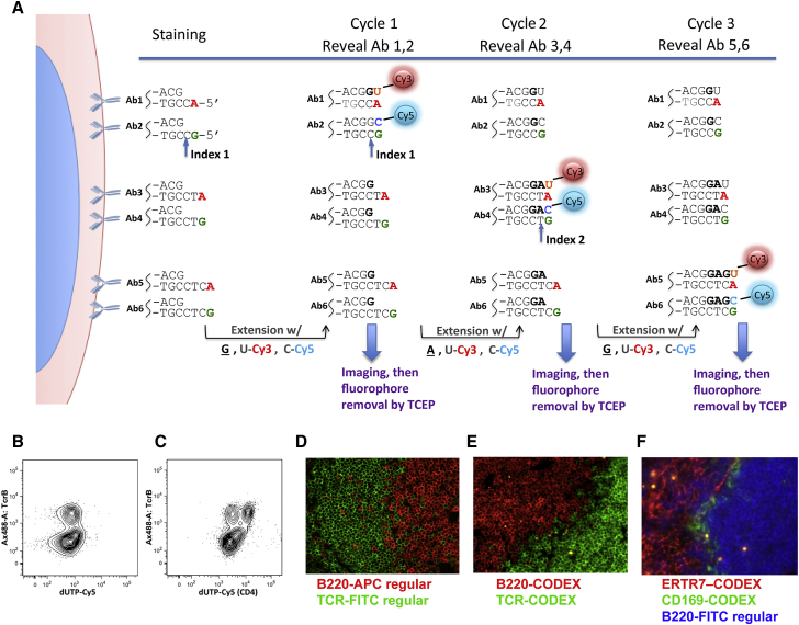
“…in which antibodies are iteratively visualized using DNA barcodes, fluorescent dNTP analogs, and an in situ polymerization-based indexing procedure 151 ; for other cyclic microscopy methods, see Elhanani et al. 152 Such emerging techniques provide the precision of IHC and similar optical imaging…”
From morphology to single-cell molecules: high-resolution 3D histology in biomedicine
Xu, X., Su, J., Zhu, R., Li, K., Zhao, X., Fan, J., & Mao, F. (2025) · Molecular Cancer · doi:10.1186/s12943-025-02240-x · Cited by 43
“This is of great significance for understanding the complex structure of tissues, cell-to-cell interactions, and the microenvironment of diseases [156–158].”
Lou, Z., Zhang, Y., Liang, X., Cao, M., Ma, Y., Chen, P. R., & Fan, X. (2025) · Journal of the American Chemical Society · doi:10.1021/jacs.4c17879 · Cited by 42
Tools and Concepts for Interrogating and Defining Cellular Identity
McKinley, K. L., Castillo‐Azofeifa, D., & Klein, O. D. (2020) · Cell stem cell · doi:10.1016/j.stem.2020.03.015 · Cited by 37
Herck, Y. V., Antoranz, A., Andhari, M. D., Milli, G., Bechter, O. E., Smet, F. D., & Bosisio, F. M. (2021) · Frontiers in Oncology · doi:10.3389/fonc.2021.636681 · Cited by 40
“In this approach, the transcriptomic data acquired via CITE-seq (89) can be mapped to spatially resolved CODEX mIHC data (77), while retaining the single cell spatial resolution by making use of measurements of the same antigens in both methods (88).”
MethodZhu, W., Cai, E., Li, H., Wang, P., Shen, A., Popp, J., & Hu, J. (2021) · Angewandte Chemie International Edition · doi:10.1002/anie.202106136 · Cited by 39
Oligonucleotide conjugated antibody strategies for cyclic immunostaining
Jones, J. A., McMahon, N. P., Zheng, T., Eng, J. R., Chin, K., Kwon, S., Nederlof, M. A., Gray, J. W., & Gibbs, S. L. (2021) · Scientific Reports · doi:10.1038/s41598-021-03135-9 · Cited by 38
Third‐Generation Covalent TMP‐Tag for Fast Labeling and Multiplexed Imaging of Cellular Proteins
Mo, J., Chen, J., Shi, Y., Sun, J., Wu, Y., Liu, T., Zhang, J., Zheng, Y., Li, Y., & Chen, Z. (2022) · Angewandte Chemie International Edition · doi:10.1002/anie.202207905 · Cited by 40
Spatial epitope barcoding reveals clonal tumor patch behaviors
Rovira‐Clavé, X., Drainas, A. P., Jiang, S., Bai, Y., Baron, M., Zhu, B., Dallas, A. E., Lee, M. C., Chu, T. P., Holzem, A. M. E., et al. (2022) · Cancer Cell · doi:10.1016/j.ccell.2022.09.014 · Cited by 40
Spatial determinants of CD8+ T cell differentiation in cancer
Tooley, K., Escobar, G., & Anderson, A. C. (2022) · Trends in cancer · doi:10.1016/j.trecan.2022.04.003 · Cited by 38
Spatial analysis for highly multiplexed imaging data to identify tissue microenvironments
Patrick, E., Canete, N. P., Iyengar, S. S., Harman, A. N., Sutherland, G. T., & Yang, P. (2023) · Cytometry Part A · doi:10.1002/cyto.a.24729 · Cited by 37
“Alternatively, Goltsev et al. proposed an approach identifying 100 cliques of cell types from a nearest neighbor analysis showing that these captured histologically distinct regions of cell type composition [4].”
BackgroundMethodInfluentialHigh-dimensional multi-pass flow cytometry via spectrally encoded cellular barcoding
Kwok, S. J. J., Forward, S., Fahlberg, M. D., Assita, E. R., Cosgriff, S., Lee, S. H., Abbott, G. R., Zhu, H., Minasian, N. H., Vote, A. S., et al. (2023) · Nature Biomedical Engineering · doi:10.1038/s41551-023-01144-9 · Cited by 41
Wang, Y., Liu, X., Zeng, Y., Saka, S. K., Xie, W., Goldaracena, I., Kohman, R. E., Yin, P., & Church, G. M. (2024) · Nucleic Acids Research · doi:10.1093/nar/gkae592 · Cited by 38
“Techniques such as DNA exchange imaging and CODEX enable highly multiplexed imaging but do not integrate signal amplification, leading to limited sensitivity when visualizing low-abundance targets ( 1 , 3 , 4 ).”
Engineered Proteins and Chemical Tools to Probe the Cell Surface Proteome
Leung, K., Schaefer, K., Lin, Z., Yao, Z. W., & Wells, J. A. (2025) · Chemical Reviews · doi:10.1021/acs.chemrev.4c00554 · Cited by 44
Quantifying in situ adaptive immune cell cognate interactions in humans
Liarski, V. M., Sibley, A., van Panhuys, N., Ai, J., Chang, A., Kennedy, D. E., Merolle, M. I., Germain, R. N., Giger, M. L., & Clark, M. R. (2019) · Nature Immunology · doi:10.1038/s41590-019-0315-3 · Cited by 37
“Great strides have been made in multiparameter imaging of fixed-human tissue such that 36 or more markers can be assayed simultaneously 12, 13, 14, 15 .”
Techniques for Studying Decoding of Single Cell Dynamics
Jeknić, S., Kudo, T., & Covert, M. W. (2019) · Frontiers in Immunology · doi:10.3389/fimmu.2019.00755 · Cited by 40
“A similar approach, co-detection by indexing (CODEX), uses oligonucleotide conjugated antibodies (131).”
MethodProfiling Cell Signaling Networks at Single-cell Resolution
Lun, X., & Bodenmiller, B. (2020) · Molecular & Cellular Proteomics · doi:10.1074/mcp.r119.001790 · Cited by 38
“The DNA oligonucleotides conjugated to the antibodies serve as barcodes that can be sequentially detected by fluorophorelabeled dNTPs in CODEX (75) or by fluorescent probes directly and indirectly linked to complementary DNA sequence in Exchange-PAINT (74) and immune-SABER (42).”
BackgroundMethodOptimizing multiplexed imaging experimental design through tissue spatial segregation estimation
Bost, P., Schulz, D., Engler, S., Wasserfall, C. H., & Bodenmiller, B. (2022) · Nature Methods · doi:10.1038/s41592-022-01692-z · Cited by 35
Prognostic Gene Expression, Stemness and Immune Microenvironment in Pediatric Tumors
Stahl, D. J., Knoll, R., Gentles, A. J., Vokuhl, C., Buneß, A., & Gütgemann, I. (2021) · Cancers · doi:10.3390/cancers13040854 · Cited by 36
“Novel tissue sparing multiplex staining techniques [31] may be helpful in validating CIBERSORT data on a wide range of tumor types and add information as to spatial localization.”
BackgroundA Cross Entropy Test Allows Quantitative Statistical Comparison of t-SNE and UMAP Representations
Roca, C. P., Burton, O. T., Neumann, J., Tareen, S. H. K., Whyte, C. E., Gergelits, V., Veiga, R., Humblet‐Baron, S., & Liston, A. (2022) · Cell Reports Methods · doi:10.1016/j.crmeth.2022.100390 · Cited by 36
T cell-mediated curation and restructuring of tumor tissue coordinates an effective immune response
Hickey, J. W., Haist, M., Horowitz, N. B., Caraccio, C., Tan, Y., Rech, A. J., Baertsch, M., Rovira‐Clavé, X., Zhu, B., Vazquez, G., et al. (2023) · Cell Reports · doi:10.1016/j.celrep.2023.113494 · Cited by 39
“Tumors were imaged with CO-Detection by indEXing (CODEX) multiplexed imaging 27–29 with a 42-plex antibody panel at 0, 1, 3-, 5, and 12-days post-treatment (Figures 1A and 1B, n = 3–7).”
MethodXu, A. M., Haro, M., Walts, A. E., Hu, Y., John, J., Karlan, B. Y., Merchant, A. A., & Oršulić, S. (2024) · Science Advances · doi:10.1126/sciadv.adk8805 · Cited by 40
“When applied to tumor studies, these spatially resolved methods can provide an added layer of spatial context by describing the microenvironmental niches where each cell type can be found ( 29 , 30 ).”
Maurer, K., Park, C. Y., Mani, S., Borji, M., Raths, F., Gouin, K. H., Penter, L., Jin, Y., Zhang, J. Y., Shin, C. S., et al. (2025) · Science Immunology · doi:10.1126/sciimmunol.adr0782 · Cited by 42
Subcellular localization as a driver of protein function
Sigaeva, A., Hutchings, C., Cesnik, A. J., Lilley, K. S., & Lundberg, E. K. (2026) · Nature Reviews Molecular Cell Biology · doi:10.1038/s41580-026-00947-3 · Cited by 39
“…into multiple fractions without organelle isolation or purification 2,67,201,205,269,270 273 such as those used for protein localization 274–276 , for single-molecule RNA localization 277 or for both 278 , allow one to overlay and compare the distributions of multiple molecules of interest.”
Multiplexed laser particles for spatially resolved single-cell analysis
Kwok, S. J. J., Martino, N., Dannenberg, P. H., & Yun, S. H. (2019) · Light Science & Applications · doi:10.1038/s41377-019-0183-5 · Cited by 34
Cyclic Multiplexed-Immunofluorescence (cmIF), a Highly Multiplexed Method for Single-Cell Analysis
Eng, J. R., Thibault, G., Luoh, S., Gray, J. W., Chang, Y. H., & Chin, K. (2019) · Methods in molecular biology · doi:10.1007/978-1-4939-9773-2_24 · Cited by 35
Screening for new macrophage therapeutics
Rodell, C. B., Koch, P. D., & Weissleder, R. (2019) · Theranostics · doi:10.7150/thno.34421 · Cited by 35
“Other approaches involve barcoding antibodies with DNA, using the sequence information to identify levels of each marker [50, 51].”
MethodGastman, B., Agarwal, P. K., Berger, A., Boland, G. M., Broderick, S., Butterfield, L. H., Byrd, D. R., Fecci, P. E., Ferris, R. L., Fong, Y., et al. (2020) · Journal for ImmunoTherapy of Cancer · doi:10.1136/jitc-2020-001583 · Cited by 39
Bayerl, F., Bejarano, D. A., Bertacchi, G., Doffin, A., Gobbini, É., Hubert, M., Li, L., Meiser, P., Pedde, A., Posch, W., et al. (2023) · European Journal of Immunology · doi:10.1002/eji.202249923 · Cited by 36
New tools for immunologists: models of lymph node function from cells to tissues
Ozulumba, T., Montalbine, A. N., Ortiz-Cárdenas, J. E., & Pompano, R. R. (2023) · Frontiers in Immunology · doi:10.3389/fimmu.2023.1183286 · Cited by 34
“Together, these cell populations mount systemic antibody-driven responses to infection by reducing pathogen spread and provide lasting protection following vaccination (19).”
BackgroundMiheecheva, N., Postovalova, E., Lyu, Y., Ramachandran, A., Bagaev, A., Svekolkin, V., Galkin, I., Zyrin, V., Maximov, V., Lozinsky, Y., et al. (2022) · Cell Reports · doi:10.1016/j.celrep.2022.111180 · Cited by 37
Rahim, M. K., Zhao, J., Patel, H., Lagouros, H. A., Kota, R., Fernandez, I., Gratton, E., & Haun, J. B. (2022) · Analytical Chemistry · doi:10.1021/acs.analchem.2c02149 · Cited by 34
Targeting the epigenome to reinvigorate T cells for cancer immunotherapy
Xiong, D., Zhang, L., & Sun, Z. (2023) · Military Medical Research · doi:10.1186/s40779-023-00496-2 · Cited by 36
“Advances in spatial transcriptomics and imaging technologies have highlighted the dynamic interplay of immune cells in the tumor setting [115, 116] Chemokines have emerged as important cues for the establishment of intratumoral immune niches and TLSs, which contribute to the housing of stem-like…”
BackgroundUnveiling spatial complexity in solid tumor immune microenvironments through multiplexed imaging
Scheuermann, S., Kristmann, B., Engelmann, F., Nuernbergk, A., Scheuermann, D., Koloseus, M., Abed, T., Solaß, W., & Seitz, C. M. (2024) · Frontiers in Immunology · doi:10.3389/fimmu.2024.1383932 · Cited by 36
“Multiplexed tissue imaging is an emerging technology providing new and previously unprecedented insights into cellular architecture, function, and orchestration (21, 22, 24).”
BackgroundWalter, W., Pfarr, N., Meggendorfer, M., Jost, P. J., Haferlach, T., & Weichert, W. (2020) · Seminars in Cancer Biology · doi:10.1016/j.semcancer.2020.10.015 · Cited by 35
SITC cancer immunotherapy resource document: a compass in the land of biomarker discovery
Hu‐Lieskovan, S., Bhaumik, S., Dhodapkar, K. M., Grivel, J., Gupta, S. V., Hanks, B. A., Janetzki, S., Kleen, T. O., Koguchi, Y., Lund, A. W., et al. (2020) · Journal for ImmunoTherapy of Cancer · doi:10.1136/jitc-2020-000705 · Cited by 35
Single-cell high-dimensional imaging mass cytometry: one step beyond in oncology
Glasson, Y., Chépeaux, L.-A., Dumé, A., Lafont, V., Faget, J., Bonnefoy, N., & Michaud, H. (2023) · Seminars in Immunopathology · doi:10.1007/s00281-022-00978-w · Cited by 34
“, PhenoCycler, formerly known as CODEX [3], from AKOYA, or MACSima [4] from Miltenyi) have allowed increasing the number of biomarkers that can be assessed simultaneously.”
BackgroundKayvanjoo, A. H., Šplíchalová, I., Bejarano, D. A., Huang, H., Mauel, K., Makdissi, N., Heider, D., Tew, H. M., Balzer, N. R., Greto, E., et al. (2024) · eLife · doi:10.7554/elife.86493 · Cited by 34
“To determine whether LT-HSCs interact with distinct macrophage populations, we assessed cellular interactions via co-detection by indexing (CODEX)- enabled high-dimensional imaging ( Black et al., 2021 ; Frede et al., 2022 ; Goltsev et al., 2018 ; Figure 4A ).”
Profiling joint tissues at single-cell resolution: advances and insights
Pandey, A., & Bhutani, N. (2023) · Nature Reviews Rheumatology · doi:10.1038/s41584-023-01052-x · Cited by 34
SLIDE: Significant Latent Factor Interaction Discovery and Exploration across biological domains
Rahimikollu, J., Xiao, H., Rosengart, A., Rosen, A. B. I., Tabib, T., Zdinak, P. M., He, K., Bing, X., Bunea, F., Wegkamp, M. H., et al. (2024) · Nature Methods · doi:10.1038/s41592-024-02175-z · Cited by 35
“Next, we evaluated SLIDE on a wide range of other spatial data modalities and technologies—Slide-seq 34 , MERFISH 35 and CODEX 36 .”
Imaging 3D cell cultures with optical microscopy
Hsieh, H.-C., Han, Q., Brenes, D., Bishop, K. W., Wang, R., Wang, Y., Poudel, C., Glaser, A. K., Freedman, B., Vaughan, J. C., et al. (2025) · Nature Methods · doi:10.1038/s41592-025-02647-w · Cited by 38
“Alternatively, a single round of staining with barcoded antibodies, each containing unique oligonucleotide sequences, has been combined with a sequential readout of barcodes using complementary fluorescent oligonucleotides (for example, CODEX 291 , Immuno-SABER 292 ).”
Clinical and research applications of multiplexed immunohistochemistry and in situ hybridization
McGinnis, L. M., Ibarra-Lopez, V., Rost, S., & Ziai, J. M. (2021) · The Journal of Pathology · doi:10.1002/path.5663 · Cited by 32
“A recent paper combines a thorough technical validation of the technology with its application to the study of re-distribution of immune cell subsets in spleen in a mouse lupus model [9].”
MethodLabel-Free Deep Profiling of the Tumor Microenvironment
You, S., Chaney, E. J., Tu, H., Sun, Y., Sinha, S., & Boppart, S. A. (2021) · Cancer Research · doi:10.1158/0008-5472.can-20-3124 · Cited by 36
“This metric was adapted from Goltsev and colleagues’ work on labeled cell analysis of the mouse splenic architecture (35) to quantify the cellular interaction strength.”
BackgroundMethodInfluentialTissue Multiplex Analyte Detection in Anatomic Pathology – Pathways to Clinical Implementation
Wharton, K. A., Wood, D., Manesse, M., Maclean, K. H., Leiß, F., & Żuraw, A. (2021) · Frontiers in Molecular Biosciences · doi:10.3389/fmolb.2021.672531 · Cited by 35
“ISH to detect DNA is used to identify chromosomal translocations and gene copy number changes, and RNAscope (Advanced Cell Diagnostics, BioTechne Inc.) has emerged as a sensitive ISH technique to visualize RNA in FFPE tissue, offering advantages over traditional RNA ISH techniques to detect low abundance transcripts,…”
MethodInfluentialMurray, H. C., Johnson, K. R., Sedlock, A. B., Highet, B., Dieriks, B. V., Anekal, P. V., Faull, R. L. M., Curtis, M. A., Koretsky, A. P., & Maric, D. (2022) · Communications Biology · doi:10.1038/s42003-022-03032-5 · Cited by 39
Castro, L. N. G., Liu, I., & Filbin, M. G. (2022) · Neuro-Oncology · doi:10.1093/neuonc/noac211 · Cited by 34
Patel, A. G., Ashenberg, O., Collins, N. B., Segerstolpe, Å., Jiang, S., Slyper, M., Huang, X., Caraccio, C., Jin, H., Sheppard, H., et al. (2024) · bioRxiv (Cold Spring Harbor Laboratory) · doi:10.1101/2024.01.07.574538 · Cited by 39
Kiyokawa, H., Abe, M., Matsui, T., Kurashige, M., Ohshima, K., Tahara, S., Nojima, S., Ogino, T., Sekido, Y., Mizushima, T., et al. (2022) · The American Journal of Pathology · doi:10.1016/j.ajpath.2022.03.006 · Cited by 32
Spatial mapping of cancer tissues by OMICS technologies
Ahmed, R., Augustine, R., Valera, E., Ganguli, A., Mesaeli, N., Ahmad, I. S., Bashir, R., & Hasan, A. (2021) · Biochimica et Biophysica Acta (BBA) - Reviews on Cancer · doi:10.1016/j.bbcan.2021.188663 · Cited by 32
“proteins of interest in mouse-isolated splenocyte cells [133].”
BackgroundMachine learning integrative approaches to advance computational immunology
Curion, F., & Theis, F. J. (2024) · Genome Medicine · doi:10.1186/s13073-024-01350-3 · Cited by 36
AI-empowered perturbation proteomics for complex biological systems
Qian, L., Sun, R., Aebersold, R. H., Bühlmann, P., Sander, C., & Guo, T. (2024) · Cell Genomics · doi:10.1016/j.xgen.2024.100691 · Cited by 33
Zhou, Y., Ye, H., Yu, Y., Ge, C., Yin, M., Liu, Z., Shen, J., Zhou, R., Li, Y., Leong, K. W., et al. (2025) · Journal of the American Chemical Society · doi:10.1021/jacs.4c15646 · Cited by 32
Overview of Tissue Imaging Methods
Patel, S. S., & Rodig, S. J. (2019) · Methods in molecular biology · doi:10.1007/978-1-4939-9773-2_21 · Cited by 32
Functional crosstalk between T cells and monocytes in cancer and atherosclerosis
Padgett, L. E., Araujo, D. J., Hedrick, C. C., & Olingy, C. E. (2020) · Journal of Leukocyte Biology · doi:10.1002/jlb.1mir0420-076r · Cited by 32
Lindner, A. U., Salvucci, M., McDonough, E., Cho, S., Stachtea, X., O’Connell, E., Corwin, A. D., Santamaría-Pang, A., Carberry, S., Fichtner, M. W., et al. (2021) · Cell Death and Differentiation · doi:10.1038/s41418-021-00895-9 · Cited by 30
“…used as a tool for spatial analysis of tumor cell types and microenvironment over the last 10 years (Angelo et al., 2014; Gerdes et al., 2013; Goltsev et al., 2018; Gut et al., 2018; Kalra and Baker, 2017; Rashid et al., 2019; Saka et al., 2019; Tan et al., 2020) and there are increasing…”
MethodQuantifying and correcting slide-to-slide variation in multiplexed immunofluorescence images
Harris, C. R., McKinley, E. T., Roland, J. T., Liu, Q., Shrubsole, M. J., Lau, K. S., Coffey, R. J., Wrobel, J., & Vandekar, S. (2021) · Bioinformatics · doi:10.1093/bioinformatics/btab877 · Cited by 32
Blessin, N. C., Yang, C. H., Mandelkow, T., Raedler, J. B., Li, W., Bady, E., Simon, R., Vettorazzi, E., Lennartz, M., Bernreuther, C., et al. (2023) · The Journal of Pathology · doi:10.1002/path.6057 · Cited by 32
“Further improvements in mfIHC will soon enable the simultaneous analysis of 8 [55], 40 [56], and more [57] antibodies on one tissue section.”
BackgroundSong, Y., Das, D., Zhang, Y., Chen, M., Fernie, A. R., Zhu, F., & Han, J. (2023) · Trends in biotechnology · doi:10.1016/j.tibtech.2023.05.010 · Cited by 36
Learning cell communication from spatial graphs of cellsPreprint
Fischer, D. S., Schaar, A. C., & Theis, F. J. (2021) · bioRxiv (Cold Spring Harbor Laboratory) · doi:10.1101/2021.07.11.451750 · Cited by 32
Dissecting Tumor-Immune Microenvironment in Breast Cancer at a Spatial and Multiplex Resolution
Tzoras, E., Zerdes, I., Tsiknakis, N., Manikis, G. C., Mezheyeuski, A., Bergh, J. C. S., Matikas, A., & Foukakis, T. (2022) · Cancers · doi:10.3390/cancers14081999 · Cited by 30
SPACEc: a streamlined, interactive Python workflow for multiplexed image processing and analysis
Tan, Y., Kempchen, T. N., Becker, M., Haist, M., Feyaerts, D., Liu, J., Toma, M. I., Xiao, Y., Su, G., Rech, A. J., et al. (2025) · Nature Communications · doi:10.1038/s41467-025-65658-3 · Cited by 39
High-Parameter Single-Cell Analysis
Chattopadhyay, P. K., Winters, A. F., Lomas, W. E., Laino, A. S., & Woods, D. M. (2019) · Annual Review of Analytical Chemistry · doi:10.1146/annurev-anchem-061417-125927 · Cited by 30
RESTORE: Robust intEnSiTy nORmalization mEthod for multiplexed imaging
Chang, Y. H., Chin, K., Thibault, G., Eng, J. R., Burlingame, E. A., & Gray, J. W. (2020) · Communications Biology · doi:10.1038/s42003-020-0828-1 · Cited by 29
“ecent developments in multiplexed staining and imaging such as cyclic immuno fl uorescence (CyCIF) 1 – 3 , multi-plexed immunohistochemistry (IHC) 4 , CODEX 5 , and other imaging methods have greatly expanded the palette that researchers and pathologists can use to visualize and analyze tissue sections, allowing deep i…”
Background
Metadata
- Authors
- Goltsev, Y., Samusik, N., Kennedy-Darling, J., Bhate, S., Hale, M., Vazquez, G., Black, S., & Nolan, G. P.
- Affiliations
- Stanford Medicine (US)
- Corresponding author
- Nolan, Garry P.
- Venue
- Cell
- Publisher
- Elsevier BV
- Published
- 2018-08
- Volume
- 174
- Issue
- 4
- Pages
- 968-981.e15
- ISSN
- 0092-8674 (ISSN-L)
- PMC
- PMC6086938
- OpenAlex
- W2949238013
- Cited by
- 1,501
- References
- 20
- Citations by year
- 2026: 231 · 2025: 244 · 2024: 232 · 2023: 198 · 2022: 194 · 2021: 194 · 2020: 125 · 2019: 72 · 2018: 9
- FWCI
- 35.39
- Type
- journal article
- Language
- en
- Open access
- hybrid
- doi.org
- Licence
- Locations
- Cell (publishedVersion, cc-by)
- bioRxiv (Cold Spring Harbor Laboratory) (acceptedVersion)
- Europe PMC (PubMed Central) (publishedVersion, cc-by)
- PubMed Central (publishedVersion, cc-by)
- PubMed (publishedVersion)
- Funders
- US NIH (U19AI057229, 1U19AI100627, R01CA184968, 1R33CA183654-01, R33CA183692, 1R01GM10983601, 1R21CA183660, 1R01NS08953301, 5UH2AR067676, 1R01CA19665701, R01HL120724, R01HL128173)
- Bill and Melinda Gates foundation (OPP1113682)
- Department of Defense (W81XWH-14-1-0180)
- Northrop-Grumman Corporation (7500108142)
- NIH (U54-CA209971)
- Rachford & Carlotta A. Harris Endowed Chair
- Topics
- Life Sciences › Biochemistry, Genetics and Molecular Biology › Molecular Biology › Single-cell and spatial transcriptomics
- Life Sciences › Biochemistry, Genetics and Molecular Biology › Biophysics › Cell Image Analysis Techniques
- Life Sciences › Biochemistry, Genetics and Molecular Biology › Biophysics › Advanced Fluorescence Microscopy Techniques
- Keywords
- codices, splenic architecture, MRL/lpr mice, systemic lupus erythematosus, cell-cell interactions, autoimmune disease
- MeSH terms
- Animals, Antibodies, Disease Models, Animal, Female, Image Processing, Computer-Assisted, Lupus Erythematosus, Systemic, Male, Mass Spectrometry, Spleen, Oligonucleotide Probes, Mice, Inbred MRL lpr, Mice
About these checks
Registry, retraction, Cites and statistics results are computed, not judged, with one exception: a printed reference that no index lists is read once by a model before it counts as a citation error. Claims are extracted by a model and kept only when their quote appears verbatim in the text; each cited source is then checked, with a model’s help, for whether it says what the claim relies on. Those verdicts can be wrong — read the claim’s quote and the cited source before relying on one.
Statistics and arithmetic are recomputed, not judged.
Confidence bands are shown only for fields calibrated on our annotated test set. None is calibrated yet, so no band is shown.
What counts as a claim ›How we measure confidence ›How we check this ›
Are you an author, or do you see a problem with this page? Request a review
