

The DOI matches its Crossref record; 49 of the 49 references it cites were found in a registry or an index.
A Role for p53 in the Adaptation to Glutamine Starvation through the Expression of SLC1A3
Open accessAbstract
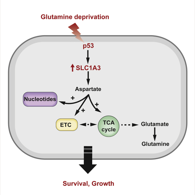
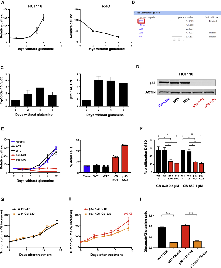
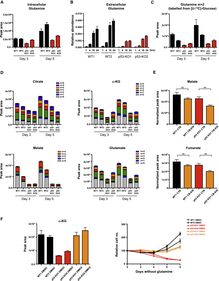
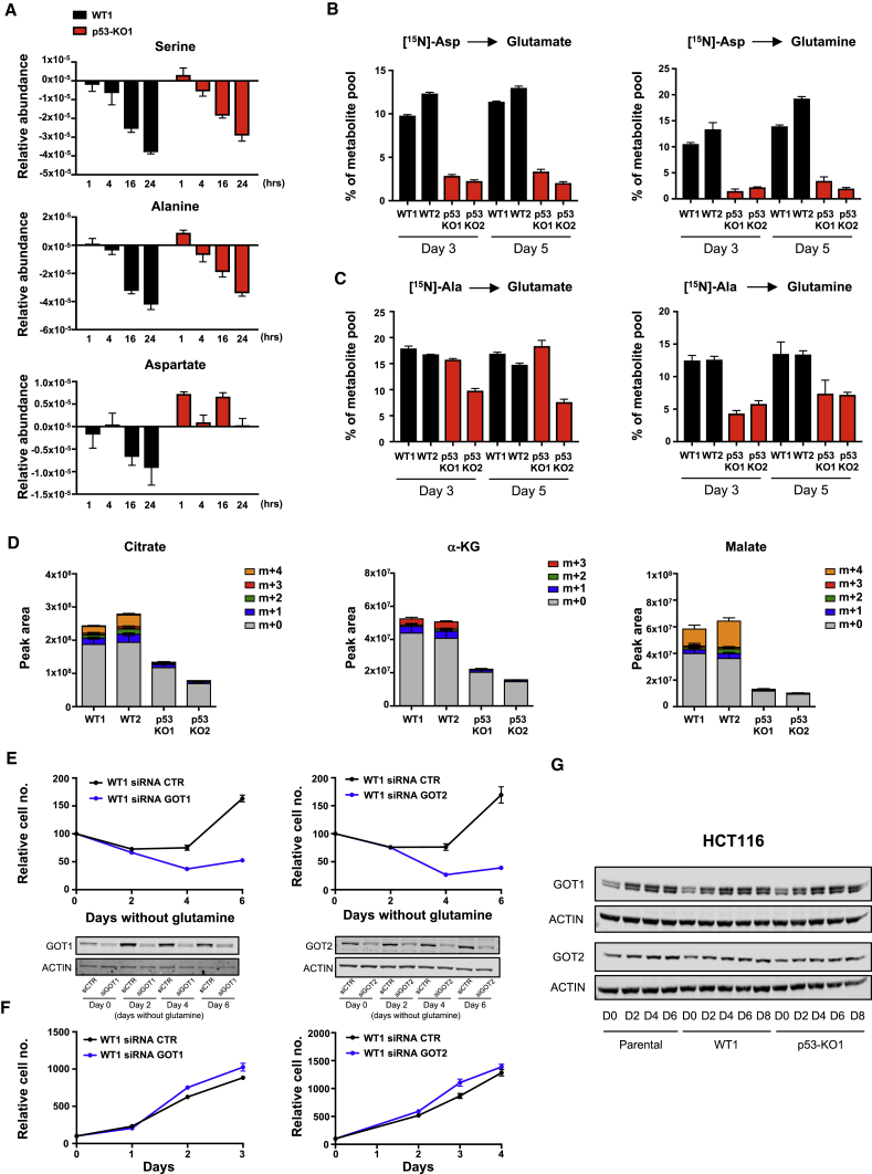
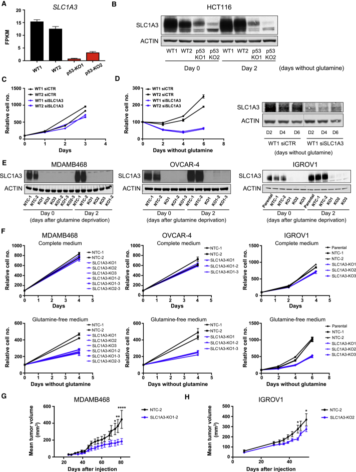
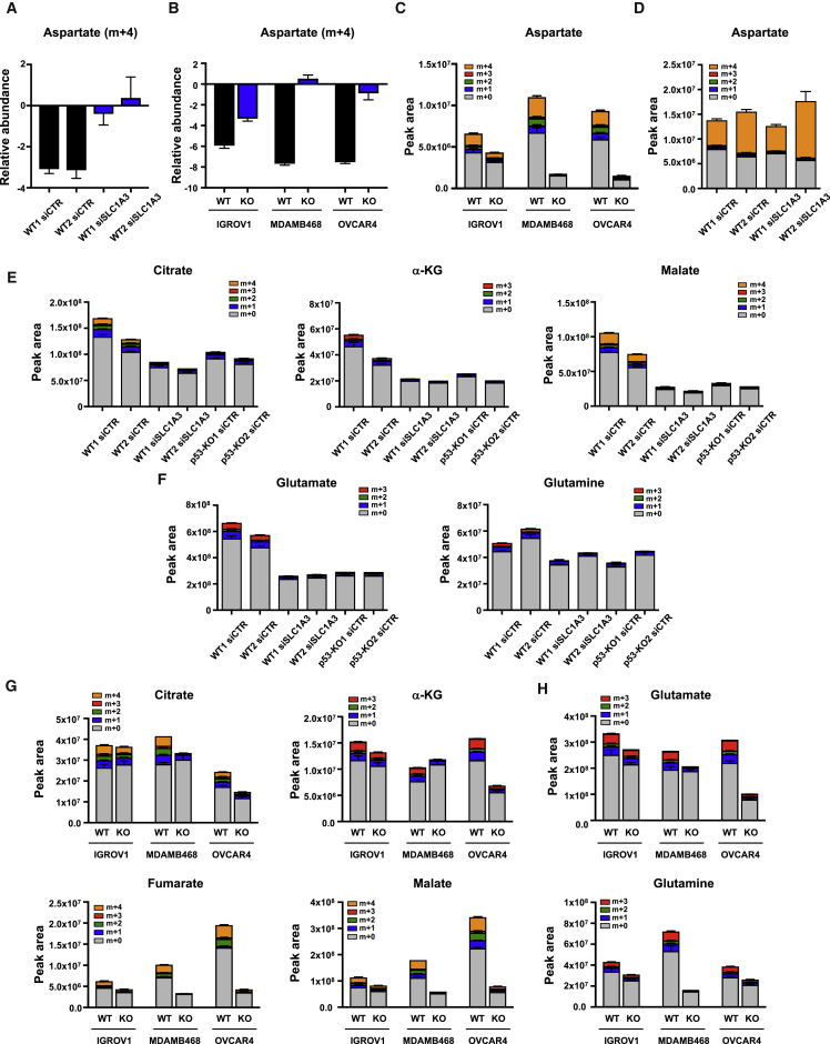
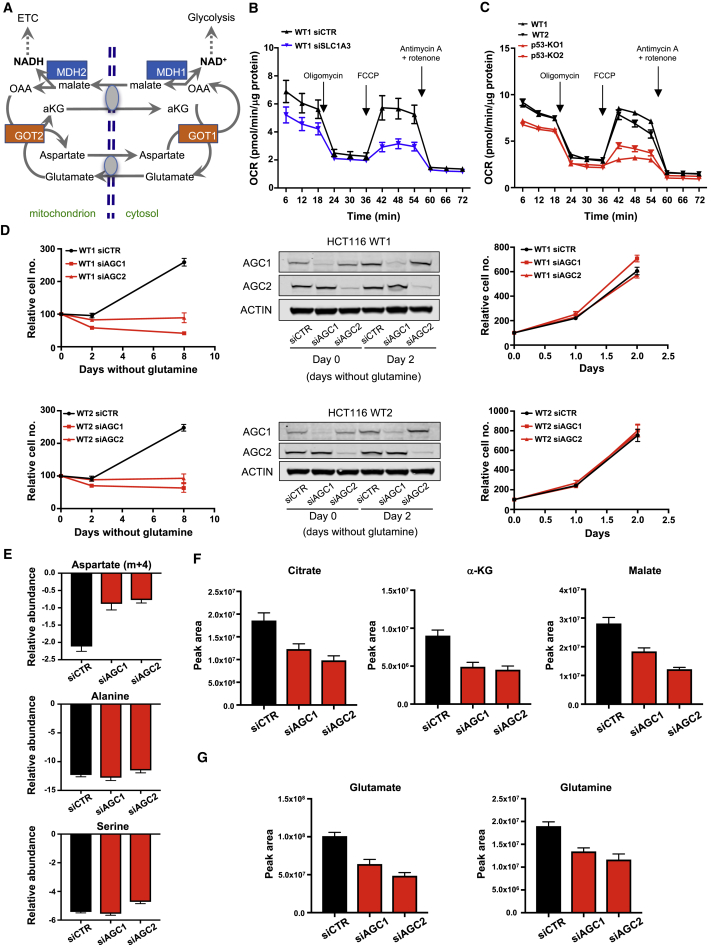
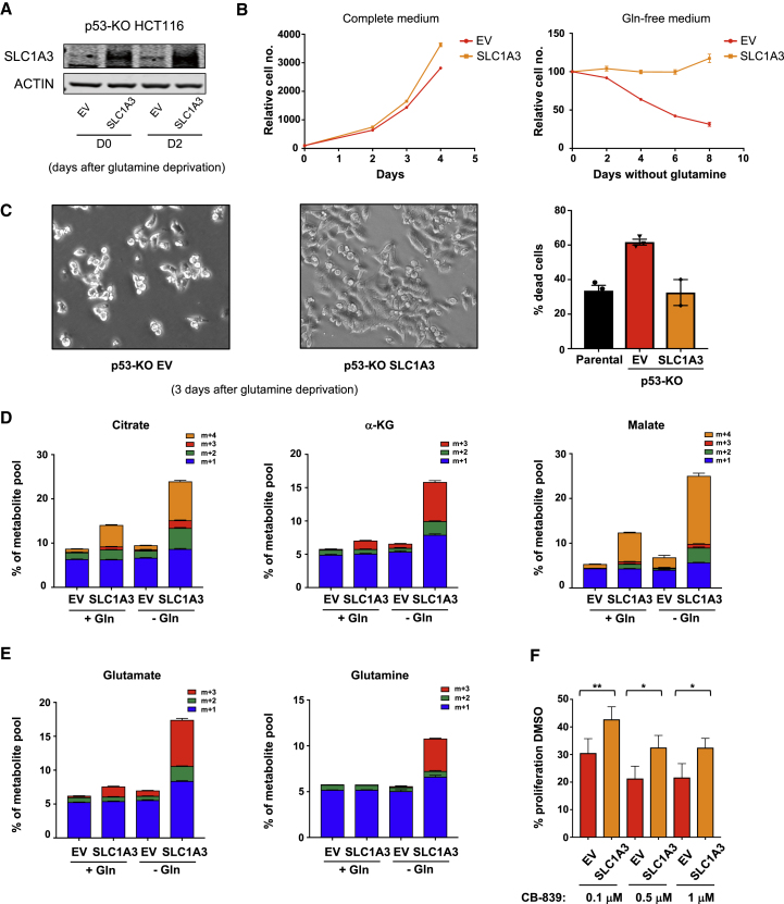
Numerous mechanisms to support cells under conditions of transient nutrient starvation have been described. Several functions of the tumor-suppressor protein p53 can contribute to the adaptation of cells to metabolic stress and help cancer cell survival under nutrient-limiting conditions. We show here that p53 promotes the expression of SLC1A3, an aspartate/glutamate transporter that allows the utilization of aspartate to support cells in the absence of extracellular glutamine. Under glutamine deprivation, SLC1A3 expression maintains electron transport chain and tricarboxylic acid cycle activity, promoting de novo glutamate, glutamine, and nucleotide synthesis to rescue cell viability. Tumor cells with high levels of SLC1A3 expression are resistant to glutamine starvation, and SLC1A3 depletion retards the growth of these cells in vitro and in vivo, suggesting a therapeutic potential for SLC1A3 inhibition.
Overview
The claims and statistics of this paper haven’t been checked yet.
Run the check — free, no account needed. It reads the open-access text and checks the sources it cites.
Integrity signals
Registry
Cites
Statistics
Figures
Verification
Claims the paper makes
The claims and statistics of this paper haven’t been checked yet.
Run the check — free, no account needed. It reads the open-access text and checks the sources it cites.
Claims other papers make about this one
Each is checked against this paper’s own text: does it say what the citing paper says it does?
In the citing paper
“Recently, SLC1A3, as a crucial effector of p53, has been shown to support cell survival and growth in the absence of glutamine149.”
From Glutamine reliance in cell metabolism (2020) · cites this paper as [149]
Self-check
Percentages, sums and reported differences re-computed from the paper’s own numbers.
The self-check needs the paper’s full text.
Citations
Cites
Moderated estimation of fold change and dispersion for RNA-seq data with DESeq2
Love, M. I., Huber, W., & Anders, S. (2014) · Genome Biology · doi:10.1186/s13059-014-0550-8 · Cited by 95,106
Multiplex Genome Engineering Using CRISPR/Cas Systems
Cong, L., Ran, F. A., Cox, D., Lin, S., Barretto, R., Habib, N., Hsu, P. D., Wu, X., Jiang, W., Marraffini, L. A., et al. (2013) · Science · doi:10.1126/science.1231143 · Cited by 15,339
Kim, D., Pertea, G., Trapnell, C., Pimentel, H., Kelley, R., & Salzberg, S. L. (2013) · Genome Biology · doi:10.1186/gb-2013-14-4-r36 · Cited by 12,668
Trapnell, C., Roberts, A., Goff, L. A., Pertea, G. M., Kim, D., Kelley, D. R., Pimentel, H., Salzberg, S. L., Rinn, J. L., & Pachter, L. S. (2012) · Nature Protocols · doi:10.1038/nprot.2012.016 · Cited by 12,145
Glutamate neurotoxicity and diseases of the nervous system
Choi, D. W. (1988) · Neuron · doi:10.1016/0896-6273(88)90162-6 · Cited by 8,188
Improved vectors and genome-wide libraries for CRISPR screening
Sanjana, N. E., Shalem, O., & Zhang, F. (2014) · Nature Methods · doi:10.1038/nmeth.3047 · Cited by 5,239
Blinded by the Light: The Growing Complexity of p53
Vousden, K. H., & Prives, C. (2009) · Cell · doi:10.1016/j.cell.2009.04.037 · Cited by 3,163
Self-citationA role for Wnt signalling in self-renewal of haematopoietic stem cells
Reya, T., Duncan, A. W., Ailles, L., Domen, J., Scherer, D. C., Willert, K., Hintz, L., Nusse, R., & Weissman, I. L. (2003) · Nature · doi:10.1038/nature01593 · Cited by 2,159
From Krebs to clinic: glutamine metabolism to cancer therapy
Altman, B. J., Stine, Z. E., & Dang, C. V. (2016) · Nature Reviews Cancer · doi:10.1038/nrc.2016.71 · Cited by 2,119
Morgenstern, J. P., & Land, H. (1990) · Nucleic Acids Research · doi:10.1093/nar/18.12.3587 · Cited by 2,099
Cited by
Sorted by citations
Glutamine reliance in cell metabolism
Yoo, H. C., Yu, Y. C., Sung, Y., & Han, J. M. (2020) · Experimental & Molecular Medicine · doi:10.1038/s12276-020-00504-8 · Cited by 1,021
Distinct Regulation of Th17 and Th1 Cell Differentiation by Glutaminase-Dependent Metabolism
Johnson, M. O., Wolf, M. M., Madden, M. Z., Andrejeva, G., Sugiura, A., Contreras, D. C., Maseda, D., Liberti, M. V., Paz, K. G., Kishton, R. J., et al. (2018) · Cell · doi:10.1016/j.cell.2018.10.001 · Cited by 729
Amino acid metabolism in health and disease
Ling, Z., Jiang, Y.-F., Ru, J.-N., Lu, J.-H., Ding, B., & Wu, J. (2023) · Signal Transduction and Targeted Therapy · doi:10.1038/s41392-023-01569-3 · Cited by 590
Targeting glutamine metabolism as a therapeutic strategy for cancer
Jin, J., Byun, J., Choi, Y., & Park, K. (2023) · Experimental & Molecular Medicine · doi:10.1038/s12276-023-00971-9 · Cited by 605
Understanding the complexity of p53 in a new era of tumor suppression
Liu, Y., Su, Z., Tavana, O., & Gu, W. J. (2024) · Cancer Cell · doi:10.1016/j.ccell.2024.04.009 · Cited by 538
New aspects of amino acid metabolism in cancer
Vettore, L. A., Westbrook, R. L., & Tennant, D. A. (2019) · British Journal of Cancer · doi:10.1038/s41416-019-0620-5 · Cited by 508
Dietary modifications for enhanced cancer therapy
Kanarek, N., Petrova, B., & Sabatini, D. M. (2020) · Nature · doi:10.1038/s41586-020-2124-0 · Cited by 418
Energy metabolism in health and diseases
Liu, H., Wang, S., Wang, J., Guo, X., Song, Y., Fu, K., Gao, Z., Liu, D., He, W., & Yang, L. (2025) · Signal Transduction and Targeted Therapy · doi:10.1038/s41392-025-02141-x · Cited by 484
Lacroix, M., Riscal, R., Arena, G., Linares, L. K., & Cam, L. L. (2019) · Molecular Metabolism · doi:10.1016/j.molmet.2019.10.002 · Cited by 376
Metabolic communication in the tumour–immune microenvironment
Kao, K., Vilbois, S., Tsai, C., & Ho, P. (2022) · Nature Cell Biology · doi:10.1038/s41556-022-01002-x · Cited by 356
p53, cancer and the immune response
Blagih, J., Buck, M. D., & Vousden, K. H. (2020) · Journal of Cell Science · doi:10.1242/jcs.237453 · Cited by 333
Self-citationp53 and Tumor Suppression: It Takes a Network
Boutelle, A. M., & Attardi, L. D. (2021) · Trends in Cell Biology · doi:10.1016/j.tcb.2020.12.011 · Cited by 335
Cuproptosis: p53-regulated metabolic cell death?
Chen, X., Ling, H., Hao, Q., & Zhou, X. Y. (2023) · Cell Death and Differentiation · doi:10.1038/s41418-023-01125-0 · Cited by 289
The complexity of p53-mediated metabolic regulation in tumor suppression
Liu, Y., & Gu, W. J. (2021) · Seminars in Cancer Biology · doi:10.1016/j.semcancer.2021.03.010 · Cited by 267
Amino acid metabolism in tumor biology and therapy
Chen, J., Cui, L., Lu, S., & Xu, S. (2024) · Cell Death and Disease · doi:10.1038/s41419-024-06435-w · Cited by 277
Metabolic Remodeling Promotes Cardiac Hypertrophy by Directing Glucose to Aspartate Biosynthesis
Ritterhoff, J., Young, S., Villet, O. M., Shao, D., Neto, F. C., Bettcher, L., Hsu, Y. A., Kolwicz, S. C., Raftery, D., & Tian, R. (2019) · Circulation Research · doi:10.1161/circresaha.119.315483 · Cited by 229
Cytosolic Aspartate Availability Determines Cell Survival When Glutamine Is Limiting
Alkan, H. F., Walter, K. E., Luengo, A., Madreiter‐Sokolowski, C. T., Stryeck, S., Lau, A. N., Zoughbi, W. A., Lewis, C. A., Thomas, C. J., Höefler, G., et al. (2018) · Cell Metabolism · doi:10.1016/j.cmet.2018.07.021 · Cited by 205
Oncogenic KRAS induces NIX-mediated mitophagy to promote pancreatic cancer
Humpton, T. J., Alagesan, B., DeNicola, G. M., Lu, D., Yordanov, G. N., Leonhardt, C. S., Yao, M. A., Alagesan, P., Zaatari, M. N., Park, Y., et al. (2019) · Cancer Discovery · doi:10.1158/2159-8290.cd-18-1409 · Cited by 198
Self-citationBott, A. J., Shen, J., Tonelli, C., Zhan, L., Sivaram, N., Jiang, Y.-P., Yu, X., Bhatt, V., Chiles, E., Zhong, H., et al. (2019) · Cell Reports · doi:10.1016/j.celrep.2019.09.056 · Cited by 185
Glutamine depletion regulates Slug to promote EMT and metastasis in pancreatic cancer
Recouvreux, M. V., Moldenhauer, M. R., Galenkamp, K. M. O., Jung, M., James, B., Zhang, Y., Lowy, A., Bagchi, A., & Commisso, C. (2020) · Journal of Experimental Medicine · doi:10.1084/jem.20200388 · Cited by 181
Adapt and conquer: Metabolic flexibility in cancer growth, invasion and evasion
Kreuzaler, P. A., Panina, Y., Segal, J., & Yuneva, M. O. (2019) · Molecular Metabolism · doi:10.1016/j.molmet.2019.08.021 · Cited by 170
Dihydroorotate dehydrogenase in oxidative phosphorylation and cancer
Boukalová, Š., Hubáčková, S., Milošević, M., Ezrova, Z., Neužil, J., & Rohlena, J. (2020) · Biochimica et Biophysica Acta (BBA) - Molecular Basis of Disease · doi:10.1016/j.bbadis.2020.165759 · Cited by 167
Exploring the multiple roles of guardian of the genome: P53
Feroz, W., & Sheikh, A. M. A. (2020) · Egyptian Journal of Medical Human Genetics · doi:10.1186/s43042-020-00089-x · Cited by 172
Wei, F., Wang, D., Wei, J., Tang, N., Tang, L. M., Xiong, F., Guo, C. J., Zhou, M., Li, X., Li, G., et al. (2020) · Cellular and Molecular Life Sciences · doi:10.1007/s00018-020-03581-0 · Cited by 128
Signaling, metabolism, and cancer: An important relationship for therapeutic intervention
Vaghari‐Tabari, M., Ferns, G. A., Qujeq, D., Andevari, A. N., Sabahi, Z., & Moein, S. (2021) · Journal of Cellular Physiology · doi:10.1002/jcp.30276 · Cited by 126
Metabolic Reprogramming in Anticancer Drug Resistance: A Focus on Amino Acids
Pranzini, E., Pardella, E., Paoli, P., Fendt, S., & Taddei, M. L. (2021) · Trends in cancer · doi:10.1016/j.trecan.2021.02.004 · Cited by 116
Starve Cancer Cells of Glutamine: Break the Spell or Make a Hungry Monster?
Jiang, J., Srivastava, S., & Zhang, J. (2019) · Cancers · doi:10.3390/cancers11060804 · Cited by 108
Targeting glutamine metabolism slows soft tissue sarcoma growth
Lee, P., Malik, D. M., Perkons, N. R., Huangyang, P., Khare, S., Rhoades, S. D., Gong, Y.-Y., Burrows, M., Finan, J. M., Nissim, I., et al. (2020) · Nature Communications · doi:10.1038/s41467-020-14374-1 · Cited by 105
Gabra, M. B. I., Yang, Y., Li, H., Senapati, P., Hanse, E. A., Lowman, X. H., Tran, T. Q., Zhang, L., Doan, L. T., Xu, X., et al. (2020) · Nature Communications · doi:10.1038/s41467-020-17181-w · Cited by 99
Pi, M., Kuang, H., Yue, C., Yang, Q., Wu, A., Li, Y., Assaraf, Y. G., Yang, D., & Wu, S. (2022) · Drug Resistance Updates · doi:10.1016/j.drup.2022.100822 · Cited by 98
Glutamine Metabolism in Brain Tumors
Natarajan, S. K., & Venneti, S. (2019) · Cancers · doi:10.3390/cancers11111628 · Cited by 99
Defining a metabolic landscape of tumours: genome meets metabolism
Nanda, C. S., Venkateswaran, S. V., Patani, N., & Yuneva, M. O. (2019) · British Journal of Cancer · doi:10.1038/s41416-019-0663-7 · Cited by 95
Glutamine addiction in tumor cell: oncogene regulation and clinical treatment
Li, X., Peng, X., Li, Y., Wei, S., He, G., Liu, J., Li, X., Yang, S., Dai, L., Lin, W., et al. (2024) · Cell Communication and Signaling · doi:10.1186/s12964-023-01449-x · Cited by 103
Yuan, M., Tu, B., Li, H., Pang, H., Zhang, N., Fan, M., Bai, J., Wang, W., Shu, Z., DuFort, C. C., et al. (2022) · Nature Cancer · doi:10.1038/s43018-022-00426-6 · Cited by 98
Wang, R., Cao, L., Thorne, R. F., Zhang, X. D., Li, J., Shao, F., Zhang, L., & Wu, M. (2021) · Science Advances · doi:10.1126/sciadv.abe5708 · Cited by 95
Kaymak, I., Maier, C. R., Schmitz, W., Campbell, A. D., Dankworth, B., Ade, C. P., Walz, S., Paauwe, M., Kalogirou, C., Marouf, H., et al. (2019) · Cancer Research · doi:10.1158/0008-5472.can-19-0650 · Cited by 90
Advancing Cancer Treatment by Targeting Glutamine Metabolism—A Roadmap
Halama, A., & Suhre, K. (2022) · Cancers · doi:10.3390/cancers14030553 · Cited by 93
Li, J., Ye, Y., Liu, Z., Zhang, G., Dai, H., Li, J., Zhou, B., Li, Y., Zhao, Q., Huang, J., et al. (2022) · Nature Cancer · doi:10.1038/s43018-022-00354-5 · Cited by 87
Targeting extracellular nutrient dependencies of cancer cells
García‐Bermúdez, J., Williams, R. T., Guarecuco, R., & Birsoy, K. (2019) · Molecular Metabolism · doi:10.1016/j.molmet.2019.11.011 · Cited by 93
Li, M., Thorne, R. F., Shi, R., Zhang, X. D., Li, J., Li, J., Zhang, Q., Wu, M., & Liu, L. (2021) · Advanced Science · doi:10.1002/advs.202003732 · Cited by 81
Zhou, Q., Lin, W., Wang, C., Sun, F., Ju, S., Chen, Q., Wang, Y., Chen, Y., Li, H., Wang, L., et al. (2022) · Nature Communications · doi:10.1038/s41467-022-30559-2 · Cited by 80
Sestrins at the Interface of ROS Control and Autophagy Regulation in Health and Disease
Cordani, M., Sánchez‐Álvarez, M., Strippoli, R., Bazhin, A. V., & Donadelli, M. (2019) · Oxidative Medicine and Cellular Longevity · doi:10.1155/2019/1283075 · Cited by 76
Liao, Q., Deng, J., Tong, J., Gan, Y., Hong, W., Dong, H., Cao, M., Chen, X., Chen, Y., Xie, B., et al. (2024) · Molecular Cell · doi:10.1016/j.molcel.2024.11.013 · Cited by 80
Xu, R., Yang, J., Ren, B., Wang, H., Yang, G., Chen, Y., You, L., & Zhao, Y. (2020) · Frontiers in Oncology · doi:10.3389/fonc.2020.572722 · Cited by 74
Yokoyama, Y., Estok, T. M., & Wild, R. (2022) · Molecular Cancer Therapeutics · doi:10.1158/1535-7163.mct-22-0282 · Cited by 74
SLC1A3 contributes to L‐asparaginase resistance in solid tumors
Sun, J., Nagel, R., Zaal, E. A., Ugalde, A. P., Han, R., Proost, N., Song, J., Pataskar, A., Burylo, A. M., Fu, H., et al. (2019) · The EMBO Journal · doi:10.15252/embj.2019102147 · Cited by 69
Deregulated transcription factors in cancer cell metabolisms and reprogramming
Kant, R., Manne, R., Anas, M., Penugurti, V., Chen, T., Pan, B., Hsu, C.-C., & Lin, H. (2022) · Seminars in Cancer Biology · doi:10.1016/j.semcancer.2022.10.001 · Cited by 76
Regulation of Ferroptosis by Amino Acid Metabolism in Cancer
Yang, J., Dai, X., Xu, H., Tang, Q., & Bi, F. (2022) · International Journal of Biological Sciences · doi:10.7150/ijbs.64982 · Cited by 70
Determinants of nutrient limitation in cancer
Sullivan, M. R., & Heiden, M. G. V. (2019) · Critical Reviews in Biochemistry and Molecular Biology · doi:10.1080/10409238.2019.1611733 · Cited by 70
Freidman, N., Chen, I., Wu, Q., Briot, C., Holst, J., Font, J. S., Vandenberg, R. J., & Ryan, R. M. (2020) · Neurochemical Research · doi:10.1007/s11064-019-02934-x · Cited by 68
RETRACTED: SLC1A3 promotes gastric cancer progression via the PI3K/AKT signalling pathway
Xu, L., Chen, J., Jia, L., Chen, X., Moumin, F. A., & Cai, J. (2020) · Journal of Cellular and Molecular Medicine · doi:10.1111/jcmm.16060 · Cited by 62
Retractedp53‐mediated adaptation to serine starvation is retained by a common tumour‐derived mutant
Humpton, T. J., Hock, A. K., Maddocks, O. D. K., & Vousden, K. H. (2018) · Cancer & Metabolism · doi:10.1186/s40170-018-0191-6 · Cited by 58
Self-citationThe p53 Pathway and Metabolism: The Tree That Hides the Forest
Lahalle, A., Lacroix, M., Blasio, C. D., Cissé, M. Y., Linares, L. K., & Cam, L. L. (2021) · Cancers · doi:10.3390/cancers13010133 · Cited by 69
Wu, S., Guo, B., Zhang, L., Zhu, X., Zhao, P., Deng, J., Zheng, J., Li, F., Wang, Y., Zhang, S., et al. (2022) · Oncogene · doi:10.1038/s41388-022-02229-6 · Cited by 60
Context is everything: extrinsic signalling and gain-of-function p53 mutants
Amelio, I., & Melino, G. (2020) · Cell Death Discovery · doi:10.1038/s41420-020-0251-x · Cited by 66
A PROTAC‐Based Cuproptosis Sensitizer in Lung Cancer Therapy
Wang, Y., Yao, X., Lu, Y., Ruan, J., Yang, Z., Wang, C., Yang, N., Gao, Y., & Shi, S. (2025) · Advanced Materials · doi:10.1002/adma.202501435 · Cited by 59
Mutant p53 in cell-cell interactionsPreprint
Pilley, S. E., Rodríguez, T. A., & Vousden, K. H. (2021) · Genes & Development · doi:10.1101/gad.347542.120 · Cited by 54
Self-citationSupply and demand: Cellular nutrient uptake and exchange in cancer
Papalazarou, V., & Maddocks, O. D. K. (2021) · Molecular Cell · doi:10.1016/j.molcel.2021.08.026 · Cited by 53
He, W., Hu, Y., Chen, D., Li, Y., Ye, D., Zhao, Q., Li, L., Shi, X., Lu, L., Yin, Z., et al. (2022) · Clinical and Translational Medicine · doi:10.1002/ctm2.800 · Cited by 55
Sestrins as a Therapeutic Bridge between ROS and Autophagy in Cancer
Sánchez‐Álvarez, M., Strippoli, R., Donadelli, M., Bazhin, A. V., & Cordani, M. (2019) · Cancers · doi:10.3390/cancers11101415 · Cited by 52
Glutamine Metabolism in Cancer Stem Cells: A Complex Liaison in the Tumor Microenvironment
Pacifico, F., Leonardi, A., & Crescenzi, E. (2023) · International Journal of Molecular Sciences · doi:10.3390/ijms24032337 · Cited by 53
Transporters at the Interface between Cytosolic and Mitochondrial Amino Acid Metabolism
Hewton, K. G., Johal, A. S., & Parker, S. J. (2021) · Metabolites · doi:10.3390/metabo11020112 · Cited by 52
The emerging relationship between metabolism and DNA repair
Cucchi, D., Gibson, A., & Martin, S. A. (2021) · Cell Cycle · doi:10.1080/15384101.2021.1912889 · Cited by 55
Contribution of adipocytes in the tumor microenvironment to breast cancer metabolism
Lyu, X., Zhang, Q., Fares, H. M., Wang, Y., Han, Y., & Sun, L. (2022) · Cancer Letters · doi:10.1016/j.canlet.2022.215616 · Cited by 48
Helenius, I. T., Madala, H. R., & Yeh, J.-R. J. (2021) · Biomolecules · doi:10.3390/biom11111666 · Cited by 43
A CRISPRi/a screening platform to study cellular nutrient transport in diverse microenvironments
Chidley, C., Darnell, A. M., Gaudio, B. L., Lien, E. C., Barbeau, A. M., Heiden, M. G. V., & Sorger, P. K. (2024) · Nature Cell Biology · doi:10.1038/s41556-024-01402-1 · Cited by 45
Lactic acidosis induces resistance to the pan-Akt inhibitor uprosertib in colon cancer cells
Barnes, E., Xu, Y., Benito, A., Herendi, L., Siskos, A., Aboagye, E. O., Nijhuis, A. M., & Keun, H. C. (2020) · British Journal of Cancer · doi:10.1038/s41416-020-0777-y · Cited by 49
Emerging Roles of the Tumor Suppressor p53 in Metabolism
Yu, L., Wu, M. Y., Zhu, G., & Xu, Y. (2022) · Frontiers in Cell and Developmental Biology · doi:10.3389/fcell.2021.762742 · Cited by 49
Selective advantage of epigenetically disrupted cancer cells via phenotypic inertia
Loukas, I., Simeoni, F., Milan, M., Inglese, P., Patel, H., Goldstone, R. J., East, P., Strohbuecker, S., Mitter, R., Talsania, B., et al. (2022) · Cancer Cell · doi:10.1016/j.ccell.2022.10.002 · Cited by 46
Nutrient transporters: connecting cancer metabolism to therapeutic opportunities
Nwosu, Z. C., Song, M. G., di Magliano, M. P., Lyssiotis, C. A., & Kim, S. E. (2023) · Oncogene · doi:10.1038/s41388-023-02593-x · Cited by 51
Zheng, L., Liu, C., & Yu, K. (2023) · Journal of Hematology & Oncology · doi:10.1186/s13045-023-01522-5 · Cited by 50
Restall, I. J., Cseh, O., Richards, L. M., Pugh, T. J., Luchman, H. A., & Weiss, S. (2020) · Cancer Research · doi:10.1158/0008-5472.can-19-3923 · Cited by 42
The crisscross between p53 and metabolism in cancer
Mao, Y., & Jiang, P. (2023) · Acta Biochimica et Biophysica Sinica · doi:10.3724/abbs.2023109 · Cited by 45
Harnessing the Co-vulnerabilities of Amino Acid-Restricted Cancers
Pathria, G., & Ronai, Z. A. (2021) · Cell Metabolism · doi:10.1016/j.cmet.2020.12.009 · Cited by 41
Linder, S. J., Bernasocchi, T., Martínez-Pastor, B., Sullivan, K. D., Galbraith, M. D., Lewis, C. A., Ferrer, C. M., Boon, R., da Silveira, G. G., Cho, H. M., et al. (2023) · Nature Metabolism · doi:10.1038/s42255-023-00919-3 · Cited by 40
Fan, Y., Xue, H., Li, Z., Huo, M., Gao, H., & Guan, X. (2024) · Frontiers in Pharmacology · doi:10.3389/fphar.2024.1345522 · Cited by 40
Humpton, T. J., Hall, H., Kiourtis, C., Nixon, C., Clark, W. A., Hedley, A., Shaw, R., Bird, T. G., Blyth, K., & Vousden, K. H. (2021) · Cell Death and Differentiation · doi:10.1038/s41418-021-00871-3 · Cited by 42
Self-citationSun, H.-W., Wu, W., Chen, H., Xu, Y.-T., Yang, Y., Chen, J., Yu, X.-J., Wang, Z., Shuang, Z., & Zheng, L. (2021) · Frontiers in Immunology · doi:10.3389/fimmu.2020.616367 · Cited by 40
Malic enzyme 2 maintains protein stability of mutant p53 through 2-hydroxyglutarate
Zhao, M., Yao, P., Mao, Y., Wu, J., Wang, W., Geng, C., Cheng, J., Du, W., & Jiang, P. (2022) · Nature Metabolism · doi:10.1038/s42255-022-00532-w · Cited by 40
Hsu, D. J., Gao, J., Yamaguchi, N., Pinzaru, A. M., Wu, Q., Nandan, M. O., Liberti, M. V., Heissel, S., Alwaseem, H., Tavazoie, S., et al. (2023) · Science Advances · doi:10.1126/sciadv.ade9120 · Cited by 39
Ingaramo, M., Sánchez, J. A., Perrimon, N., & Dekanty, A. (2020) · Cell Reports · doi:10.1016/j.celrep.2020.108321 · Cited by 40
Metabolic regulation by p53: Implications for cancer therapy
Koo, K. Y., Moon, K., He, S., & Lee, M.-S. (2025) · Molecules and Cells · doi:10.1016/j.mocell.2025.100198 · Cited by 45
Tackling cuproptosis: from metabolic rewiring to therapeutic exploitation in cancer
Hao, Q., Gan, Y., & Zhou, X. (2026) · Cellular and Molecular Immunology · doi:10.1038/s41423-026-01387-x · Cited by 43
Han, Y., Pu, Y., Liu, X., Liu, Z., Chen, Y., Tang, L., Zhou, J., Song, Q., & Ji, Q. (2024) · Cancer Letters · doi:10.1016/j.canlet.2024.217186 · Cited by 36
Hashimoto, N., Nagano, H., & Tanaka, T. (2019) · Endocrine Journal · doi:10.1507/endocrj.ej18-0565 · Cited by 37
Cell Intrinsic and Systemic Metabolism in Tumor Immunity and Immunotherapy
Coleman, M. F., Cozzo, A. J., Pfeil, A. J., Etigunta, S. K., & Hursting, S. D. (2020) · Cancers · doi:10.3390/cancers12040852 · Cited by 37
Glutamine promotes escape from therapy-induced senescence in tumor cells
Pacifico, F., Badolati, N., Mellone, S., Stornaiuolo, M., Leonardi, A., & Crescenzi, E. (2021) · Aging · doi:10.18632/aging.203495 · Cited by 37
Non-Coding RNAs as Key Regulators of Glutaminolysis in Cancer
Ortiz-Pedraza, Y., Muñoz-Bello, J. O., Olmedo‐Nieva, L., Contreras‐Paredes, A., Martínez-Ramírez, I., Langley, E., & Lizano, M. (2020) · International Journal of Molecular Sciences · doi:10.3390/ijms21082872 · Cited by 34
Take Advantage of Glutamine Anaplerosis, the Kernel of the Metabolic Rewiring in Malignant Gliomas
Martins, F., Gonçalves, L. G., Pojo, M., & Serpa, J. (2020) · Biomolecules · doi:10.3390/biom10101370 · Cited by 35
The role of autophagy in colorectal cancer: Impact on pathogenesis and implications in therapy
Mahgoub, E., Taneera, J., Sulaiman, N. D., & Saber-Ayad, M. M. (2022) · Frontiers in Medicine · doi:10.3389/fmed.2022.959348 · Cited by 33
Kong, Y., Wu, M., Wan, X., Sun, M., Zhang, Y., Wu, Z., Li, C., Liang, X., Gao, L., Ma, C., et al. (2023) · Redox Biology · doi:10.1016/j.redox.2023.102732 · Cited by 35
Immunosurveillance encounters cancer metabolism
Chuang, Y., Tzeng, S., Ho, P., & Tsai, C. (2024) · EMBO Reports · doi:10.1038/s44319-023-00038-w · Cited by 33
Cellular metabolism in pancreatic cancer as a tool for prognosis and treatment (Review)
Zuzčák, M., & Trnka, J. (2022) · International Journal of Oncology · doi:10.3892/ijo.2022.5383 · Cited by 33
Chen, C., Ye, L., Yi, J., Liu, T., & Li, Z. (2023) · Breast Cancer Research and Treatment · doi:10.1007/s10549-023-07032-9 · Cited by 30
Lemberg, K. M., Zhao, L., Wu, Y., Veeravalli, V., Alt, J., Aguilar, J. M. H., Dash, R. P., Lam, J., Tenora, L., Rodriguez, C., et al. (2019) · Molecular Cancer Therapeutics · doi:10.1158/1535-7163.mct-19-0319 · Cited by 28
Li, G., Zhou, X., Jiang, W., Wu, P., Liu, Y., Jiang, J., Kuang, S., Tang, L. Y., Shi, H.-Q., & Feng, L. (2020) · Aquaculture Research · doi:10.1111/are.14777 · Cited by 30
Fan, H., Wang, K., Zhao, X., Song, B., Yao, T., Liu, T., Gao, G., Lu, W., & Liu, C. (2024) · Frontiers in Aging Neuroscience · doi:10.3389/fnagi.2024.1335122 · Cited by 33
SLC1A3 facilitates Newcastle disease virus replication by regulating glutamine catabolism
Liu, P.-R., Tang, N., Meng, C., Yin, Y.-C., Qiu, X., Tan, L., Sun, Y.-J., Song, C.-P., Liu, W., Liao, Y., et al. (2022) · Virulence · doi:10.1080/21505594.2022.2112821 · Cited by 25
PRMT1 promotes Warburg effect by regulating the PKM2/PKM1 ratio in non-small cell lung cancer
Lu, P., Zhao, Y., Tan, J., Hou, J., Jin, X., Liu, D., Huang, B., & Lü, J. (2024) · Cell Death and Disease · doi:10.1038/s41419-024-06898-x · Cited by 28
Wang, C., Wang, Z., Liu, W., & Ai, Z. (2019) · Biochemical and Biophysical Research Communications · doi:10.1016/j.bbrc.2019.02.023 · Cited by 28
Metadata
- Authors
- Tajan, M., Hock, A. K., Blagih, J., Robertson, N. A., Labuschagne, C. F., Kruiswijk, F., Humpton, T. J., Adams, P. D., & Vousden, K. H.
- Affiliations
- The Francis Crick Institute (GB)
- Cancer Research UK Scotland Institute (GB)
- University of Glasgow (GB)
- Sanford Burnham Prebys Medical Discovery Institute (US)
- Corresponding author
- Vousden, Karen H.
- ORCID
- Venue
- Cell Metabolism
- Publisher
- Elsevier BV
- Published
- 2018-11
- Volume
- 28
- Issue
- 5
- Pages
- 721-736.e6
- ISSN
- 1550-4131 (ISSN-L)
- PMC
- PMC6224545
- OpenAlex
- W2885597648
- Cited by
- 230
- References
- 49
- Citations by year
- 2026: 19 · 2025: 21 · 2024: 31 · 2023: 29 · 2022: 40 · 2021: 26 · 2020: 35 · 2019: 24 · 2018: 4
- FWCI
- 6.73
- Type
- journal article
- Language
- en
- Open access
- hybrid
- doi.org
- Licence
- Locations
- Cell Metabolism (publishedVersion, cc-by)
- Europe PMC (PubMed Central) (publishedVersion, cc-by)
- PubMed Central (publishedVersion, cc-by)
- PubMed (publishedVersion)
- Figshare (submittedVersion, cc-by)
- Funders
- Wellcome Trust
- Cancer Research UK (C10652/A16566, C596/A10419, C596/A26855, WO/2017/144877)
- Medical Research Council
- Francis Crick Institute
- Astex Pharmaceuticals
- European Research Council (322842-METABOp53)
- Topics
- Life Sciences › Biochemistry, Genetics and Molecular Biology › Cancer Research › Cancer, Hypoxia, and Metabolism
- Life Sciences › Biochemistry, Genetics and Molecular Biology › Biochemistry › Amino Acid Enzymes and Metabolism
- Life Sciences › Biochemistry, Genetics and Molecular Biology › Molecular Biology › Metabolism, Diabetes, and Cancer
- Keywords
- p53, GLAST, glutamine starvation, glutamate synthesis, nucleotide synthesis, electron transport chain, TCA cycle, metabolic stress, tumor cell survival, cancer therapy
- MeSH terms
- Adaptation, Physiological, Animals, Cell Survival, Citric Acid Cycle, Female, Glutamine, Humans, Mice, Inbred BALB C, Neoplasms, Starvation, Tumor Suppressor Protein p53, Excitatory Amino Acid Transporter 1, Cell Line, Tumor
About these checks
Registry, retraction, Cites and statistics results are computed, not judged, with one exception: a printed reference that no index lists is read once by a model before it counts as a citation error. Claims are extracted by a model and kept only when their quote appears verbatim in the text; each cited source is then checked, with a model’s help, for whether it says what the claim relies on. Those verdicts can be wrong — read the claim’s quote and the cited source before relying on one.
Statistics and arithmetic are recomputed, not judged.
Confidence bands are shown only for fields calibrated on our annotated test set. None is calibrated yet, so no band is shown.
What counts as a claim ›How we measure confidence ›How we check this ›
Are you an author, or do you see a problem with this page? Request a review
