

The DOI matches its Crossref record; 108 of the 111 references it cites were found in a registry or an index.
Machine Learning Methods for Histopathological Image Analysis
Open accessAbstract
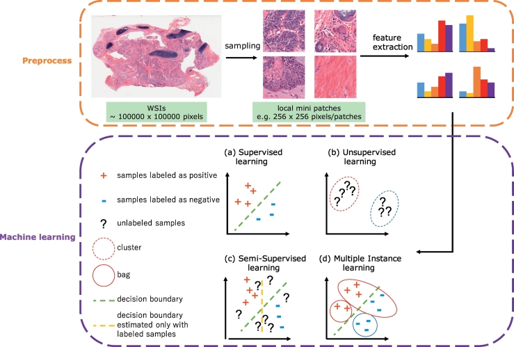
Abundant accumulation of digital histopathological images has led to the increased demand for their analysis, such as computer-aided diagnosis using machine learning techniques. However, digital pathological images and related tasks have some issues to be considered. In this mini-review, we introduce the application of digital pathological image analysis using machine learning algorithms, address some problems specific to such analysis, and propose possible solutions.
Overview
The claims and statistics of this paper haven’t been checked yet.
Run the check — free, no account needed. It reads the open-access text and checks the sources it cites.
Integrity signals
Registry
Cites
Statistics
Figures
Verification
Claims the paper makes
The claims and statistics of this paper haven’t been checked yet.
Run the check — free, no account needed. It reads the open-access text and checks the sources it cites.
Claims other papers make about this one
Each is checked against this paper’s own text: does it say what the citing paper says it does?
The source backs part of the claim, not all of it — or backs it for a different population, condition or comparison.
In the citing paper
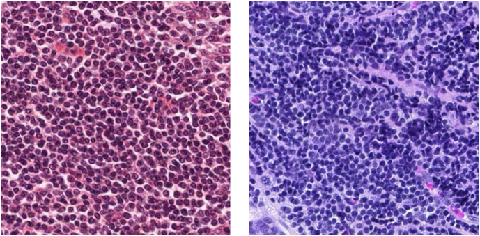
“In histopathology applications-studying tissue slides under a microscope-this variation can arise from sources like differences in the patient population or in slide staining and image acquisition (Veta et al., 2016; Komura and Ishikawa, 2018; Tellez et al., 2019).”
From WILDS: A Benchmark of in-the-Wild Distribution Shifts (2020) · cites this paper as Komura and Ishikawa, 2018
Experimental — accuracy not yet measuredThe source backs part of the claim, not all of it — or backs it for a different population, condition or comparison.
In the citing paper
“In histopathology applications-studying tissue slides under a microscope-this variation can arise from sources like differences in the patient population or in slide staining and image acquisition (Veta et al., 2016; Komura and Ishikawa, 2018; Tellez et al., 2019).”
From WILDS: A Benchmark of in-the-Wild Distribution Shifts (2020) · cites this paper as Komura and Ishikawa, 2018
Experimental — accuracy not yet measured
Self-check
Percentages, sums and reported differences re-computed from the paper’s own numbers.
The self-check needs the paper’s full text.
Citations
Cites
ImageNet classification with deep convolutional neural networks
Krizhevsky, A., Sutskever, I., & Hinton, G. E. (2017) · Communications of the ACM · doi:10.1145/3065386 · Cited by 112,155
ImageNet Large Scale Visual Recognition Challenge
Russakovsky, O., Deng, J., Su, H., Krause, J., Satheesh, S., Ma, S., Huang, Z., Karpathy, A., Khosla, A., Bernstein, M., et al. (2015) · International Journal of Computer Vision · doi:10.1007/s11263-015-0816-y · Cited by 39,380
A survey on deep learning in medical image analysis
Litjens, G., Kooi, T., Bejnordi, B. E., Setio, A. A. A., Ciompi, F., Ghafoorian, M., van der Laak, J. A. W. M., van Ginneken, B., & Sánchez, C. I. (2017) · Medical Image Analysis · doi:10.1016/j.media.2017.07.005 · Cited by 15,332
The cancer genome atlas pan-cancer analysis project
The Cancer Genome Atlas Research Network, Weinstein, J. N., Collisson, E. A., Mills, G. B., Shaw, K. R. M., Ozenberger, B. A., Ellrott, K., Shmulevich, I., Sander, C., & Stuart, J. M. (2013) · Nature Genetics · doi:10.1038/ng.2764 · Cited by 9,092
A comparative study of texture measures with classification based on featured distributions
Ojala, T., Pietikäinen, M., & Harwood, D. (1996) · Pattern Recognition · doi:10.1016/0031-3203(95)00067-4 · Cited by 6,882
Deep learning in medical image analysis
Shen, D., Wu, G., & Suk, H.-I. (2017) · Annual Review of Biomedical Engineering · doi:10.1146/annurev-bioeng-071516-044442 · Cited by 4,968
Solving the multiple instance problem with axis-parallel rectangles
Dietterich, T. G., Lathrop, R. H., & Lozano-Pérez, T. (1997) · Artificial Intelligence · doi:10.1016/s0004-3702(96)00034-3 · Cited by 2,866
Virtual Adversarial Training: A Regularization Method for Supervised and Semi-Supervised Learning
Miyato, T., Maeda, S., Koyama, M., & Ishii, S. (2018) · IEEE Transactions on Pattern Analysis and Machine Intelligence · doi:10.1109/tpami.2018.2858821 · Cited by 2,860
Codella, N. C. F., Gutman, D., Celebi, M. E., Helba, B., Marchetti, M. A., Dusza, S. W., Kalloo, A., Liopyris, K., Mishra, N., Kittler, H., et al. (2018) · 2018 IEEE 15th International Symposium on Biomedical Imaging (ISBI 2018) · doi:10.1109/isbi.2018.8363547 · Cited by 2,620
Bilinear CNN Models for Fine-Grained Visual Recognition
Lin, T., RoyChowdhury, A., & Maji, S. (2015) · IEEE/CVF International Conference on Computer Vision (ICCV) · doi:10.1109/iccv.2015.170 · Cited by 2,234
Research works printed but not linked: 5
- Barry J.D., Fagny M., Paulson J.N., Aerts H., Platig J. and Quackenbush J.: Histopathological image QTL discovery of thyroid autoimmune disease variants. In: BioRxiv. : 2017.
- ScanNet: a fast and dense scanning framework for metastatic breast cancer detection from whole-slide images - semantic scholar. https://www.semanticscholar.org/paper/ScanNet-A-Fast-and-Dense-Scanning-Framework-for-Me-Lin-Chen/9484287f4d5d52d10b5d362c462d4d6955655f8e.
- MICCAI grand challenge: tumor proliferation assessment challenge (TUPAC16). MICCAI Gd Chall Tumor Prolif Assess Chall TUPAC16. http://tupac.tue-image.nl/.
- KimiaPath24: dataset for retrieval and classification in digital pathology. : KIMIA Lab 2017.
- Tong S. and Koller D.: Support vector machine active learning with applications to text classification. J Mach Learn Res 2001; 2: 45.
Cited by
Sorted by citations
Items 701–800 of 940
Reshma, I. A., Franchet, C., Gaspard, M., Ionescu, R. T., Mothe, J., Cussat‐Blanc, S., Luga, H., & Brousset, P. (2022) · Journal of Imaging Informatics in Medicine · doi:10.1007/s10278-022-00618-7 · Cited by 2
A Multi-Feature Fusion Network for Pathological Identification of Tumor Cells
Lu, Z., Zhao, J., Sun, Y., Xu, F., & Ma, X. (2022) · IEEE Access · doi:10.1109/access.2022.3160290 · Cited by 1
Optimizing machine learning hyperparameters in two-photon FLIM image analysis
Mbogo, B. P., Siller, K. H., Zhang, J., Wallrabe, H. K., Alam, S. R., & Periasamy, A. (2023) · Multiphoton Microscopy in the Biomedical Sciences · doi:10.1117/12.2666216 · Cited by 1
Artificial Intelligence-Based Lung Nodule Detection: A Survey
Shah, S., & Mahajan, A. (2023) · Lecture notes in electrical engineering · doi:10.1007/978-981-19-9876-8_2 · Cited by 1
Color-SIFT Features for Histopathological Image Analysis
Ouddai, G., Hamdi, I., & Ghézala, H. B. (2023) · Studies in computational intelligence · doi:10.1007/978-3-031-26135-0_5 · Cited by 1
Machine Learning in Tissue Polarimetry
Makkithaya, K. N., Melanthota, S. K., Kistenev, Y. V., Bykov, A. V., Novikova, T., Meglinski, I. V., & Mazumder, N. (2023) · Biological and medical physics, biomedical engineering · doi:10.1007/978-3-031-31852-8_14 · Cited by 1
Prabu, S., Aravind, M. L., Aravindh, J. P. M., Maruthupandiyan, K., & Yokeshwaran, M. B. (2023) · International Conference on Applied Intelligence and Sustainable Computing (ICAISC) · doi:10.1109/icaisc58445.2023.10199859 · Cited by 1
Hamamoto, S., Oura, M., Shundo, A., Kawaguchi, D., Yamamoto, S., Takano, H., Uesugi, M., Takeuchi, A., Iwai, T., Seto, Y., et al. (2023) · Science and Technology of Advanced Materials Methods · doi:10.1080/27660400.2023.2270529 · Cited by 1
Güler, R., & Şentürk, Z. K. (2023) · doi:10.1109/ubmk59864.2023.10286650 · Cited by 1
Kumar, P., & Sharma, M. K. (2024) · Advances in medical diagnosis, treatment, and care (AMDTC) book series · doi:10.4018/979-8-3693-3218-4.ch008 · Cited by 1
Detection and Classification of Brain Tumor using Deep Learning Techniques
Gundabatini, S. G. (2024) · International Journal for Research in Applied Science and Engineering Technology · doi:10.22214/ijraset.2024.59343 · Cited by 1
Liu, W., Qu, A., Yuan, J., Wang, L., Chen, J., Zhang, X., Wang, H., Han, Z., & Li, Y. (2024) · PubMed · doi:10.14670/hh-18-715 · Cited by 1
Machine Learning in Medical Diagnosis of Cancer
Raj, R., Jyoti, J., Singh, A., & Kumar, K. (2024) · IEEE International Conference for Convergence in Technology (I2CT) · doi:10.1109/i2ct61223.2024.10544354 · Cited by 1
Lee, Y. C. (2024) · Korean Journal of Otorhinolaryngology - Head and Neck Surgery · doi:10.3342/kjorl-hns.2024.00010 · Cited by 1
Artificial Intelligence in Cancer: A SWOT Analysis
Torkay, G., Fadlallah, N., Karagöz, A., Canlı, M., Saydam, E., Mete, A., Kızılışık, F., Darıcı, H., & YEŞİL, Y. (2024) · Journal of AI · doi:10.61969/jai.1469589 · Cited by 2
Reza, M. T., Alam, M. G. R., Rahman, R., & Dipto, S. M. (2024) · PLoS ONE · doi:10.1371/journal.pone.0303541 · Cited by 1
AI-Based Vision Screening Tool for Keratoconus
Rahman, R., Kumar, P., Sharma, Y. K., Sharma, M. K., & Challa, N. K. (2024) · Lecture notes in electrical engineering · doi:10.1007/978-981-97-5231-7_15 · Cited by 1
Dijital Patoloji ve Yapay Zekâ: Gelişimi, Uygulama Örnekleri ve Gelecek Yönelimler
Çalık, N., Gökcan, M. T., Topuz, Y., & Varlı, S. (2024) · Türkiye Sağlık Enstitüleri Başkanlığı Dergisi · doi:10.5578/tusebdergisi.20240338 · Cited by 1
Histopathology Image Augmentation Through StyleGAN2-ADA
Muñoz, B., Raquel Pezoa, R., & Gutierrez, H. (2025) · Communications in computer and information science · doi:10.1007/978-3-031-80084-9_15 · Cited by 1
Gobinath, A., Rajeswari, P., Anandan, M., & Kumar, N. S. (2025) · Advances in computational intelligence and robotics book series · doi:10.4018/979-8-3373-1275-0.ch003 · Cited by 1
Karuppasamy, A., Zidoum, H. M., Al-Rashdi, M. S. S., & Al-Bahri, M. (2025) · Intelligence-Based Medicine · doi:10.1016/j.ibmed.2025.100248 · Cited by 1
Mohamadi, Z., Partovifar, P., Ahmadzadeh, H., Ahmadi, E. A., Ghanbari, A., Feyzipour, S., Atefat, F., Jahanpeyma, N., asl, F. H., ZarinKhat, A., et al. (2025) · Intelligence-Based Medicine · doi:10.1016/j.ibmed.2025.100250 · Cited by 2
A Hybrid ViT-GRU Model for Breast Cancer Detection: Addressing Class Imbalance Challenges
Zobair, M. J., Rahman, M. A., Hossain, M., Khan, N., Akash, M. A. A. K., & Bijoy, M. H. I. (2025) · International Conference on Electrical, Computer and Communication Engineering · doi:10.1109/ecce64574.2025.11013095 · Cited by 1
Extraction of Features from Breast Cancer Mammogram Image Using Some Image Processing Techniques
Pati, D. P., & Panda, S. (2018) · International Conference on Parallel, Distributed and Grid Computing (PDGC) · doi:10.1109/pdgc.2018.8745891 · Cited by 1
Implementation of Machine Learning-Aided Imaging Analytics for Histopathological Image Diagnosis
Yu, Y., Wang, J., Chun, H. E., Xu, Y., Fong, E. L. S., Wee, A., & Yu, H. (2020) · Elsevier eBooks · doi:10.1016/b978-0-12-801238-3.11388-1 · Cited by 1
The Axis “Human Papillomavirus - Anal Squamous Cell Carcinoma”: A Review
Monteiro, A. C. B., Khelassi, A., Tananska, V. T., Iano, Y., Arthur, R., & França, R. P. (2020) · Medical Technologies Journal · doi:10.26415/2572-004x-vol3iss4p471-484 · Cited by 1
Machine Learning Cancer Diagnosis Based on Medical Image Size and Modalities
Al-Dhabyani, W., & Fahmy, A. (2020) · Studies in computational intelligence · doi:10.1007/978-3-030-52067-0_9 · Cited by 1
Breast Cancer Detection Using Texture Features and KNN Algorithm
Murugan, T. D., & Kanojia, M. G. (2021) · Advances in intelligent systems and computing · doi:10.1007/978-3-030-73050-5_77 · Cited by 1
Ramos, R. T. D. M., de Santana, M. A., Sousa, P. A., Ferreira, M. R. A., dos Santos, W. P., & Soares, L. A. L. (2021) · Journal of Liquid Chromatography & Related Technologies · doi:10.1080/10826076.2021.1932521 · Cited by 1
Learning Techniquesforpre Malignancy Detection in Human Cells a Review
Jumnake, G. F., Mahalle, P. N., & Shinde, G. R. (2020) · International Journal of Engineering and Advanced Technology · doi:10.35940/ijeat.f1622.089620 · Cited by 1
Improvement of Automatic Diagnosis of Soft Tissue Tumours Using ML
Charitharyan, P. (2021) · International Journal for Research in Applied Science and Engineering Technology · doi:10.22214/ijraset.2021.38981 · Cited by 1
Diagnostics and Personalized Medicine
Zanders, E. D. (2020) · Springer eBooks · doi:10.1007/978-3-030-57814-5_14 · Cited by 1
Customized EfficientNet for Histopathology Image Representation
(2022) · IEEE Symposium Series on Computational Intelligence (SSCI) · doi:10.1109/ssci51031.2022.10022191 · Cited by 1
Artificial intelligence in breast cancer: An opportunity for early diagnosis
Malla, R. R., & Katneni, V. (2023) · Elsevier eBooks · doi:10.1016/b978-0-443-15280-1.00004-2 · Cited by 1
Highly Efficient Follicular Segmentation in Thyroid Cytopathological Whole Slide ImagePreprint
Tao, S., Guo, Y., Zhu, C., Chen, H., Zhang, Y., Yang, J., & Liu, J. (2019) · arXiv (Cornell University) · doi:10.48550/arxiv.1902.05431 · Cited by 1
Automated acquisition of knowledge beyond pathologistsPreprint
Yamamoto, Y., Tsuzuki, T., Akatsuka, J., Ueki, M., Morikawa, H., Numata, Y., Takahara, T., Tsuyuki, T., Shimizu, A., Maeda, I., et al. (2019) · bioRxiv (Cold Spring Harbor Laboratory) · doi:10.1101/539791 · Cited by 1
Not again! Data Leakage in Digital PathologyPreprint
Bussola, N., Marcolini, A., Maggio, V., Jurman, G., & Furlanello, C. (2019) · arXiv (Cornell University) · Cited by 1
Gu, H., Xu, Y., Haeri, M., & Chen, X. (2020) · arXiv (Cornell University) · Cited by 1
Serdiuk, A. A., Urman, R. D., & Engwall, S. (2020) · Journal of Clinical Anesthesia · doi:10.1016/j.jclinane.2020.109965 · Cited by 1
MORPHIOUS: A Machine Learning Workflow to Naively Detect the Activation of Microglia and AstrocytesPreprint
Silburt, J., & Aubert, I. (2020) · bioRxiv (Cold Spring Harbor Laboratory) · doi:10.1101/2020.08.17.251843 · Cited by 1
Deep learning from multiple experts improves identification of amyloid neuropathologiesPreprint
Wong, D., & Keiser, M. J. (2021) · bioRxiv (Cold Spring Harbor Laboratory) · doi:10.17605/osf.io/xh2jd · Cited by 1
Automated Segmentation of Complex Biological Structures in Histopathology Images
Salsabili, S. (2022) · doi:10.22215/etd/2022-14880 · Cited by 1
Kara, A. (2022) · Research Square · doi:10.21203/rs.3.rs-2193871/v1 · Cited by 1
A Bladder Cancer Grading System Using Deep Neural Network Architectures
Habibi, K. (2023) · doi:10.32920/24043230 · Cited by 1
A Bladder Cancer Grading System Using Deep Neural Network Architectures
Habibi, K. (2023) · doi:10.32920/24043230.v1 · Cited by 1
Histopathological Deep Learning: Exploring Ovarian Cancer Subtypes with Image AnalysisPreprint
Ramanathan, L., Rachel, K., Namineni, S. L., & Phaneendra, B. H. (2025) · SSRN Electronic Journal · doi:10.2139/ssrn.5130082 · Cited by 1
Utilizing Machine Learning Techniques to Rapidly Identify MUC2 Expression in Colon Cancer Tissues
Periyakoil, P. K. (2018) · CaltechTHESIS (California Institute of Technology) · doi:10.7907/sapn-r691
A Cinematic View of Tissue Microbiology in the Live Infected Host
Richter‐Dahlfors, A., & Melican, K. (2019) · Microbiology Spectrum · doi:10.1128/microbiolspec.bai-0007-2019
Deep Learning for Estimating Synaptic Health of Primary Neuronal Cell CulturePreprint
Kormilitzin, A., Yang, X., Stone, W. H., Woffindale, C., Nicholls, F. J., Ribé, E. M., Nevado‐Holgado, A. J., & Buckley, N. J. (2019) · arXiv (Cornell University) · doi:10.48550/arxiv.1908.11399
López-Pérez, M., Colomer, A., Sales, M. A., Molina, R., & Naranjo, V. (2019) · Lecture notes in computer science · doi:10.1007/978-3-030-33607-3_5
A simple computer vision algorithm as a clinical aid for the pathologistPreprint
Lázaro-Guevara, J. M., Robles, B. J. F., Murga, A. E., & Garrido, K. A. (2019) · bioRxiv (Cold Spring Harbor Laboratory) · doi:10.1101/853325
Lyashenko, V. V., Kobylin, O., & Шафроненко, А. (2019) · CAOL . International Conference on Advanced Optoelectronics and Lasers · doi:10.1109/caol46282.2019.9019575
Ortega-Ruíz, M. A., Karabağ, C., Garduño, V. G., & Reyes‐Aldasoro, C. C. (2020) · bioRxiv (Cold Spring Harbor Laboratory) · doi:10.1101/2020.04.09.034348
Hinders, M. K. (2020) · Springer eBooks · doi:10.1007/978-3-030-49395-0_1
Rony, J. (2020) · Espace ÉTS (ETS)
Polarized Light Microscopy of Human Breast Cancer
Westreich, J. (2019) · TSpace (University of Toronto)
Ramírez, L., & Sebastián, J. J. (2020)
Synthetic Generation of Three-Dimensional Cancer Cell Models from Histopathological ImagesPreprint
Alon, Y., Yu, X., & Zhou, H. (2021) · arXiv (Cornell University) · doi:10.48550/arxiv.2101.11600
Collaborative General Purpose Convolutional Neural Networks
Ogawa, K., & Hartono, P. (2021) · Journal of Signal Processing · doi:10.2299/jsp.25.53
Machine Learning Approaches Pertinent to Glioma Classification
Amin, S. B. (2021) · Molecular pathology library · doi:10.1007/978-3-030-69170-7_4
Gerard, G., & Piastra, M. (2021) · arXiv (Cornell University) · doi:10.48550/arxiv.2105.14338
Moyes, A., Gault, R., Zhang, K., Ming, J., Crookes, D., & Wang, J. (2021) · arXiv (Cornell University) · doi:10.48550/arxiv.2107.07271
Plasmodium falciparum Detection in Cell Images Using Convolutional Neural Network
Ghose, S., Datta, S., Malathy, C., & Gayathri, M. K. (2021) · Smart innovation, systems and technologies · doi:10.1007/978-981-16-1502-3_40
Performance of a machine learning model in recognition of normal tissue from histological sectionsPreprint
Sanyal, P., Paul, S., & Das, A. (2021) · medRxiv · doi:10.1101/2021.07.20.21260573
Abbet, C., Studer, L., Fischer, A., Dawson, H., Zlobec, I., Bozorgtabar, B., & Thiran, J. (2021) · arXiv (Cornell University)
Exploration of deep-learning based classification with human SNP image graphsPreprint
Chen, C.-H., Tung, K.-F., & Lin, W. (2021) · bioRxiv (Cold Spring Harbor Laboratory) · doi:10.1101/2021.10.01.462710
Manavalan, M. (2020) · Engineering International · doi:10.18034/ei.v8i2.574 · Cited by 1
Transferability of Deep Learning Models for Focus Quality Assessment in Digital PathologyPreprint
Miles, A., Hosseini, M. S., Tang, S., Wang, Z., Damaskinos, S., & Plataniotis, K. N. (2021) · Research Square · doi:10.21203/rs.3.rs-1120682/v1
Automated Cell Segmentation for Phase-Contrast Images of Adhesion Cell Culture
Ye, G., & Kaya, M. (2021) · International Conference on Bio-engineering for Smart Technologies (BioSMART) · doi:10.1109/biosmart54244.2021.9677717
Das, D., & Mahanta, L. B. (2022) · Springer eBooks · doi:10.1007/978-3-030-64573-1_189
Villalba-Diez, J., & Ordieres Meré, J. (2022) · MDPI eBooks · doi:10.3390/books978-3-0365-3813-6
A Cinematic View of Tissue Microbiology in the Live Infected Host
Richter‐Dahlfors, A., & Melican, K. (2020) · John Wiley & Sons, Inc. eBooks · doi:10.1128/9781683670261.ch22
A Digital Pathology Platform for Artificial Intelligence Data Sharing (Preprint)Preprint
Kang, Y., Kim, Y. J., Park, S., Ro, G., Hong, C., Jang, H., Cho, S., Hong, W., Kang, D. U., Kim, E., et al. (2020) · doi:10.2196/preprints.21968
Survey on Machine learning Techniques used for Information Security
Soni, S. (2022) · International Conference on Emerging Trends in Artificial Intelligence and Smart Systems (THEETAS) · doi:10.4108/eai.16-4-2022.2318143
Medical Image Analysis With Intelligent Techniques
Ganapathy, J., Purushothaman, R., Sathishkumar, M., & Vishal, L. (2022) · Advances in medical technologies and clinical practice book series · doi:10.4018/978-1-6684-4671-3.ch003
White, B. S., Woo, X., Koc, S., Sheridan, T., Neuhauser, S. B., Wang, S., Evrard, Y. A., Landua, J. D., Mashl, R. J., Davies, S. R., et al. (2022) · bioRxiv (Cold Spring Harbor Laboratory) · doi:10.1101/2022.10.26.512745
Learning from limited labelled data: contributions to weak, few-shot, and unsupervised learning
Rodríguez, J. J. S. (2022) · RiuNet (Universitat Politècnica de València) · doi:10.4995/thesis/10251/190633
Freitas, D. D. C., & Reis, D. L. (2022) · Brazilian Journal of Development · doi:10.34117/bjdv8n11-320
Fathalla, K. M., Eltelwany, A. S., & Saad, A. A. (2022) · Indian Journal of Computer Science and Engineering · doi:10.21817/indjcse/2022/v13i6/221306030
Fares, M. A., Tarek, K., & Rahmoun, D. E. (2023) · bioRxiv (Cold Spring Harbor Laboratory) · doi:10.1101/2023.01.25.525505
Park, J., Jang, B. G., Kim, Y. W., Park, H., Kim, B., Kim, M. J., Ko, H., Gwak, J. M., Lee, E. J., Chung, Y. R., et al. (2023) · doi:10.1158/1078-0432.c.6530208
Medical image classification in breast sentinel lymph node detection
Dayo, A., & Omodele (2023) · World Journal of Biology Pharmacy and Health Sciences · doi:10.30574/wjbphs.2023.14.1.0153
Ebrahiminia, A., Radman, M., & SAMIMI, P. A. (2023) · Journal of Mechanics in Medicine and Biology · doi:10.1142/s0219519423500392
Early Detection of Breast Cancer using An Improved Deep-Learning Model
Yousif, M., Alrababah, H., Atif, M., Ahmad, M., & Khan, M. A. (2023) · International Conference on Business Analytics for Technology and Security (ICBATS) · doi:10.1109/icbats57792.2023.10111184
Super-resolution histology of paraffin-embedded samples via photonic chip-based microscopyPreprint
Villegas-Hernandéz, L. E., Dubey, V. K., Mao, H., Pradhan, M., Tinguely, J., Hansen, D. H., Acuña, S., Zapotoczny, B., Agarwal, K., Nystad, M., et al. (2023) · bioRxiv (Cold Spring Harbor Laboratory) · doi:10.1101/2023.06.14.544765
Diagnosing Soft Tissue Tumors using Machine Learning Techniques: A Survey
Ghanim, N. Y., & Alneamy, J. S. M. (2023) · Mağallaẗ al-tarbiyaẗ wa-al-ʻilm · doi:10.33899/edusj.2023.137706.1316
Machine learning for medical processing: algorithms and analysis
Zhang, Z. (2023) · International Conference on Modern Medicine and Global Health (ICMMGH) · doi:10.1117/12.2692341
SSANet: spatial stain attention network for pathological images classification
Xie, Y., Zhang, Y., Hou, J., Chen, D., & Guan, G. (2023) · Multimedia Tools and Applications · doi:10.1007/s11042-023-16313-w · Cited by 1
Wu, Y., Wang, Y., Tang, Y., Li, B., Guan, C., Huang, Z., Wang, S., Xue, J., Li, S., Chen, J., et al. (2023) · SSRN Electronic Journal · doi:10.2139/ssrn.4610005
Bioinformatics, Digital Pathology, and Computational Pathology for Surgical Pathologists
Mohanty, S. K., Shafi, S., & Parwani, A. V. (2023) · Springer eBooks · doi:10.1007/978-3-031-35118-1_2
Wani, J. A., Sharma, N., Rakhra, M., Singh, A. P., & Reena, R. (2023) · Transactions on computer systems and networks · doi:10.1007/978-981-99-4518-4_16
Kim, J., Jee, S., & Ro, S.-H. (2023) · Sustainability · doi:10.3390/su152316495 · Cited by 1
Biloborodova, T., Brosnan, B., Skarga-Bandurova, I., & Strauß, D. J. (2023) · Lecture notes in computer science · doi:10.1007/978-3-031-49011-8_19
Chaudhary, M., & Agrawal, H. (2023) · International Conference on Informatics (ICI) · doi:10.1109/ici60088.2023.10421032
Metadata
- Authors
- Komura, D., & Ishikawa, S.
- Affiliations
- Tokyo Medical and Dental University (JP)
- Corresponding author
- Komura, Daisuke
- Venue
- Computational and Structural Biotechnology Journal
- Published
- 2018-01
- Volume
- 16
- Pages
- 34-42
- ISSN
- 2001-0370 (ISSN-L)
- PMC
- PMC6158771
- OpenAlex
- W2751723768
- Cited by
- 937
- References
- 111
- Citations by year
- 2026: 95 · 2025: 111 · 2024: 104 · 2023: 122 · 2022: 114 · 2021: 140 · 2020: 146 · 2019: 88 · 2018: 17
- FWCI
- 11.44
- Type
- journal article
- Language
- en
- Open access
- gold
- doi.org
- Licence
- Locations
- Computational and Structural Biotechnology Journal (publishedVersion, cc-by)
- Europe PMC (PubMed Central) (publishedVersion, cc-by)
- PubMed Central (publishedVersion, cc-by)
- PubMed (publishedVersion)
- arXiv (Cornell University) (submittedVersion)
- DOAJ (DOAJ: Directory of Open Access Journals) (submittedVersion, cc-by-sa)
- arXiv (Cornell University) (cc-by)
- Funders
- JSPS (25710020)
- Topics
- Physical Sciences › Computer Science › Artificial Intelligence › AI in cancer detection
- Health Sciences › Medicine › Radiology, Nuclear Medicine and Imaging › Radiomics and Machine Learning in Medical Imaging
- Life Sciences › Biochemistry, Genetics and Molecular Biology › Biophysics › Cell Image Analysis Techniques
- Keywords
- digital histopathology, histopathology image analysis, computer-aided diagnosis, machine learning
About these checks
Registry, retraction, Cites and statistics results are computed, not judged, with one exception: a printed reference that no index lists is read once by a model before it counts as a citation error. Claims are extracted by a model and kept only when their quote appears verbatim in the text; each cited source is then checked, with a model’s help, for whether it says what the claim relies on. Those verdicts can be wrong — read the claim’s quote and the cited source before relying on one.
Statistics and arithmetic are recomputed, not judged.
Confidence bands are shown only for fields calibrated on our annotated test set. None is calibrated yet, so no band is shown.
What counts as a claim ›How we measure confidence ›How we check this ›
Are you an author, or do you see a problem with this page? Request a review
