

The DOI matches its Crossref record; 108 of the 111 references it cites were found in a registry or an index.
Machine Learning Methods for Histopathological Image Analysis
Open accessAbstract
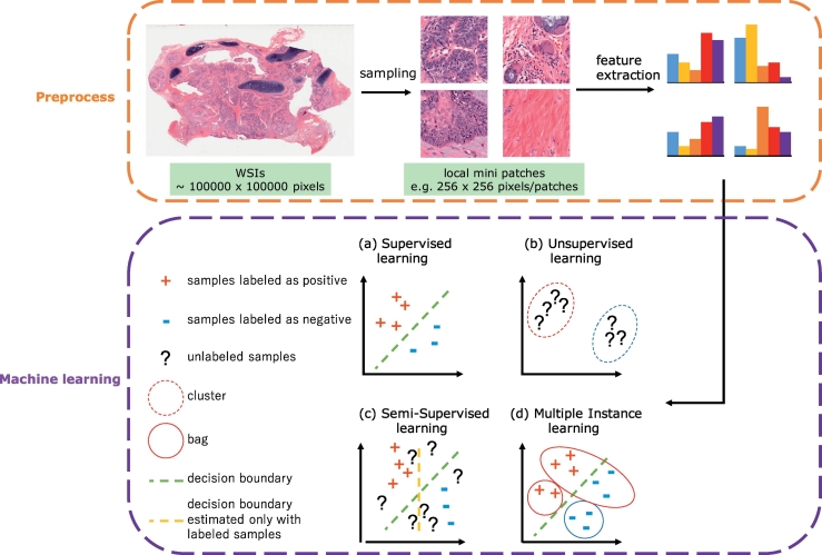
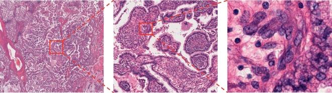
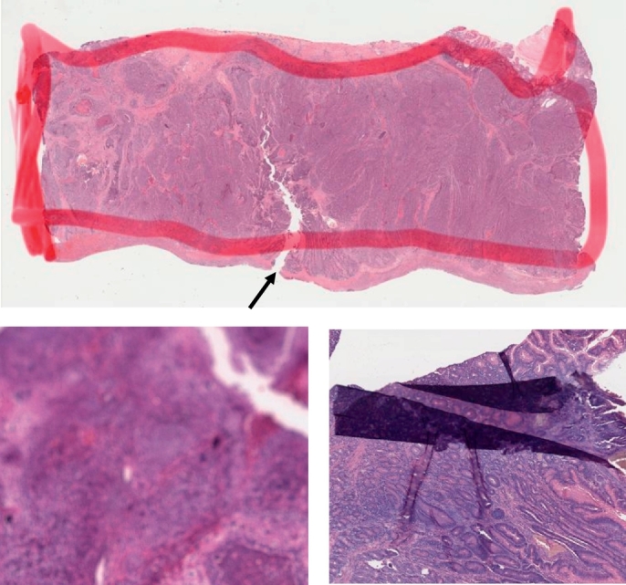
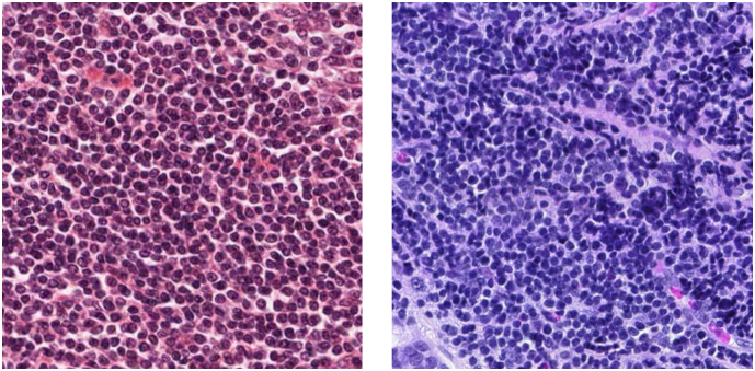
Abundant accumulation of digital histopathological images has led to the increased demand for their analysis, such as computer-aided diagnosis using machine learning techniques. However, digital pathological images and related tasks have some issues to be considered. In this mini-review, we introduce the application of digital pathological image analysis using machine learning algorithms, address some problems specific to such analysis, and propose possible solutions.
Overview
The claims and statistics of this paper haven’t been checked yet.
Run the check — free, no account needed. It reads the open-access text and checks the sources it cites.
Integrity signals
Registry
Cites
Statistics
Figures
Verification
Claims the paper makes
The claims and statistics of this paper haven’t been checked yet.
Run the check — free, no account needed. It reads the open-access text and checks the sources it cites.
Claims other papers make about this one
Each is checked against this paper’s own text: does it say what the citing paper says it does?
The source backs part of the claim, not all of it — or backs it for a different population, condition or comparison.
In the citing paper
“In histopathology applications-studying tissue slides under a microscope-this variation can arise from sources like differences in the patient population or in slide staining and image acquisition (Veta et al., 2016; Komura and Ishikawa, 2018; Tellez et al., 2019).”
From WILDS: A Benchmark of in-the-Wild Distribution Shifts (2020) · cites this paper as Komura and Ishikawa, 2018
Experimental — accuracy not yet measuredThe source backs part of the claim, not all of it — or backs it for a different population, condition or comparison.
In the citing paper
“In histopathology applications-studying tissue slides under a microscope-this variation can arise from sources like differences in the patient population or in slide staining and image acquisition (Veta et al., 2016; Komura and Ishikawa, 2018; Tellez et al., 2019).”
From WILDS: A Benchmark of in-the-Wild Distribution Shifts (2020) · cites this paper as Komura and Ishikawa, 2018
Experimental — accuracy not yet measured
Self-check
Percentages, sums and reported differences re-computed from the paper’s own numbers.
The self-check needs the paper’s full text.
Citations
Cites
ImageNet classification with deep convolutional neural networks
Krizhevsky, A., Sutskever, I., & Hinton, G. E. (2017) · Communications of the ACM · doi:10.1145/3065386 · Cited by 112,155
ImageNet Large Scale Visual Recognition Challenge
Russakovsky, O., Deng, J., Su, H., Krause, J., Satheesh, S., Ma, S., Huang, Z., Karpathy, A., Khosla, A., Bernstein, M., et al. (2015) · International Journal of Computer Vision · doi:10.1007/s11263-015-0816-y · Cited by 39,380
A survey on deep learning in medical image analysis
Litjens, G., Kooi, T., Bejnordi, B. E., Setio, A. A. A., Ciompi, F., Ghafoorian, M., van der Laak, J. A. W. M., van Ginneken, B., & Sánchez, C. I. (2017) · Medical Image Analysis · doi:10.1016/j.media.2017.07.005 · Cited by 15,332
The cancer genome atlas pan-cancer analysis project
The Cancer Genome Atlas Research Network, Weinstein, J. N., Collisson, E. A., Mills, G. B., Shaw, K. R. M., Ozenberger, B. A., Ellrott, K., Shmulevich, I., Sander, C., & Stuart, J. M. (2013) · Nature Genetics · doi:10.1038/ng.2764 · Cited by 9,092
A comparative study of texture measures with classification based on featured distributions
Ojala, T., Pietikäinen, M., & Harwood, D. (1996) · Pattern Recognition · doi:10.1016/0031-3203(95)00067-4 · Cited by 6,882
Deep learning in medical image analysis
Shen, D., Wu, G., & Suk, H.-I. (2017) · Annual Review of Biomedical Engineering · doi:10.1146/annurev-bioeng-071516-044442 · Cited by 4,968
Solving the multiple instance problem with axis-parallel rectangles
Dietterich, T. G., Lathrop, R. H., & Lozano-Pérez, T. (1997) · Artificial Intelligence · doi:10.1016/s0004-3702(96)00034-3 · Cited by 2,866
Virtual Adversarial Training: A Regularization Method for Supervised and Semi-Supervised Learning
Miyato, T., Maeda, S., Koyama, M., & Ishii, S. (2018) · IEEE Transactions on Pattern Analysis and Machine Intelligence · doi:10.1109/tpami.2018.2858821 · Cited by 2,860
Codella, N. C. F., Gutman, D., Celebi, M. E., Helba, B., Marchetti, M. A., Dusza, S. W., Kalloo, A., Liopyris, K., Mishra, N., Kittler, H., et al. (2018) · 2018 IEEE 15th International Symposium on Biomedical Imaging (ISBI 2018) · doi:10.1109/isbi.2018.8363547 · Cited by 2,620
Bilinear CNN Models for Fine-Grained Visual Recognition
Lin, T., RoyChowdhury, A., & Maji, S. (2015) · IEEE/CVF International Conference on Computer Vision (ICCV) · doi:10.1109/iccv.2015.170 · Cited by 2,234
Histopathological Image Analysis: A Review
Gurcan, M. N., Boucheron, L. E., Can, A., Madabhushi, A., Rajpoot, N. M., & Yener, B. (2009) · IEEE Reviews in Biomedical Engineering · doi:10.1109/rbme.2009.2034865 · Cited by 2,052
A sequential algorithm for training text classifiersPreprint
Lewis, D., & Gale, W. A. (1994) · arXiv (Cornell University) · doi:10.5555/188490.188495 · Cited by 1,838
Understanding Black-box Predictions via Influence Functions
Koh, P. W., & Liang, P. (2017) · arXiv · doi:10.48550/arxiv.1703.04730 · Cited by 1,514
Deep learning for digital pathology image analysis: A comprehensive tutorial with selected use cases
Janowczyk, A. R., & Madabhushi, A. (2016) · Journal of Pathology Informatics · doi:10.4103/2153-3539.186902 · Cited by 1,387
A Sequential Algorithm for Training Text Classifiers
Lewis, D. D., & Gale, W. A. (1994) · SIGIR ’94 · doi:10.1007/978-1-4471-2099-5_1 · Cited by 1,208
Evaluating the Visualization of What a Deep Neural Network Has Learned
Samek, W., Binder, A., Montavon, G., Lapuschkin, S., & Muller, K.-R. (2017) · IEEE Transactions on Neural Networks and Learning Systems · doi:10.1109/tnnls.2016.2599820 · Cited by 1,164
A Dataset and a Technique for Generalized Nuclear Segmentation for Computational Pathology
Kumar, N., Verma, R., Sharma, S., Bhargava, S., Vahadane, A., & Sethi, A. (2017) · IEEE Transactions on Medical Imaging · doi:10.1109/tmi.2017.2677499 · Cited by 1,160
Deep learning as a tool for increased accuracy and efficiency of histopathological diagnosis
Litjens, G., Sánchez, C. I., Timofeeva, N., Hermsen, M., Nagtegaal, I., Kovacs, I., Hulsbergen - van de Kaa, C., Bult, P., van Ginneken, B., & van der Laak, J. (2016) · Scientific Reports · doi:10.1038/srep26286 · Cited by 1,069
Classification of breast cancer histology images using Convolutional Neural Networks
Araújo, T., Aresta, G., Castro, E., Rouco, J., Aguiar, P., Eloy, C., Polónia, A., & Campilho, A. (2017) · PLoS ONE · doi:10.1371/journal.pone.0177544 · Cited by 1,044
Deep Learning for Identifying Metastatic Breast CancerPreprint
Wang, D., Khosla, A., Gargeya, R., Irshad, H., & Beck, A. H. (2016) · arXiv.org · doi:10.48550/arxiv.1606.05718 · Cited by 1,028
Breast cancer histopathological image classification using Convolutional Neural Networks
Spanhol, F. A., Oliveira, L. S., Petitjean, C., & Heutte, L. (2016) · International Joint Conference on Neural Networks (IJCNN) · doi:10.1109/ijcnn.2016.7727519 · Cited by 1,028
Gland segmentation in colon histology images: The glas challenge contest
Sirinukunwattana, K., Pluim, J. P. W., Chen, H., Qi, X., Heng, P., Guo, Y. B., Wang, L. Y., Matuszewski, B. J., Bruni, E., Sanchez, U., et al. (2016) · Medical Image Analysis · doi:10.1016/j.media.2016.08.008 · Cited by 1,014
Yu, K.-H., Zhang, C., Berry, G. J., Altman, R. B., Ré, C., Rubin, D. L., & Snyder, M. (2016) · Nature Communications · doi:10.1038/ncomms12474 · Cited by 972
Semi-Supervised Learning with Ladder NetworksPreprint
Rasmus, A., Harri Valpola, B., Honkala, M., Berglund, M., & Raiko, T. (2015) · arXiv (Cornell University) · doi:10.48550/arxiv.1507.02672 · Cited by 941
Patch-Based Convolutional Neural Network for Whole Slide Tissue Image Classification
Hou, L., Samaras, D., Kurç, T., Gao, Y., Davis, J., & Saltz, J. H. (2016) · IEEE Computer Society Conference on Computer Vision and Pattern Recognition/Proceedings - IEEE Computer Society Conference on Computer Vision and Pattern Recognition/Proceedings · doi:10.1109/cvpr.2016.266 · Cited by 921
Gao, Y., Beijbom, O., Zhang, N., & Darrell, T. (2016) · IEEE Computer Society Conference on Computer Vision and Pattern Recognition/Proceedings - IEEE Computer Society Conference on Computer Vision and Pattern Recognition/Proceedings · doi:10.1109/cvpr.2016.41 · Cited by 919
Systematic Analysis of Breast Cancer Morphology Uncovers Stromal Features Associated with Survival
Beck, A. H., Sangoi, A. R., Leung, S. C. Y., Marinelli, R. J., Nielsen, T. O., van de Vijver, M. J., West, R. B., van de Rijn, M., & Koller, D. (2011) · Science Translational Medicine · doi:10.1126/scitranslmed.3002564 · Cited by 780
Breast Cancer Multi-classification from Histopathological Images with Structured Deep Learning Model
Han, Z., Wei, B., Zheng, Y., Yin, Y., Li, K., & Li, S. (2017) · Scientific Reports · doi:10.1038/s41598-017-04075-z · Cited by 696
DCAN: Deep Contour-Aware Networks for Accurate Gland Segmentation
Chen, H., Qi, X., Yu, L., & Heng, P. (2016) · IEEE Computer Society Conference on Computer Vision and Pattern Recognition/Proceedings - IEEE Computer Society Conference on Computer Vision and Pattern Recognition/Proceedings · doi:10.1109/cvpr.2016.273 · Cited by 671
Multi-class texture analysis in colorectal cancer histology
Kather, J. N., Weis, C.-A., Bianconi, F., Melchers, S. M., Schad, L. R., Gaiser, T., Marx, A., & Zöllner, F. G. (2016) · Scientific Reports · doi:10.1038/srep27988 · Cited by 578
Xing, F., & Yang, L. (2016) · IEEE Reviews in Biomedical Engineering · doi:10.1109/rbme.2016.2515127 · Cited by 527
Khan, A. M., Rajpoot, N., Treanor, D. E., & Magee, D. (2014) · IEEE Transactions on Biomedical Engineering · doi:10.1109/tbme.2014.2303294 · Cited by 522
Detecting Cancer Metastases on Gigapixel Pathology Images
Liu, Y., Gadepalli, K., Norouzi, M., Dahl, G. E., Kohlberger, T., Boyko, A., Venugopalan, S., Timofeev, A., Nelson, P. Q., Corrado, G. S., et al. (2017) · arXiv · doi:10.48550/arxiv.1703.02442 · Cited by 506
Visualizing Deep Neural Network Decisions: Prediction Difference Analysis
Zintgraf, L. M., Cohen, T. S., Adel, T., & Welling, M. (2017) · arXiv · doi:10.48550/arxiv.1702.04595 · Cited by 487
Xu, J., Luo, X., Wang, G., Gilmore, H. L., & Madabhushi, A. (2016) · Neurocomputing · doi:10.1016/j.neucom.2016.01.034 · Cited by 461
Xu, Y. L., Jia, Z., Wang, L.-B., Ai, Y., Zhang, F., Lai, M., & Chang, E. (2017) · BMC Bioinformatics · doi:10.1186/s12859-017-1685-x · Cited by 428
Bilinear Convolutional Neural Networks for Fine-Grained Visual Recognition
Lin, T., RoyChowdhury, A., & Maji, S. (2017) · IEEE Transactions on Pattern Analysis and Machine Intelligence · doi:10.1109/tpami.2017.2723400 · Cited by 408
Mitosis detection in breast cancer histological images An ICPR 2012 contest
Roux, L., Racoceanu, D., Loménie, N., Kulikova, M. S., Irshad, H., Jacques, K., Capron, F., Genestie, C., Naour, G. L., & Gurcan Metin, N. (2013) · Journal of Pathology Informatics · doi:10.4103/2153-3539.112693 · Cited by 303
Learning Discriminative Reconstructions for Unsupervised Outlier Removal
Xia, Y., Cao, X., Wen, F., Hua, G., & Sun, J. Z. (2015) · IEEE/CVF International Conference on Computer Vision (ICCV) · doi:10.1109/iccv.2015.177 · Cited by 301
Stain Specific Standardization of Whole-Slide Histopathological Images
Bejnordi, B. E., Litjens, G. J. S., Timofeeva, N., Otte‐Höller, I., Homeyer, A., Karssemeijer, N., & van der Laak, J. A. W. M. (2015) · IEEE Transactions on Medical Imaging · doi:10.1109/tmi.2015.2476509 · Cited by 291
Weakly supervised histopathology cancer image segmentation and classification
Xu, Y. L., Zhu, J.-Y., Chang, E., Lai, M., & Tu, Z. (2014) · Medical Image Analysis · doi:10.1016/j.media.2014.01.010 · Cited by 286
Comprehensive Histologic Analysis of ALK-Rearranged Lung Carcinomas
Yoshida, A., Tsuta, K., Nakamura, H., Kohno, T., Takahashi, F., Asamura, H., Sekine, I., Fukayama, M., Shibata, T., Furuta, K., et al. (2011) · The American Journal of Surgical Pathology · doi:10.1097/pas.0b013e3182233e06 · Cited by 281
Song, Y., Zhang, L., Chen, S., Ni, D., Lei, B., & Wang, T. (2015) · IEEE Transactions on Biomedical Engineering · doi:10.1109/tbme.2015.2430895 · Cited by 272
Basavanhally, A. N., Ganesan, S., Agner, S. C., Monaco, J., Feldman, M. D., Tomaszewski, J. E., Bhanot, G., & Madabhushi, A. (2009) · IEEE Transactions on Biomedical Engineering · doi:10.1109/tbme.2009.2035305 · Cited by 272
The Genotype-Tissue Expression (GTEx) project
Lonsdale, J., Thomas, J., Salvatore, M., Phillips, R., Lo, E., Shad, S., Hasz, R., Walters, G., Garcia, F., Young, N., et al. (2013) · Nature Genetics · doi:10.1038/ng.2653 · Cited by 263
Digital images and the future of digital pathology
Pantanowitz, L. (2010) · Journal of Pathology Informatics · doi:10.4103/2153-3539.68332 · Cited by 242
Constrained deep weak supervision for histopathology image segmentation
Jia, Z., Huang, X., Chang, E. I.-C., & Xu, Y. (2017) · IEEE Transactions on Medical Imaging · doi:10.1109/tmi.2017.2724070 · Cited by 242
AUTOMATED GRADING OF PROSTATE CANCER USING ARCHITECTURAL AND TEXTURAL IMAGE FEATURES
Doyle, S. T., Hwang, M. E., Shah, K., Madabhushi, A., Feldman, M., & Tomaszeweski, J. E. (2007) · IEEE International Symposium on Biomedical Imaging (ISBI) · doi:10.1109/isbi.2007.357094 · Cited by 235
Towards Large-Scale Histopathological Image Analysis: Hashing-Based Image Retrieval
Zhang, X., Liu, W., Dündar, M., Badve, S., & Zhang, S. (2014) · IEEE Transactions on Medical Imaging · doi:10.1109/tmi.2014.2361481 · Cited by 224
Ciompi, F., Geessink, O. G. F., Bejnordi, B. E., de Souza, G. S., Baidoshvili, A., Litjens, G. J. S., van Ginneken, B., Nagtegaal, I. D., & van der Laak, J. A. W. M. (2017) · IEEE International Symposium on Biomedical Imaging (ISBI) · doi:10.1109/isbi.2017.7950492 · Cited by 218
Digital pathology image analysis: opportunities and challenges
Madabhushi, A. (2009) · Imaging in Medicine · doi:10.2217/iim.09.9 · Cited by 212
Collaborative analysis of multi-gigapixel imaging data using Cytomine
Marée, R., Rollus, L., Stévens, B., Hoyoux, R., Louppe, G., Vandaele, R., Begon, J.-M., Kainz, P., Geurts, P., & Wehenkel, L. A. (2016) · Bioinformatics · doi:10.1093/bioinformatics/btw013 · Cited by 196
Multiparametric immune profiling in HPV– oral squamous cell cancer
Feng, Z., Bethmann, D., Kappler, M., Ballesteros‐Merino, C., Eckert, A., Bell, R. B., Cheng, A., Bui, T. G., Leidner, R. S., Urba, W. J., et al. (2017) · JCI Insight · doi:10.1172/jci.insight.93652 · Cited by 177
Molin, M. D., Matthaei, H., Wu, J., Blackford, A. L., Debeljak, M., Rezaee, N., Wolfgang, C. L., Butturini, G., Salvia, R., Bassi, C., et al. (2013) · Annals of Surgical Oncology · doi:10.1245/s10434-013-3096-1 · Cited by 170
Domain-Adversarial Neural Networks to Address the Appearance Variability of Histopathology Images
Lafarge, M. W., Pluim, J. P. W., Eppenhof, K. A. J., Moeskops, P., & Veta, M. (2017) · Lecture notes in computer science · doi:10.1007/978-3-319-67558-9_10 · Cited by 161
Evaluation and benchmark for biological image segmentation
Gelasca, E. D., Byun, J., Obara, B., & Manjunath, B. S. (2008) · Proceedings - International Conference on Image Processing · doi:10.1109/icip.2008.4712130 · Cited by 161
Kieffer, B., Babaie, M., Kalra, S., & Tizhoosh, H. R. (2017) · International Conference on Image Processing Theory, Tools and Applications (IPTA) · doi:10.1109/ipta.2017.8310149 · Cited by 161
Identification of tumor epithelium and stroma in tissue microarrays using texture analysis
Linder, N., Konsti, J., Turkki, R., Rahtu, E., Lundin, M., Nordling, S., Haglund, C. H., Ahonen, T., Pietikäinen, M., & Lundin, J. E. (2012) · Diagnostic Pathology · doi:10.1186/1746-1596-7-22 · Cited by 158
Emerging Themes in Image Informatics and Molecular Analysis for Digital Pathology
Bhargava, R., & Madabhushi, A. (2016) · Annual Review of Biomedical Engineering · doi:10.1146/annurev-bioeng-112415-114722 · Cited by 149
Breast Cancer Histopathological Image Classification: Is Magnification Important?
Gupta, V., & Bhavsar, A. V. (2017) · IEEE Conference on Computer Vision and Pattern Recognition Workshops (CVPRW) · doi:10.1109/cvprw.2017.107 · Cited by 128
One-class kernel subspace ensemble for medical image classification
Zhang, Y., Zhang, B., Coenen, F., Xiao, J., & Lu, W. (2014) · EURASIP Journal on Advances in Signal Processing · doi:10.1186/1687-6180-2014-17 · Cited by 126
Machine learning approaches to analyze histological images of tissues from radical prostatectomies
Gertych, A., Ing, N., Ma, Z., Fuchs, T. J., Salman, S., Mohanty, S. K., Bhele, S., Velásquez, A., Amin, M. B., & Knudsen, B. S. (2015) · Computerized Medical Imaging and Graphics · doi:10.1016/j.compmedimag.2015.08.002 · Cited by 117
Turkki, R., Linder, N., Kovanen, P. E., Pellinen, T. S., & Lundin, J. E. (2016) · Journal of Pathology Informatics · doi:10.4103/2153-3539.189703 · Cited by 109
Histopathological image classification with bilinear convolutional neural networks
Wang, C., Shi, J., Zhang, Q., & Ying, S. (2017) · Annual International Conference of the IEEE Engineering in Medicine and Biology Society (EMBC) · doi:10.1109/embc.2017.8037745 · Cited by 106
Neural Stain-Style Transfer Learning using GAN for Histopathological ImagesPreprint
Cho, H., Lim, S., Choi, G., & Min, H. (2017) · arXiv (Cornell University) · doi:10.48550/arxiv.1710.08543 · Cited by 106
A Boosting Cascade for Automated Detection of Prostate Cancer from Digitized Histology
Doyle, S. T., Madabhushi, A., Feldman, M. D., & Tomaszeweski, J. E. (2006) · Lecture notes in computer science · doi:10.1007/11866763_62 · Cited by 106
Transfer Learning for Cell Nuclei Classification in Histopathology Images
Bayramoğlu, N., & Heikkilä, J. (2016) · Lecture notes in computer science · doi:10.1007/978-3-319-49409-8_46 · Cited by 103
The Stanford Tissue Microarray Database
Marinelli, R. J., Montgomery, K. D., Liu, C. L., Shah, N. H., Prapong, W., Nitzberg, M., Zachariah, Z. K., Sherlock, G. J., Natkunam, Y., West, R. B., et al. (2007) · Nucleic Acids Research · doi:10.1093/nar/gkm861 · Cited by 103
A comparative study of CNN, BoVW and LBP for classification of histopathological images
Kumar, M. D., Babaie, M., Zhu, S., Kalra, S., & Tizhoosh, H. R. (2017) · IEEE Symposium Series on Computational Intelligence (SSCI) · doi:10.1109/ssci.2017.8285162 · Cited by 96
Eye Movements as an Index of Pathologist Visual Expertise: A Pilot Study
Brunyé, T. T., Carney, P. A., Allison, K. H., Shapiro, L. G., Weaver, D. L., & Elmore, J. G. (2014) · PLoS ONE · doi:10.1371/journal.pone.0103447 · Cited by 94
Kothari, S., Phan, J. H., & Wang, M. D. (2013) · Journal of Pathology Informatics · doi:10.4103/2153-3539.117448 · Cited by 83
H&E-stained Whole Slide Image Deep Learning Predicts SPOP Mutation State in Prostate CancerPreprint
Schaumberg, A. J., Rubin, M. A., & Fuchs, T. J. (2016) · bioRxiv (Cold Spring Harbor Laboratory) · doi:10.1101/064279 · Cited by 79
Kather, J. N., Marx, A., Reyes‐Aldasoro, C. C., Schad, L. R., Zöllner, F. G., & Weis, C. (2015) · Oncotarget · doi:10.18632/oncotarget.4383 · Cited by 72
Mercan, E., Aksoy, S., Shapiro, L. G., Weaver, D. L., Brunyé, T. T., & Elmore, J. G. (2016) · Journal of Imaging Informatics in Medicine · doi:10.1007/s10278-016-9873-1 · Cited by 71
A structured latent model for ovarian carcinoma subtyping from histopathology slides
BenTaieb, A., Li-Chang, H. H., Huntsman, D. D., & Hamarneh, G. (2017) · Medical Image Analysis · doi:10.1016/j.media.2017.04.008 · Cited by 69
Doyle, S. T., Monaco, J., Feldman, M., Tomaszewski, J., & Madabhushi, A. (2011) · BMC Bioinformatics · doi:10.1186/1471-2105-12-424 · Cited by 69
Interactive phenotyping of large-scale histology imaging data with HistomicsML
Nalisnik, M., Amgad, M. T., Lee, S., Halani, S. H., Vega, J. E. V., Brat, D. J., Gutman, D. A., & Cooper, L. (2017) · Scientific Reports · doi:10.1038/s41598-017-15092-3 · Cited by 67
Sheikhzadeh, F., Ward, R. K., van Niekerk, D., & Guillaud, M. D. (2018) · PLoS ONE · doi:10.1371/journal.pone.0190783 · Cited by 63
Content-based histopathology image retrieval using a kernel-based semantic annotation framework
Caicedo, J. C., Gonzalez, F. A., & Romero, E. (2011) · Journal of Biomedical Informatics · doi:10.1016/j.jbi.2011.01.011 · Cited by 58
Classification and Retrieval of Digital Pathology Scans: A New Dataset
Babaie, M., Kalra, S., Sriram, A., Mitcheltree, C., Zhu, S., Khatami, A., Rahnamayan, S., & Tizhoosh, H. R. (2017) · IEEE/CVF Conference on Computer Vision and Pattern Recognition Workshops (CVPRW) · doi:10.1109/cvprw.2017.106 · Cited by 56
Caie, P. D., Turnbull, A., Farrington, S. M., Oniscu, A., & Harrison, D. J. (2014) · Journal of Translational Medicine · doi:10.1186/1479-5876-12-156 · Cited by 53
Content-based histopathology image retrieval using CometCloud
Qi, X., Wang, D., Ivan Rodero, A., Diaz‐Montes, J., Gensure, R. H., Xing, F., Zhong, H., Goodell, L. A., Parashar, M., Foran, D. J., et al. (2014) · BMC Bioinformatics · doi:10.1186/1471-2105-15-287 · Cited by 44
Deep learning assessment of tumor proliferation in breast cancer histological images
Shah, M., Wang, D., Rubadue, C. A., Suster, D. I., & Beck, A. H. (2017) · IEEE International Conference on Bioinformatics and Biomedicine (BIBM) · doi:10.1109/bibm.2017.8217719 · Cited by 42
Mungle, T., Tewary, S., Das, D. K., Arun, I., Basak, B., Agarwal, S., Ahmed, R., Chatterjee, S., & CHAKRABORTY, C. (2017) · Journal of Microscopy · doi:10.1111/jmi.12552 · Cited by 39
Mehta, N., Alomari, R. S., & Chaudhary, V. (2009) · Annual International Conference of the IEEE Engineering in Medicine and Biology Society (EMBC) · doi:10.1109/iembs.2009.5334811 · Cited by 37
Raghunath, V., Braxton, M. O., Gagnon, S. A., Brunyé, T. T., Allison, K. H., Reisch, L. M., Weaver, D. L., Elmore, J. G., & Shapiro, L. G. (2012) · Journal of Pathology Informatics · doi:10.4103/2153-3539.104905 · Cited by 36
Exploring automatic prostate histopathology image gleason grading via local structure modeling
Wang, D., Foran, D. J., Ren, J., Zhong, H., Kim, I. Y., & Qi, X. (2015) · Annual International Conference of the IEEE Engineering in Medicine and Biology Society (EMBC) · doi:10.1109/embc.2015.7318936 · Cited by 34
Content-based image retrieval of digitized histopathology in boosted spectrally embedded spaces
Sridhar, A., Doyle, S., & Madabhushi, A. (2015) · Journal of Pathology Informatics · doi:10.4103/2153-3539.159441 · Cited by 34
Detection of blur artifacts in histopathological whole-slide images of endomyocardial biopsies
Wu, H., Phan, J. H., Bhatia, A. K., Cundiff, C. A., Shehata, B. M., & Wang, M. D. (2015) · Annual International Conference of the IEEE Engineering in Medicine and Biology Society (EMBC) · doi:10.1109/embc.2015.7318465 · Cited by 33
Ma, Z., Shiao, S. L., Yoshida, E. J., Swartwood, S., Huang, F., Doche, M. E., Chung, A. P., Knudsen, B. S., & Gertych, A. (2017) · Diagnostic Pathology · doi:10.1186/s13000-017-0658-8 · Cited by 31
Saito, A., Numata, Y., Hamada, T., Horisawa, T., Cosatto, E., Graf, H.-P., Kuroda, M., & Yamamoto, Y. (2016) · Journal of Pathology Informatics · doi:10.4103/2153-3539.189699 · Cited by 29
Sparks, R. E., & Madabhushi, A. (2016) · Scientific Reports · doi:10.1038/srep27306 · Cited by 27
Detection of tissue folds in whole slide images
Bautista, P. A., & Yagi, Y. (2009) · Annual International Conference of the IEEE Engineering in Medicine and Biology Society (EMBC) · doi:10.1109/iembs.2009.5334529 · Cited by 27
An Active Learning Approach for Rapid Characterization of Endothelial Cells in Human Tumors
Padmanabhan, R., Somasundar, V. H., Griffith, S. D., Zhu, J., Samoyedny, D., Tan, K. S., Hu, J., Liao, X., Carin, L., Yoon, S. S., et al. (2014) · PLoS ONE · doi:10.1371/journal.pone.0090495 · Cited by 26
Parallel multiple instance learning for extremely large histopathology image analysis
Xu, Y. L., Li, Y., Shen, Z., Wu, Z., Gao, T. F., Fan, Y., Lai, M., & Chang, E. (2017) · BMC Bioinformatics · doi:10.1186/s12859-017-1768-8 · Cited by 25
Automatic Lymphocyte Detection in H&E Images with Deep Neural NetworksPreprint
Chen, J., & Srinivas, C. (2016) · arXiv (Cornell University) · doi:10.48550/arxiv.1612.03217 · Cited by 24
Rexhepaj, E., Agnarsdóttir, M., Bergman, J., Edqvist, P.-H. D., Bergqvist, M., Uhlén, M., Gallagher, W. M., Lundberg, E. K., & Pontén, F. (2013) · PLoS ONE · doi:10.1371/journal.pone.0062070 · Cited by 24
Scalable Histopathological Image Analysis via Active Learning
Zhu, Y., Zhang, S., Liu, W., & Metaxas, D. (2014) · Lecture notes in computer science · doi:10.1007/978-3-319-10443-0_47 · Cited by 21
Histopathological Image QTL Discovery of Immune Infiltration Variants
Barry, J. D., Fagny, M., Paulson, J. N., Aerts, H. J. W. L., Platig, J., & Quackenbush, J. F. (2018) · iScience · doi:10.1016/j.isci.2018.07.001 · Cited by 19
Automated Training Data Generation for Microscopy Focus Classification
Gao, D., Padfield, D. R., Rittscher, J., & McKay, R. R. (2010) · Lecture notes in computer science · doi:10.1007/978-3-642-15745-5_55 · Cited by 15
Intraoperative neuropathology of glioma recurrence: cell detection and classification
Abas, F. S., Gokozan, H. N., Goksel, B., Otero, J. J., & Gürcan, M. N. (2016) · Proceedings of SPIE, the International Society for Optical Engineering/Proceedings of SPIE · doi:10.1117/12.2216448 · Cited by 15
Peikari, M., Zubovits, J. T., Clarke, G. M., & Martel, A. L. (2015) · Lecture notes in computer science · doi:10.1007/978-3-319-24888-2_32 · Cited by 15
Unsupervised feature learning for content-based histopathology image retrieval
Vanegas, J. A., Arévalo, J., & Gonzalez, F. A. (2014) · International Workshop on Content-Based Multimedia Indexing (CBMI) · doi:10.1109/cbmi.2014.6849815 · Cited by 12
Automatic breast cancer grading in lymph nodes using a deep neural networkPreprint
Wollmann, T., & Rohr, K. (2017) · arXiv (Cornell University) · doi:10.48550/arxiv.1707.07565 · Cited by 12
A Brief History of the Discovery of Helicobacter pylori
Marshall, B. J. (2016) · Springer eBooks · doi:10.1007/978-4-431-55705-0_1 · Cited by 11
A discriminant multi-scale histopathology descriptor using dictionary learning
Romo, D., García‐Arteaga, J. D., Arbeláez, P., & Romero, E. (2014) · Proceedings of SPIE, the International Society for Optical Engineering/Proceedings of SPIE · doi:10.1117/12.2043935 · Cited by 10
Sheikhzadeh, F., Guillaud, M. D., & Ward, R. K. (2016) · arXiv (Cornell University) · doi:10.48550/arxiv.1612.09420 · Cited by 9
Histopathological image QTL discovery of immune infiltration variantsPreprint
Barry, J. D., Fagny, M., Paulson, J. N., Aerts, H. J. W. L., Platig, J., & Quackenbush, J. F. (2017) · bioRxiv (Cold Spring Harbor Laboratory) · doi:10.1101/126730 · Cited by 3
Research works printed but not linked: 5
- Barry J.D., Fagny M., Paulson J.N., Aerts H., Platig J. and Quackenbush J.: Histopathological image QTL discovery of thyroid autoimmune disease variants. In: BioRxiv. : 2017.
- ScanNet: a fast and dense scanning framework for metastatic breast cancer detection from whole-slide images - semantic scholar. https://www.semanticscholar.org/paper/ScanNet-A-Fast-and-Dense-Scanning-Framework-for-Me-Lin-Chen/9484287f4d5d52d10b5d362c462d4d6955655f8e.
- MICCAI grand challenge: tumor proliferation assessment challenge (TUPAC16). MICCAI Gd Chall Tumor Prolif Assess Chall TUPAC16. http://tupac.tue-image.nl/.
- KimiaPath24: dataset for retrieval and classification in digital pathology. : KIMIA Lab 2017.
- Tong S. and Koller D.: Support vector machine active learning with applications to text classification. J Mach Learn Res 2001; 2: 45.
Cited by
Sorted by citations
Causability and explainability of artificial intelligence in medicine
Holzinger, A., Langs, G., Denk, H., Zatloukal, K., & Müller, H. (2019) · WIREs Data Mining and Knowledge Discovery · doi:10.1002/widm.1312 · Cited by 1,885
Eleven grand challenges in single-cell data science
Lähnemann, D., Köster, J., Szczurek, E., McCarthy, D. J., Hicks, S. C., Robinson, M. D., Vallejos, C. A., Campbell, K. R., Beerenwinkel, N., Mahfouz, A., et al. (2020) · Genome Biology · doi:10.1186/s13059-020-1926-6 · Cited by 1,327
Digital pathology and artificial intelligence
Niazi, M. K. K., Parwani, A. V., & Gurcan, M. N. (2019) · The Lancet Oncology · doi:10.1016/s1470-2045(19)30154-8 · Cited by 1,232
Redefining Radiology: A Review of Artificial Intelligence Integration in Medical Imaging
Najjar, R. (2023) · Diagnostics · doi:10.3390/diagnostics13172760 · Cited by 1,025
Tellez, D., Litjens, G., Bándi, P., Bulten, W., Bokhorst, J.-M., Ciompi, F., & van der Laak, J. (2019) · Medical Image Analysis · doi:10.1016/j.media.2019.101544 · Cited by 649
Metadata
- Authors
- Komura, D., & Ishikawa, S.
- Affiliations
- Tokyo Medical and Dental University (JP)
- Corresponding author
- Komura, Daisuke
- Venue
- Computational and Structural Biotechnology Journal
- Published
- 2018-01
- Volume
- 16
- Pages
- 34-42
- ISSN
- 2001-0370 (ISSN-L)
- PMC
- PMC6158771
- OpenAlex
- W2751723768
- Cited by
- 937
- References
- 111
- Citations by year
- 2026: 95 · 2025: 111 · 2024: 104 · 2023: 122 · 2022: 114 · 2021: 140 · 2020: 146 · 2019: 88 · 2018: 17
- FWCI
- 11.44
- Type
- journal article
- Language
- en
- Open access
- gold
- doi.org
- Licence
- Locations
- Computational and Structural Biotechnology Journal (publishedVersion, cc-by)
- Europe PMC (PubMed Central) (publishedVersion, cc-by)
- PubMed Central (publishedVersion, cc-by)
- PubMed (publishedVersion)
- arXiv (Cornell University) (submittedVersion)
- DOAJ (DOAJ: Directory of Open Access Journals) (submittedVersion, cc-by-sa)
- arXiv (Cornell University) (cc-by)
- Funders
- JSPS (25710020)
- Topics
- Physical Sciences › Computer Science › Artificial Intelligence › AI in cancer detection
- Health Sciences › Medicine › Radiology, Nuclear Medicine and Imaging › Radiomics and Machine Learning in Medical Imaging
- Life Sciences › Biochemistry, Genetics and Molecular Biology › Biophysics › Cell Image Analysis Techniques
- Keywords
- digital histopathology, histopathology image analysis, computer-aided diagnosis, machine learning
About these checks
Registry, retraction, Cites and statistics results are computed, not judged, with one exception: a printed reference that no index lists is read once by a model before it counts as a citation error. Claims are extracted by a model and kept only when their quote appears verbatim in the text; each cited source is then checked, with a model’s help, for whether it says what the claim relies on. Those verdicts can be wrong — read the claim’s quote and the cited source before relying on one.
Statistics and arithmetic are recomputed, not judged.
Confidence bands are shown only for fields calibrated on our annotated test set. None is calibrated yet, so no band is shown.
What counts as a claim ›How we measure confidence ›How we check this ›
Are you an author, or do you see a problem with this page? Request a review
