

The DOI matches its Crossref record; 4 of the 4 references it cites were found in a registry or an index.
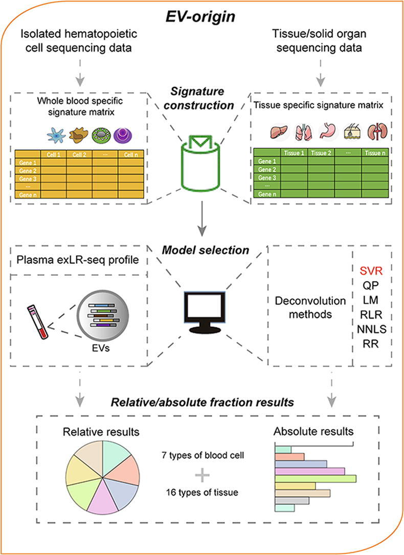
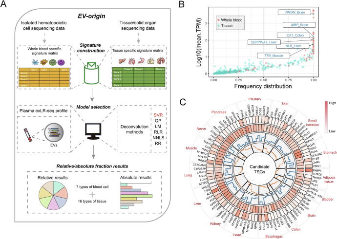
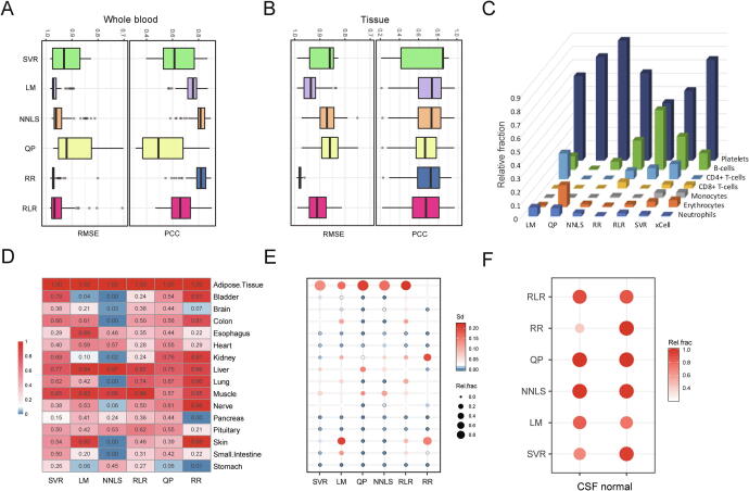
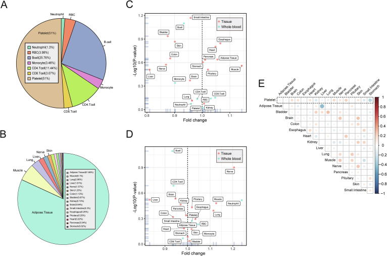
EV-origin: Enumerating the tissue-cellular origin of circulating extracellular vesicles using exLR profile
Overview
The claims and statistics of this paper haven’t been checked yet.
Run the check — free, no account needed. It reads the open-access text and checks the sources it cites.
Integrity signals
Registry
Cites
Statistics
Figures
Verification
Claims the paper makes
The claims and statistics of this paper haven’t been checked yet.
Run the check — free, no account needed. It reads the open-access text and checks the sources it cites.
Claims other papers make about this one
Each is checked against this paper’s own text: does it say what the citing paper says it does?
The cited source doesn’t support the claim as written.
In the citing paper
“Li and colleagues have developed an algorithm to define the origin of extracellular vesicles, based on membrane markers: their results indicated that only 0.65% of the vesicles present in blood come from the brain [52].”
From miR-92a-3p and miR-320a are Upregulated in Plasma Neuron-Derived Extracellular Vesicles of Patients with Frontotemporal Dementia (2025) · cites this paper as [52]
Checked by a reasoning model — may be wrong.
Experimental — accuracy not yet measured
Self-check
Percentages, sums and reported differences re-computed from the paper’s own numbers.
The self-check needs the paper’s full text.
Citations
Cites
The biology , function , and biomedical applications of exosomes
Kalluri, R., & LeBleu, V. S. (2020) · Science · doi:10.1126/science.aau6977 · Cited by 10,770
xCell: digitally portraying the tissue cellular heterogeneity landscape
Aran, D., Hu, Z., & Butte, A. J. (2017) · Genome Biology · doi:10.1186/s13059-017-1349-1 · Cited by 4,397
Self-citationRNA delivery by extracellular vesicles in mammalian cells and its applications
O’Brien, K., Breyne, K., Ughetto, S., Laurent, L. C., & Breakefield, X. O. (2020) · Nature Reviews Molecular Cell Biology · doi:10.1038/s41580-020-0251-y · Cited by 1,811
Adipose-derived circulating miRNAs regulate gene expression in other tissues
Thomou, T., Mori, M. A., Dreyfuss, J. M., Konishi, M., Sakaguchi, M., Wolfrum, C., Rao, T. N., Winnay, J. N., Garcia-Martin, R., Grinspoon, S. K., et al. (2017) · Nature · doi:10.1038/nature21365 · Cited by 1,539
Research works printed but not linked: 46
- 10.1038/nrm.2017.125 · DOI · Paper page
- 10.1016/j.cmet.2019.07.011 · DOI
- 10.1016/j.cell.2019.03.023 · DOI
- 10.1056/NEJMra1704286 · DOI
- Barcelo M., Mata A., Bassas L. and Larriba S.: Exosomal microRNAs in seminal plasma are markers of the origin of azoospermia and can predict the presence of sperm in testicular tissue. Hum Reprod 2018; 33: 1087. · DOI
- Dou Y.Q., Kong P., Li C.L., Sun H.X., Li W.W., Yu Y. et al: Smooth muscle SIRT1 reprograms endothelial cells to suppress angiogenesis after ischemia. Theranostics 2020; 10: 1197. · DOI
- Saenz-Pipaon G., San Martin P., Planell N., Maillo A., Ravassa S., Vilas-Zornoza A. et al: Functional and transcriptomic analysis of extracellular vesicles identifies calprotectin as a new prognostic marker in peripheral arterial disease (PAD). J Extracell Vesicl 2020; 9: 1729646. · DOI
- Santamaria-Martos F., Benitez I.D., Latorre J., Lluch A., Moreno-Navarrete J.M., Sabater M. et al: Comparative and functional analysis of plasma membrane-derived extracellular vesicles from obese vs. nonobese women. Clin Nutr 2019; .
- Zhang J.T., Qin H., Man Cheung F.K., Su J., Zhang D.D., Liu S.Y. et al: Plasma extracellular vesicle microRNAs for pulmonary ground-glass nodules. J. Extracell. Vesicl. 2019; 8: 1663666. · DOI
- 10.1038/s41586-018-0392-8 · DOI · Paper page
- 10.1016/j.immuni.2019.01.010 · DOI
- Zhou R., Chen K.K., Zhang J., Xiao B., Huang Z., Ju C. et al: The decade of exosomal long RNA species: an emerging cancer antagonist. Molecular Cancer 2018; 17: 75. · DOI
- Cheng J., Meng J., Zhu L. and Peng Y.: Exosomal noncoding RNAs in Glioma: biological functions and potential clinical applications. Molecular cancer 2020; 19: 66. · DOI
- Saugstad J.A., Lusardi T.A., Van Keuren-Jensen K.R., Phillips J.I., Lind B., Harrington C.A. et al: Analysis of extracellular RNA in cerebrospinal fluid. Journal of Extracellular Vesicles 2017; 6: 1317577. · DOI
- Yang L., Han B., Zhang Z., Wang S., Bai Y., Zhang Y. et al: Extracellular Vesicle-Mediated Delivery of CircSCMH1 Promotes Functional Recovery in Rodent and Nonhuman Primate Ischemic Stroke Models. Circulation 2020; .
- 10.1016/j.cell.2017.06.031 · DOI
- Li Y., Zhao J., Yu S., Wang Z., He X., Su Y. et al: Extracellular Vesicles Long RNA Sequencing Reveals Abundant mRNA, circRNA, and lncRNA in Human Blood as Potential Biomarkers for Cancer Diagnosis. Clin Chem 2019; 65: 798. · DOI
- 10.1136/gutjnl-2019-318860 · DOI · Paper page
- de Rond L., Libregts S., Rikkert L.G., Hau C.M., van der Pol E., Nieuwland R. et al: Refractive index to evaluate staining specificity of extracellular vesicles by flow cytometry. Journal of Extracellular Vesicles 2019; 8: 1643671. · DOI
- 10.1002/pros.23764 · DOI
- Shang M., Ji J.S., Song C., Gao B.J., Jin J.G., Kuo W.P. et al: Extracellular Vesicles: A Brief Overview and Its Role in Precision Medicine. Methods Mol Biol 2017; 1660: 1. · DOI
- Burrello J., Bolis S., Balbi C., Burrello A., Provasi E., Caporali E. et al: An extracellular vesicle epitope profile is associated with acute myocardial infarction. J Cell Mol Med 2020; .
- Castellani C., Burrello J., Fedrigo M., Burrello A., Bolis S., Di Silvestre D. et al: Circulating extracellular vesicles as non-invasive biomarker of rejection in heart transplant. The Journal of Heart and Lung Transplantation : The Official Publication of the International Society for Heart Transplantation 2020; 39: 1136. · DOI
- 10.1080/09537104.2017.1280602 · DOI
- Vacchi E., Burrello J., Di Silvestre D., Burrello A., Bolis S., Mauri P. et al: Immune profiling of plasma-derived extracellular vesicles identifies Parkinson disease. Neurology(R) Neuroimmunology & Neuroinflammation 2020; 7: (6).
- 10.1016/j.biomaterials.2018.10.034 · DOI
- 10.1186/s13059-016-1070-5 · DOI
- 10.3389/fimmu.2018.01286 · DOI
- 10.1038/nmeth.3337 · DOI · Paper page
- Sturm G., Finotello F. and List M.: Immunedeconv: An R Package for Unified Access to Computational Methods for Estimating Immune Cell Fractions from Bulk RNA-Sequencing Data. Methods Mol Biol 2020; 2120: 223. · DOI
- 10.1093/bioinformatics/bty019 · DOI
- 10.1371/journal.pone.0006098 · DOI
- Gong T., Hartmann N., Kohane I.S., Brinkmann V., Staedtler F., Letzkus M. et al: Optimal deconvolution of transcriptional profiling data using quadratic programming with application to complex clinical blood samples. PLoS ONE 2011; 6: e27156. · DOI
- Kim H., Bi Y., Pal S., Gupta R. and Davuluri R.V.: IsoformEx: isoform level gene expression estimation using weighted non-negative least squares from mRNA-Seq data. BMC Bioinf 2011; 12: 305. · DOI
- Liebner D.A., Huang K. and Parvin J.D.: MMAD: microarray microdissection with analysis of differences is a computational tool for deconvoluting cell type-specific contributions from tissue samples. Bioinformatics 2014; 30: 682. · DOI
- Akat K.M., Lee Y.A., Hurley A., Morozov P., Max K.E., Brown M. et al: Detection of circulating extracellular mRNAs by modified small-RNA-sequencing analysis. JCI Insight 2019; 5.
- van Ravenzwaaij D., Cassey P. and Brown S.D.: A simple introduction to Markov Chain Monte-Carlo sampling. Psychon Bull Rev 2018; 25: 143. · DOI
- 10.1142/S0219720017500172 · DOI
- Baglio S.R., Rooijers K., Koppers-Lalic D., Verweij F.J., Perez Lanzon M., Zini N. et al: Human bone marrow- and adipose-mesenchymal stem cells secrete exosomes enriched in distinctive miRNA and tRNA species. Stem Cell Res Ther 2015; 6: 127. · DOI
- Kong J., Tian H., Zhang F., Zhang Z., Li J., Liu X. et al: Extracellular vesicles of carcinoma-associated fibroblasts creates a pre-metastatic niche in the lung through activating fibroblasts. Molecular Cancer 2019; 18: 175. · DOI
- Li S., Li Y., Chen B., Zhao J., Yu S., Tang Y. et al: exoRBase: a database of circRNA, lncRNA and mRNA in human blood exosomes. Nucleic Acids Res 2018; 46: D106. · DOI
- 10.1016/j.bbcan.2018.06.002 · DOI
- Tao S.C., Guo S.C. and Zhang C.Q.: Platelet-derived Extracellular Vesicles: An Emerging Therapeutic Approach. Int J Biol Sci 2017; 13: 828. · DOI
- 10.1016/j.cell.2019.03.024 · DOI
- Turchinovich A., Drapkina O. and Tonevitsky A.: Transcriptome of Extracellular Vesicles: State-of-the-Art. Front Immunol 2019; 10: 202. · DOI
- 10.1186/1471-2164-14-319 · DOI · Paper page
Cited by
Sorted by citations
Extracellular vesicles: New horizons in neurodegeneration
Chen, J., Tian, C., Xiong, X., Yang, Y., & Zhang, J. (2025) · eBioMedicine · doi:10.1016/j.ebiom.2025.105605 · Cited by 53
Manzini, V., Cappelletti, P., Orefice, N. S., Brentari, I., Rigby, M. J., Lo Giudice, M., Feligioni, M., Rivabene, R., Crestini, A., Manfredi, F., et al. (2025) · Molecular Neurobiology · doi:10.1007/s12035-024-04386-z · Cited by 16
Metadata
- Authors
- Li, Y., He, X., Li, Q., Lai, H., Zhang, H., Hu, Z., Li, Y., & Huang, S.
- Affiliations
- Department of Integrative Oncology, Fudan University Shanghai Cancer Center, and the Shanghai Key Laboratory of Medical Epigenetics, the International Co-laboratory of Medical Epigenetics and Metabolism, Ministry of Science and Technology, Institutes of Biomedical Sciences, Fudan University, Shanghai 200032, China
- Department of Hepatic Surgery, Fudan University Shanghai Cancer Center, Shanghai Medical College, Fudan University, Shanghai 200032, China
- Venue
- Computational and Structural Biotechnology Journal
- Published
- 2020-01
- Volume
- 18
- Pages
- 2851-2859
- ISSN
- 2001-0370
- PMC
- PMC7588739
- Cited by
- 197
- References
- 4
- Language
- en
- Licence
- Funders
- Science and Technology Innovation Plan Of Shanghai Science and Technology Commission (20JC1419000)
- National Natural Science Foundation of China (81672779, 81871939, 81872294)
About these checks
Registry, retraction, Cites and statistics results are computed, not judged, with one exception: a printed reference that no index lists is read once by a model before it counts as a citation error. Claims are extracted by a model and kept only when their quote appears verbatim in the text; each cited source is then checked, with a model’s help, for whether it says what the claim relies on. Those verdicts can be wrong — read the claim’s quote and the cited source before relying on one.
Statistics and arithmetic are recomputed, not judged.
Confidence bands are shown only for fields calibrated on our annotated test set. None is calibrated yet, so no band is shown.
What counts as a claim ›How we measure confidence ›How we check this ›
Are you an author, or do you see a problem with this page? Request a review
How to cite
Cite CoChat for the checks on this page, and the paper for its own findings.
- This page
- CoChat. (2026). Citation check: EV-origin: Enumerating the tissue-cellular origin of circulating extracellular vesicles using exLR profile. https://app.cochat.ai/papers/10.1016/j.csbj.2020.10.002
- The paper
- Li, Y., He, X., Li, Q., Lai, H., Zhang, H., Hu, Z., Li, Y., & Huang, S. (2020). EV-origin: Enumerating the tissue-cellular origin of circulating extracellular vesicles using exLR profile. Computational and Structural Biotechnology Journal. https://doi.org/10.1016/j.csbj.2020.10.002
