

The DOI matches its Crossref record; 48 of the 48 references it cites were found in a registry or an index.
The mitochondria-targeted anti-oxidant MitoQ decreases ischemia-reperfusion injury in a murine syngeneic heart transplant model
Open accessAbstract
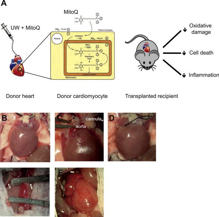
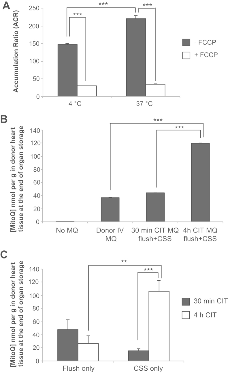
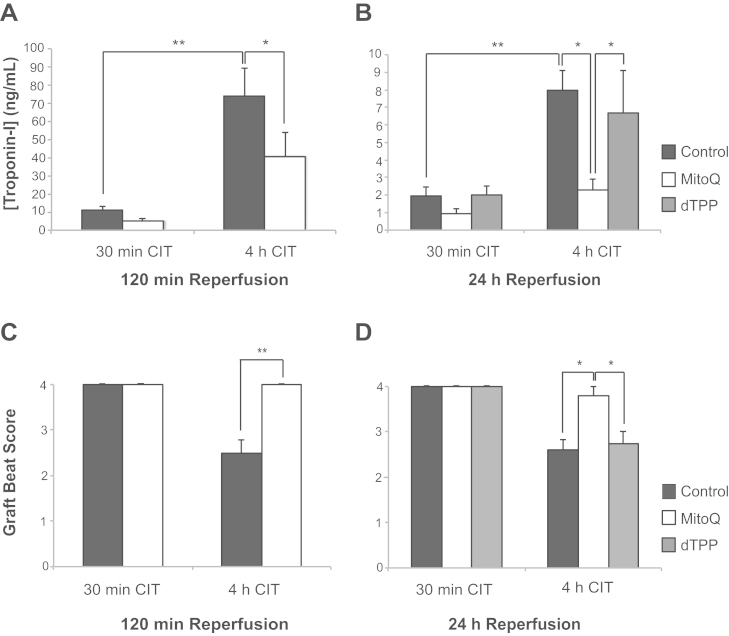
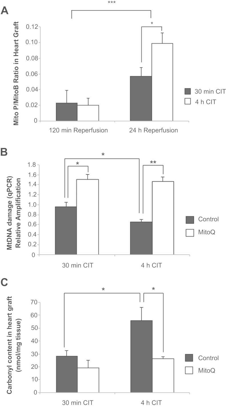
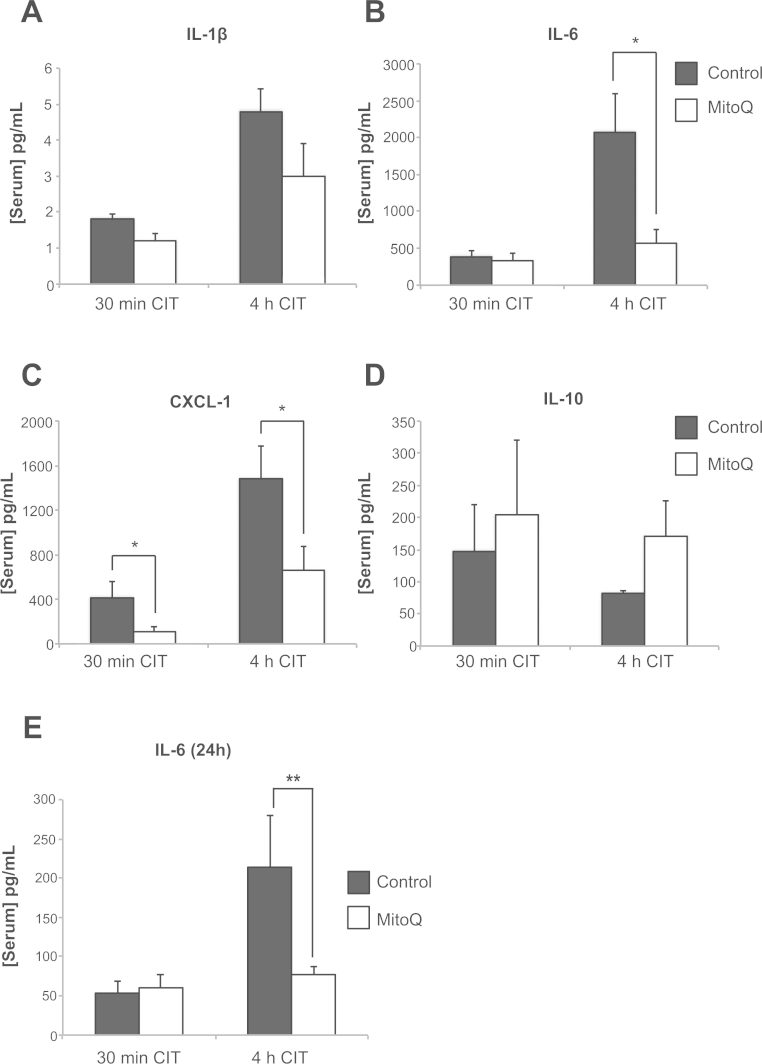
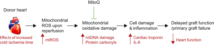
BACKGROUND: Free radical production and mitochondrial dysfunction during cardiac graft reperfusion is a major factor in post-transplant ischemia-reperfusion (IR) injury, an important underlying cause of primary graft dysfunction. We therefore assessed the efficacy of the mitochondria-targeted anti-oxidant MitoQ in reducing IR injury in a murine heterotopic cardiac transplant model. METHODS: Hearts from C57BL/6 donor mice were flushed with storage solution alone, solution containing the anti-oxidant MitoQ, or solution containing the non-anti-oxidant decyltriphenylphosphonium control and exposed to short (30 minutes) or prolonged (4 hour) cold preservation before transplantation. Grafts were transplanted into C57BL/6 recipients and analyzed for mitochondrial reactive oxygen species production, oxidative damage, serum troponin, beating score, and inflammatory markers 120 minutes or 24 hours post-transplant. RESULTS: MitoQ was taken up by the heart during cold storage. Prolonged cold preservation of donor hearts before IR increased IR injury (troponin I, beating score) and mitochondrial reactive oxygen species, mitochondrial DNA damage, protein carbonyls, and pro-inflammatory cytokine release 24 hours after transplant. Administration of MitoQ to the donor heart in the storage solution protected against this IR injury by blocking graft oxidative damage and dampening the early pro-inflammatory response in the recipient. CONCLUSIONS: IR after heart transplantation results in mitochondrial oxidative damage that is potentiated by cold ischemia. Supplementing donor graft perfusion with the anti-oxidant MitoQ before transplantation should be studied further to reduce IR-related free radical production, the innate immune response to IR injury, and subsequent donor cardiac injury.
Overview
The claims and statistics of this paper haven’t been checked yet.
Run the check — free, no account needed. It reads the open-access text and checks the sources it cites.
Integrity signals
Registry
Cites
Statistics
Figures
Verification
Claims the paper makes
The claims and statistics of this paper haven’t been checked yet.
Run the check — free, no account needed. It reads the open-access text and checks the sources it cites.
Claims other papers make about this one
Each is checked against this paper’s own text: does it say what the citing paper says it does?
In the citing paper
“MitoQ has been used extensively in animal models of disease and been shown to be protective against ischemia/reperfusion injury, cardiac hypertrophy, and hypertension (210, 211) among other pathologies.”
From Sources of Vascular Nitric Oxide and Reactive Oxygen Species and Their Regulation (2019) · cites this paper as [211]
Self-check
Percentages, sums and reported differences re-computed from the paper’s own numbers.
The self-check needs the paper’s full text.
Citations
Cites
A role for mitochondria in nlrp3 inflammasome activation
Zhou, R., Yazdi, A. S., Menu, P., & Tschopp, J. (2011) · Nature · doi:10.1038/nature09663 · Cited by 5,602
Circulating mitochondrial DAMPs cause inflammatory responses to injury
Zhang, Q., Raoof, M., Chen, Y., Sumi, Y., Sursal, T., Junger, W., Brohi, K., Itagaki, K., & Hauser, C. J. (2010) · Nature · doi:10.1038/nature08780 · Cited by 3,812
Mitochondria in innate immune responses
West, A. P., Shadel, G. S., & Ghosh, S. (2011) · Nature Reviews Immunology · doi:10.1038/nri2975 · Cited by 1,376
Halestrap, A. P. (2003) · Cardiovascular Research · doi:10.1016/s0008-6363(03)00533-9 · Cited by 1,244
Selective Targeting of a Redox-active Ubiquinone to Mitochondria within Cells
Kelso, G. F., Porteous, C. M., Coulter, C. V., Hughes, G., Porteous, W. K., Ledgerwood, E. C., Smith, R. A. J., & Murphy, M. P. (2001) · Journal of Biological Chemistry · doi:10.1074/jbc.m009093200 · Cited by 1,091
Self-citationKawaguchi, M., Takahashi, M., Hata, T., Kashima, Y., Usui, F., Morimoto, H., Izawa, A., Takahashi, Y., Masumoto, J., Koyama, J., et al. (2011) · Circulation · doi:10.1161/circulationaha.110.982777 · Cited by 884
The mitochondrial permeability transition from in vitro artifact to disease target
Bernardi, P., Krauskopf, A., Basso, E., Petronilli, V., Blalchy‐Dyson, E., Di Lisa, F., & Forte, M. A. (2006) · The FEBS Journal · doi:10.1111/j.1742-4658.2006.05213.x · Cited by 627
Targeting an antioxidant to mitochondria decreases cardiac ischemia-reperfusion injury
Adlam, V. J., Harrison, J. C., Porteous, C. M., James, A. M., Smith, R. A. J., Murphy, M. P., & Sammut, I. A. (2005) · The FASEB Journal · doi:10.1096/fj.05-3718com · Cited by 618
Self-citationSnow, B. J., Rolfe, F. L., Lockhart, M. M., Frampton, C. M., O'Sullivan, J. D., Fung, V., Smith, R. A. J., Murphy, M. P., Taylor, K. M., & On behalf of the Protect Study Group (2010) · Movement Disorders · doi:10.1002/mds.23148 · Cited by 606
Self-citationAnimal and human studies with the mitochondria‐targeted antioxidant MitoQ
Smith, R. A. J., & Murphy, M. P. (2010) · Annals of the New York Academy of Sciences · doi:10.1111/j.1749-6632.2010.05627.x · Cited by 560
Self-citation
Cited by
Sorted by citations
Brand, M. D. (2016) · Free Radical Biology and Medicine · doi:10.1016/j.freeradbiomed.2016.04.001 · Cited by 1,063
Mitochondria as a therapeutic target for common pathologies
Murphy, M. P., & Hartley, R. C. (2018) · Nature Reviews Drug Discovery · doi:10.1038/nrd.2018.174 · Cited by 846
Self-citationA Unifying Mechanism for Mitochondrial Superoxide Production during Ischemia-Reperfusion Injury
Chouchani, E. T., Pell, V. R., James, A. M., Work, L. M., Saeb-Parsy, K., Frezza, C., Krieg, T., & Murphy, M. P. (2016) · Cell Metabolism · doi:10.1016/j.cmet.2015.12.009 · Cited by 752
Self-citationSources of Vascular Nitric Oxide and Reactive Oxygen Species and Their Regulation
Tejero, J., Shiva, S., & Gladwin, M. T. (2019) · Physiological Reviews · doi:10.1152/physrev.00036.2017 · Cited by 541
Mitochondria in acute myocardial infarction and cardioprotection
Ramachandra, C. J. A., Hernández‐Reséndiz, S., Crespo-Avilan, G. E., Lin, Y., & Hausenloy, D. J. (2020) · EBioMedicine · doi:10.1016/j.ebiom.2020.102884 · Cited by 348
Ischemia and Reperfusion Injury in Kidney Transplantation: Relevant Mechanisms in Injury and Repair
Nieuwenhuijs‐Moeke, G. J., Pischke, S. E., Berger, S. P., Sanders, J., Pol, R. A., Struys, M. M. R. F., Ploeg, R. J., & Leuvenink, H. G. D. (2020) · Journal of Clinical Medicine · doi:10.3390/jcm9010253 · Cited by 336
Hypothermic oxygenated perfusion protects from mitochondrial injury before liver transplantation
Schlegel, A. A., Muller, X., Mueller, M., Степанова, А., Kron, P., de Rougemont, O., Muiesan, P., Clavien, P., Galkin, A. S., Meierhofer, D., et al. (2020) · EBioMedicine · doi:10.1016/j.ebiom.2020.103014 · Cited by 254
Hu, Q., Ren, J., Li, G., Wu, J., Wu, X., Wang, G., Gu, G., Ren, H., Hong, Z., & Li, J. (2018) · Cell Death and Disease · doi:10.1038/s41419-018-0436-x · Cited by 183
Succinate accumulation drives ischaemia-reperfusion injury during organ transplantation
Martin, J. L., Costa, A. S. H., Gruszczyk, A. V., Beach, T. E., Allen, F. M., Prag, H. A., Hinchy, E. C., Mahbubani, K. T. A., Hamed, M. O., Tronci, L., et al. (2019) · Nature Metabolism · doi:10.1038/s42255-019-0115-y · Cited by 166
Self-citationParadies, G., Paradies, V., Ruggiero, F. M., & Petrosillo, G. (2018) · American Journal of Physiology-Heart and Circulatory Physiology · doi:10.1152/ajpheart.00028.2018 · Cited by 144
The mitochondrial permeability transition pore in AD 2016: An update
Biasutto, L., Azzolini, M., Szabó, I., & Zoratti, M. (2016) · Biochimica et Biophysica Acta (BBA) - Molecular Cell Research · doi:10.1016/j.bbamcr.2016.02.012 · Cited by 131
Brand, M. D. (2020) · Critical Reviews in Biochemistry and Molecular Biology · doi:10.1080/10409238.2020.1828258 · Cited by 123
Mitochondrial ROS and mitochondria-targeted antioxidants in the aged heart
Bou‐Teen, D., Kaludercic, N., Weissman, D., Turan, B., Maack, C., Lisa, F. D., & Ruiz‐Meana, M. (2021) · Free Radical Biology and Medicine · doi:10.1016/j.freeradbiomed.2021.02.043 · Cited by 119
Escribano‐López, I., Díaz‐Morales, N., Rovira‐Llopis, S., Marañón, A., Orden, S., Álvarez, Á., Bañuls, C., Rocha, M., Murphy, M. P., Hernández‐Mijares, A., et al. (2016) · Redox Biology · doi:10.1016/j.redox.2016.10.017 · Cited by 107
Self-citationMoving Forwards by Blocking Back-Flow
Pell, V. R., Chouchani, E. T., Murphy, M. P., Brookes, P. S., & Krieg, T. (2016) · Circulation Research · doi:10.1161/circresaha.115.306569 · Cited by 104
Self-citationMitochondrial Dysfunction in Cardiovascular Diseases: Potential Targets for Treatment
Yang, J., Guo, Q., Feng, X., Liu, Y., & Zhou, Y. (2022) · Frontiers in Cell and Developmental Biology · doi:10.3389/fcell.2022.841523 · Cited by 105
Cellular Senescence, Mitochondrial Dysfunction, and Their Link to Cardiovascular Disease
Camacho-Encina, M., Booth, L. K., Redgrave, R. E., Folaranmi, O., Spyridopoulos, I., & Richardson, G. D. (2024) · Cells · doi:10.3390/cells13040353 · Cited by 103
Nuzzo, A. M., Camm, E. J., Sferruzzi-Perri, A. N., Ashmore, T. J., Yung, H. W., Cindrová‐Davies, T., Spiroski, A., Sutherland, M. R., Logan, A., Austin‐Williams, S., et al. (2018) · American Journal Of Pathology · doi:10.1016/j.ajpath.2018.07.027 · Cited by 94
Self-citationMitochondria as Therapeutic Targets in Transplantation
Saeb‐Parsy, K., Martin, J. L., Summers, D. M., Watson, C. J. E., Krieg, T., & Murphy, M. P. (2020) · Trends in Molecular Medicine · doi:10.1016/j.molmed.2020.08.001 · Cited by 88
Self-citationMitochondrial mechanisms and therapeutics in ischaemia reperfusion injury
Martin, J. L., Gruszczyk, A. V., Beach, T. E., Murphy, M. P., & Saeb‐Parsy, K. (2018) · Pediatric Nephrology · doi:10.1007/s00467-018-3984-5 · Cited by 88
Self-citationCui, L., Zhou, Q., Zheng, X., Sun, B., & Zhao, S. (2020) · Free Radical Biology and Medicine · doi:10.1016/j.freeradbiomed.2020.09.028 · Cited by 88
Chen, S., Wang, Y., Zhang, H., Chen, R., Lv, F., Li, Z., Jiang, T., Lin, D., Zhang, H., Yang, L., et al. (2019) · International Journal of Biological Sciences · doi:10.7150/ijbs.30193 · Cited by 84
Coenzyme Q10 and Cardiovascular Diseases
Gutierrez-Mariscal, F. M., de la Cruz, S., Torres-Peña, J. D., Alcala-Diaz, J. F., Yubero‐Serrano, E. M., & López‐Miranda, J. (2021) · Antioxidants · doi:10.3390/antiox10060906 · Cited by 82
Mitochondria and ischemia reperfusion injury
Panconesi, R., Widmer, J. D., Carvalho, M. F., Eden, J., Dondossola, D. E., Dutkowski, P., & Schlegel, A. A. (2022) · Current Opinion in Organ Transplantation · doi:10.1097/mot.0000000000001015 · Cited by 81
Preservation Solutions for Kidney Transplantation: History, Advances and Mechanisms
Chen, Y., Shi, J., Xia, T. C., Xu, R., He, X., & Xia, Y. (2019) · Cell Transplantation · doi:10.1177/0963689719872699 · Cited by 77
Zhang, J., Culp, M. L., Craver, J., & Darley-Usmar, V. M. (2018) · Journal of Neurochemistry · doi:10.1111/jnc.14308 · Cited by 74
Covarrubias, A. E., Lecarpentier, É., Lo, A. S., Salahuddin, S., Gray, K. J., Karumanchi, S. A., & Zsengellér, Z. K. (2018) · American Journal Of Pathology · doi:10.1016/j.ajpath.2018.09.007 · Cited by 77
Liver ischemia reperfusion injury: Mechanisms, cellular pathways, and therapeutic approaches
Oliveira, T. H., & Gonçalves, G. K. N. (2025) · International Immunopharmacology · doi:10.1016/j.intimp.2025.114299 · Cited by 79
NADH/NAD+ Redox Imbalance and Diabetic Kidney Disease
Yan, L. (2021) · Biomolecules · doi:10.3390/biom11050730 · Cited by 69
Mitochondrial-Targeted Catalase
Dai, D., Chiao, Y. A., Martin, G. M., Marcinek, D. J., Basisty, N., Quarles, E. K., & Rabinovitch, P. S. (2017) · Progress in molecular biology and translational science · doi:10.1016/bs.pmbts.2016.12.015 · Cited by 65
Mitochondrial redox and TCA cycle metabolite signaling in the heart
Vujić, A., Koo, A., Prag, H. A., & Krieg, T. (2021) · Free Radical Biology and Medicine · doi:10.1016/j.freeradbiomed.2021.02.041 · Cited by 62
Neutrophil-Related Oxidants Drive Heart and Brain Remodeling After Ischemia/Reperfusion Injury
Carbone, F., Bonaventura, A., & Montecucco, F. (2020) · Frontiers in Physiology · doi:10.3389/fphys.2019.01587 · Cited by 62
Mitochondria-Targeted Drug Delivery in Cardiovascular Disease: A Long Road to Nano-Cardio Medicine
Forini, F., Canale, P., Nicolini, G., & Iervasi, G. (2020) · Pharmaceutics · doi:10.3390/pharmaceutics12111122 · Cited by 63
Role of Oxidative DNA Damage and Repair in Atrial Fibrillation and Ischemic Heart Disease
Hu, L., Wang, Z., Carmone, C., Keijer, J., & Zhang, D. (2021) · International Journal of Molecular Sciences · doi:10.3390/ijms22083838 · Cited by 52
Kazzi, M. E., Rayner, B. S., Chami, B., Dennis, J. M., Thomas, S. R., & Witting, P. K. (2020) · Antioxidants and Redox Signaling · doi:10.1089/ars.2019.7928 · Cited by 53
Perspectives on mitochondrial relevance in cardiac ischemia/reperfusion injury
Pedriali, G., Ramaccini, D., Bouhamida, E., Wieckowski, M. R., Giorgi, C., Tremoli, E., & Pinton, P. (2022) · Frontiers in Cell and Developmental Biology · doi:10.3389/fcell.2022.1082095 · Cited by 55
Kadlec, A. O., Chabowski, D. S., Ait‐Aissa, K., Hockenberry, J. C., Otterson, M. F., Durand, M. J., Freed, J. K., Beyer, A., & Gutterman, D. D. (2017) · Hypertension · doi:10.1161/hypertensionaha.117.09289 · Cited by 49
Lung Ischaemia–Reperfusion Injury: The Role of Reactive Oxygen Species
Pak, O., Sydykov, A. S., Kosanovic, D., SCHERMULY, R. T., Dietrich, A., Schröder, K., Brandes, R. P., Gudermann, T. W., Sommer, N., & Weißmann, N. (2017) · Advances in experimental medicine and biology · doi:10.1007/978-3-319-63245-2_12 · Cited by 53
Oxidative Stress after Surgery on the Immature Heart
Fudulu, D. P., & Angelini, G. D. (2016) · Oxidative Medicine and Cellular Longevity · doi:10.1155/2016/1971452 · Cited by 49
Pros and Cons of Use of Mitochondria-Targeted Antioxidants
Plotnikov, E. Y., & Zorov, D. B. (2019) · Antioxidants · doi:10.3390/antiox8080316 · Cited by 44
Arndt, S., Baeza-Garza, C. D., Logan, A., Rosa, T., Wedmann, R., Prime, T. A., Martin, J. L., Saeb‐Parsy, K., Krieg, T., Filipović, M. R., et al. (2017) · Journal of Biological Chemistry · doi:10.1074/jbc.m117.784678 · Cited by 38
Self-citationAnti-ageing interventions for the treatment of cardiovascular disease
Abdellatif, M., Schmid, S., Fuerlinger, A., & Kroemer, G. (2024) · Cardiovascular Research · doi:10.1093/cvr/cvae177 · Cited by 44
Hamed, M. O., Logan, A., Gruszczyk, A. V., Beach, T. E., James, A. M., Dare, A. J., Barlow, A. D., Martin, J. L., Georgakopoulos, N., Gane, A., et al. (2021) · British journal of surgery · doi:10.1093/bjs/znab108 · Cited by 37
Self-citationDJ-1: A promising therapeutic candidate for ischemia-reperfusion injury
Lazzari, F. D., Prag, H. A., Gruszczyk, A. V., Whitworth, A. J., & Bisaglia, M. (2021) · Redox Biology · doi:10.1016/j.redox.2021.101884 · Cited by 36
Toldo, S. A., Quader, M. A., Salloum, F. N., Mezzaroma, E., & Abbate, A. (2016) · International Journal of Molecular Sciences · doi:10.3390/ijms17060958 · Cited by 38
Yang, M.-Y., Fan, Z., Zhang, Z., & Fan, J. (2020) · Journal of Pharmacological Sciences · doi:10.1016/j.jphs.2020.10.007 · Cited by 34
Mao, H., Zhang, Y., Xiong, Y., Zhu, Z., Wang, L., & Liu, X. (2022) · Oxidative Medicine and Cellular Longevity · doi:10.1155/2022/2213503 · Cited by 35
Lin, L., Zhou, Y., Li, Y., Wang, L., Sun, L., Zhou, L., Arai, H., Qi, Y., & Yang, X. (2017) · Biomedicine & Pharmacotherapy · doi:10.1016/j.biopha.2017.02.013 · Cited by 34
Yuan, S., Che, Y., Wang, Z., Xing, K., Xie, X., & Chen, Y. (2023) · Journal of Translational Medicine · doi:10.1186/s12967-023-04763-7 · Cited by 31
Key hepatoprotective roles of mitochondria in liver regeneration
Lamanilao, G. G., Doğan, M., Patel, P. S., Azim, S., Patel, D. S., Bhattacharya, S. K., Eason, J. D., Kuscu, C., Kuscu, C., & Bajwa, A. (2023) · American Journal of Physiology-Gastrointestinal and Liver Physiology · doi:10.1152/ajpgi.00220.2022 · Cited by 30
Bao, Y. W., Hu, C., Wang, B., Liu, X., Wu, Q., Xu, D., Shi, Z., & Sun, C. (2025) · Biology · doi:10.3390/biology14091140 · Cited by 33
Ding, S., Kim, Y., Huang, K., Um, D. J., Jung, Y., & Kong, H. (2024) · Nano Convergence · doi:10.1186/s40580-024-00423-8 · Cited by 30
Chullo, G., Rosello, A. P., Marquez, N., Colmenero, J., Brunet, M., Pera-Román, M., Roselló‐Catafau, J., Bataller, R. A., García‐Valdecasas, J. C., & Fundora, Y. (2024) · International Journal of Molecular Sciences · doi:10.3390/ijms25021117 · Cited by 27
Mitochondria-Targeted Antioxidants for the Treatment of Cardiovascular Disorders
Kim, H. K., & Han, J. (2017) · Advances in experimental medicine and biology · doi:10.1007/978-3-319-55330-6_32 · Cited by 27
Renal Mitochondrial Response to Low Temperature in Non-Hibernating and Hibernating Species
Dugbartey, G. J., Hardenberg, M. C., Kok, W. F., Boerema, A. S., Carey, H. V., Staples, J. F., Henning, R. H., & Bouma, H. R. (2017) · Antioxidants and Redox Signaling · doi:10.1089/ars.2016.6705 · Cited by 25
Paillard, M., Abdellatif, M., Andreadou, I., Bär, C., Bertrand, L., Brundel, B. J. J. M., Chiva‐Blanch, G., Davidson, S. M., Dawson, D. K., Lisa, F. D., et al. (2025) · European Journal of Heart Failure · doi:10.1002/ejhf.3674 · Cited by 29
Mitochondrial oxidative stress, calcium and dynamics in cardiac ischaemia‐reperfusion injury
Rozich, E., Ozkurede, U., Pakkiriswami, S., Gemilere, R., Azarin, S. M., & Liu, J. C. (2025) · The Journal of Physiology · doi:10.1113/jp287770 · Cited by 29
Advances in Mitochondrial Dysfunction and Its Role in Cardiovascular Diseases
Qiu, Y. J., Chang, S., Zeng, Y., & Wang, X. (2025) · Cells · doi:10.3390/cells14201621 · Cited by 27
Respiratory chain signalling is essential for adaptive remodelling following cardiac ischaemia
Szibor, M., Schreckenberg, R., Gizatullina, Z. Z., Dufour, É., Wiesnet, M., Dhandapani, P. K., Dębska–Vielhaber, G., Heidler, J., Wittig, I., Nyman, T. A., et al. (2020) · Journal of Cellular and Molecular Medicine · doi:10.1111/jcmm.15043 · Cited by 24
Self-citationZhou, P., Liu, H., Liu, X., Ling, X., Xiao, Z., Zhu, P., Zhu, Y., Lu, J., & Zheng, S. (2021) · Stem Cell Research & Therapy · doi:10.1186/s13287-020-02114-7 · Cited by 24
Accelerating cryoprotectant diffusion kinetics improves cryopreservation of pancreatic islets
Doležalová, N., Gruszczyk, A. V., Barkan, K., Gamble, J. A., Galvin, S. G., Moreth, T., O’Holleran, K., Mahbubani, K. T. A., Higgins, J., Gribble, F. M., et al. (2021) · Scientific Reports · doi:10.1038/s41598-021-89853-6 · Cited by 21
Self-citationXenotopic expression of alternative oxidase (AOX) to study mechanisms of mitochondrial disease
Viscomi, C. F., Moore, A. L., Zeviani, M., & Szibor, M. (2022) · Biochimica et Biophysica Acta (BBA) - Bioenergetics · doi:10.1016/j.bbabio.2022.148947 · Cited by 18
Endothelial Cells and Mitochondria: Two Key Players in Liver Transplantation
Parente, A., Carvalho, M. F., & Schlegel, A. A. (2023) · International Journal of Molecular Sciences · doi:10.3390/ijms241210091 · Cited by 22
Tao, L., Xue, Y.-F., Sun, F., He, X., Wang, H., Tong, C.-C., Zhang, C., Xu, D., & Chen, X. (2024) · Toxicology and Applied Pharmacology · doi:10.1016/j.taap.2024.116914 · Cited by 20
Enhanced Mitochondrial DNA Repair Resuscitates Transplantable Lungs Donated After Circulatory Death
Tan, Y. B., Pastukh, V. M., Gorodnya, O. M., Mulekar, M. S., Simmons, J. D., Machuca, T., Beaver, T. M., Wilson, G. L., & Gillespie, M. N. (2019) · Journal of Surgical Research · doi:10.1016/j.jss.2019.07.057 · Cited by 20
Sanz, M., Farine, E., Niederberger, P., Méndez‐Carmona, N., Wyss, R. K., Arnold, M., Gulac, P., Fiedler, M., Gressette, M., Garnier, A., et al. (2018) · American Journal of Transplantation · doi:10.1111/ajt.15024 · Cited by 19
Gouda, E., & Babiker, F. A. (2022) · Scientific Reports · doi:10.1038/s41598-022-22097-0 · Cited by 18
Punetha, M., Saini, S., Chaudhary, S., Bala, R., Sharma, M., Kumar, P., Kumar, D., & Yadav, P. (2023) · Tissue and Cell · doi:10.1016/j.tice.2023.102067 · Cited by 20
Tan, Y., Nie, Y., Lei, Z., & Zheng, Z. (2024) · Journal of Materials Chemistry B · doi:10.1039/d4tb00573b · Cited by 17
Westhofen, S., Jelinek, M., Dreher, L., Biermann, D., Martin, J. L., Vitzthum, H., Reichenspurner, H. C., Ehmke, H., & Schwoerer, A. P. (2019) · PLoS ONE · doi:10.1371/journal.pone.0214513 · Cited by 16
Das, J., Sah, A. K., Choudhary, R. K., Elshaikh, R. H., Bhui, U., Chowdhury, S., Abbas, A. M., Shalabi, M. G., Siddique, N. A., Alshammari, R. R., et al. (2025) · Biomedicines · doi:10.3390/biomedicines13071532 · Cited by 18
Lo, S., Blaszak, R. T., & Parajuli, N. (2020) · International Journal of Molecular Sciences · doi:10.3390/ijms21103506 · Cited by 15
Azan, N. I. N., Karim, N. A., Sulaiman, N. S., Ng, M. H., Najib, A. M., Hassan, H., & Alias, E. (2026) · Biomedicines · doi:10.3390/biomedicines14010100 · Cited by 17
Pintye, D., Sziva, R. E., Mastyugin, M., Török, M., Jacas, S., Lo, A. S., Salahuddin, S., & Zsengellér, Z. K. (2023) · Antioxidants · doi:10.3390/antiox12081578 · Cited by 14
Alotaibi, K., Arulkumaran, N., Dyson, A., & Singer, M. (2025) · Clinical Science · doi:10.1042/cs20242074 · Cited by 14
Salahi, E., Amidi, F., Zahiri, Z., Aghahosseini, M., Mashayekhi, F., Abkenari, S. A., Hosseinishenatal, S., & Sobhani, A. (2021) · Archives of Gynecology and Obstetrics · doi:10.1007/s00404-021-06230-4 · Cited by 12
Li, J., Wu, B., Teng, D., Sun, X.-Y., Li, J., Li, J., Zhang, G., & Cai, J. (2018) · Molecular Medicine Reports · doi:10.3892/mmr.2018.8384 · Cited by 11
Du, T., Su, H., Cao, D., Meng, Q., Zhang, M., Liu, Z., & Li, H. (2024) · European journal of medical research · doi:10.1186/s40001-024-02181-z · Cited by 10
Wu, Z., Miao, X., Jiang, Y., Kong, D., Liu, H., Xie, W., Shi, B., & Gong, W. (2023) · Journal of Thoracic and Cardiovascular Surgery · doi:10.1016/j.jtcvs.2023.03.005 · Cited by 10
Gouda, E., & Babiker, F. A. (2020) · All Life · doi:10.1080/26895293.2020.1832921 · Cited by 10
Mitochondrial Targeted Interventions for AgingPreprint
Liu, S. Z., Chiao, Y. A., Rabinovitch, P. S., & Marcinek, D. J. (2023) · Cold Spring Harbor Perspectives in Medicine · doi:10.1101/cshperspect.a041199 · Cited by 11
Xing, Y., Xie, S.-Y., Deng, W., & Tang, Q. (2024) · Biomedicine & Pharmacotherapy · doi:10.1016/j.biopha.2024.116936 · Cited by 11
Haxhikadrija, P., Wu, J. M. F., Hübner, S., Grün, K., Kretzschmar, T., Müller, T., Gräler, M. H., Backsch, C., Weise, A., Klein, E., et al. (2025) · Stem Cell Research & Therapy · doi:10.1186/s13287-025-04340-3 · Cited by 8
Disruption of mitochondrial DNA integrity in cardiomyocyte injury upon ischemia/reperfusion
Hu, S., Tang, X., Zhu, F., Chen, L., Wang, S., Wang, H., Li, P., & Li, Y. (2024) · Genes & Diseases · doi:10.1016/j.gendis.2024.101282 · Cited by 7
Cairns, A. G., McQuaker, S. J., Murphy, M. P., & Hartley, R. C. (2021) · Methods in molecular biology · doi:10.1007/978-1-0716-1262-0_6 · Cited by 6
Self-citationCheng, C., Meng, Y., Pan, Y., Zhang, Y., He, S.-Y. R., Baral, K., & Zhao, M. (2025) · Transplantation · doi:10.1097/tp.0000000000005432 · Cited by 5
Li, G., Chen, J., Wang, Z., Fu, Q., Tian, Z., Liu, Y., Ai, X., Wang, C., Wang, J., Yang, H., et al. (2025) · Biomaterials · doi:10.1016/j.biomaterials.2025.123494 · Cited by 5
Asl, M. M., Rahbarghazi, R., Beheshti, R., Alihemmati, A., Aliparasti, M. R., & Abedelahi, A. (2021) · Europe PMC (PubMed Central) · doi:10.22074/cellj.2021.6877 · Cited by 5
Han, X., Lang, Z., Lv, X., Xing, Y., Hou, M., Tan, Z., Zhang, T., & Leng, Y. (2025) · Molecular Biology Reports · doi:10.1007/s11033-025-10894-5 · Cited by 4
Ischaemia–Reperfusion Injury in Organ Transplantation: Role of Coenzyme Q10
Mantle, D., Cufflin, N., Purcell, T. T., & Hargreaves, I. P. (2025) · Journal of Clinical Medicine · doi:10.3390/jcm14186486 · Cited by 4
Beyond Static Cold Storage: Toward the Next Generation of Tailored Organ Preservation Solutions
Fernandes, F. W., Yildirim, F. S., Horie, H., Karakaya, O. F., Jiao, C., Crasta, G. S., Eshraghi, N., Takase, K., Diwan, T., Oliveira, L., et al. (2025) · International Journal of Molecular Sciences · doi:10.3390/ijms26199515 · Cited by 4
Lu, C., Meng, Z., Tang, S., Wei, H., Hu, Y., Chen, D., Liu, D., Wen, H. M., Dong, K., Na, N., et al. (2025) · Molecular Therapy · doi:10.1016/j.ymthe.2025.02.014 · Cited by 4
Zsengellér, Z. K., Mastyugin, M., Fusco, A. R., Vlocskó, R. B., Costa, M., Ferguson, C., Pintye, D., Sziva, R. E., Salahuddin, S., Young, B. C., et al. (2025) · Current Issues in Molecular Biology · doi:10.3390/cimb47090680 · Cited by 4
Mitochondrial translational control in cardiovascular diseases: from mechanisms to therapies
Deng, Y., Guo, N., Li, D., & Zh, W. (2026) · Redox Biology · doi:10.1016/j.redox.2026.104143 · Cited by 3
Organ Preservation: Cryobiology and Beyond
Woods, E. J., & Mullen, S. F. (2016) · Current Stem Cell Reports · doi:10.1007/s40778-016-0042-8 · Cited by 3
Yang, Z., Zhao, D., Huang, Y., Han, C., Chen, M., Wang, F., Yang, S., Wang, C., & Zhang, S. (2026) · Transplantation · doi:10.1097/tp.0000000000005622 · Cited by 2
Mitochondrial DNA regulation of hepatic ischemia-reperfusion injury and intervention strategies
Shen, R., An, P., Chen, M., Lu, P., Yang, J., Wu, L., & Wang, R. (2026) · Journal of Translational Medicine · doi:10.1186/s12967-026-08155-5 · Cited by 2
Antioxidant Therapy in CVD: Hope or Hype
Nasser, S. A., & Dent, G. (2024) · Oxidative stress in applied basic research and clinical practice · doi:10.1007/978-3-031-62131-4_6 · Cited by 2
Oxidative Stress and Antioxidant Strategies in Human Diseases
Yuan, K., & Zhang, Y. (2021) · Springer eBooks · doi:10.1007/978-981-16-0522-2_1 · Cited by 2
Надеев, А. П., Евгеньевич, К. В., Волков, А. М., Фомичев, А. В., Sirota, D. A., Zhulkov, M. O., Kliver, E. E., Volchek, A. V., Kazanskaya, G. M., & Aidagulova, S. V. (2024) · Russian Journal of Archive of Pathology · doi:10.17116/patol20248605133 · Cited by 1
Metadata
- Authors
- Dare, A. J., Logan, A., Prime, T. A., Rogatti, S., Goddard, M., Bolton, E. M., Bradley, J. A., Pettigrew, G. J., Murphy, M. P., & Saeb-Parsy, K.
- Affiliations
- University of Cambridge (GB)
- National Health Service (GB)
- Corresponding author
- Murphy, Michael P.
- ORCID
- Venue
- The Journal of Heart and Lung Transplantation
- Publisher
- Elsevier BV
- Published
- 2015-11
- Volume
- 34
- Issue
- 11
- Pages
- 1471-1480
- ISSN
- 1053-2498 (ISSN-L)
- PMC
- PMC4626443
- OpenAlex
- W1788023720
- Cited by
- 119
- References
- 48
- Citations by year
- 2026: 10 · 2025: 19 · 2024: 13 · 2023: 8 · 2022: 7 · 2021: 14 · 2020: 13 · 2019: 7 · 2018: 11 · 2017: 8 · 2016: 9
- FWCI
- 9.41
- Type
- journal article
- Language
- en
- Open access
- hybrid
- doi.org
- Licence
- Locations
- The Journal of Heart and Lung Transplantation (publishedVersion, cc-by)
- PubMed Central (publishedVersion, cc-by)
- PubMed (publishedVersion)
- Topics
- Health Sciences › Medicine › Surgery › Transplantation: Methods and Outcomes
- Health Sciences › Medicine › Cardiology and Cardiovascular Medicine › Cardiac Ischemia and Reperfusion
- Health Sciences › Medicine › Surgery › Organ Transplantation Techniques and Outcomes
- Keywords
- MitoQ, mitochondria-targeted antioxidants, ischemia-reperfusion injury, heart transplantation, primary graft dysfunction, oxidative stress, mitochondrial reactive oxygen species, cold ischemia, donor heart preservation, mitochondrial DNA damage, inflammatory cytokines
- MeSH terms
- Animals, Antioxidants, Disease Models, Animal, Female, Male, Mice, Inbred C57BL, Mitochondria, Heart, Organ Preservation, Organophosphorus Compounds, Ubiquinone, Reperfusion Injury, Heart Transplantation, Free Radical Scavengers, Rats, Wistar, Reactive Oxygen Species, Oxidative Stress, Micronutrients, Mice, Rats, Primary Graft Dysfunction
About these checks
Registry, retraction, Cites and statistics results are computed, not judged, with one exception: a printed reference that no index lists is read once by a model before it counts as a citation error. Claims are extracted by a model and kept only when their quote appears verbatim in the text; each cited source is then checked, with a model’s help, for whether it says what the claim relies on. Those verdicts can be wrong — read the claim’s quote and the cited source before relying on one.
Statistics and arithmetic are recomputed, not judged.
Confidence bands are shown only for fields calibrated on our annotated test set. None is calibrated yet, so no band is shown.
What counts as a claim ›How we measure confidence ›How we check this ›
Are you an author, or do you see a problem with this page? Request a review
