

The DOI matches its Crossref record; 6 of the 6 references it cites were found in a registry or an index.
In vivo effects: Methodologies and biokinetics of inhaled nanomaterials
Overview
The claims and statistics of this paper haven’t been checked yet.
Run the check — free, no account needed. It reads the open-access text and checks the sources it cites.
Integrity signals
Registry
Cites
Statistics
Figures

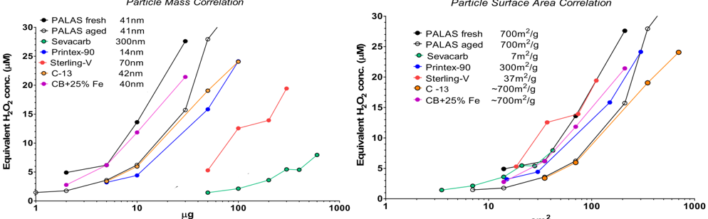
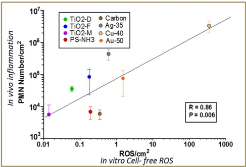
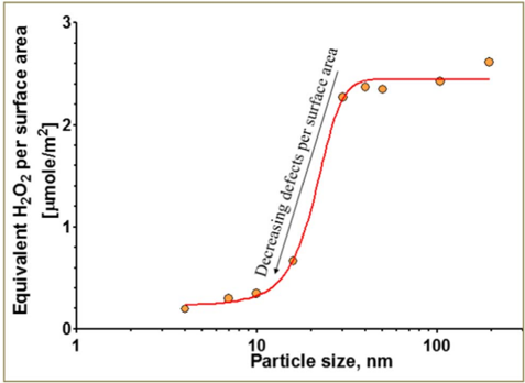
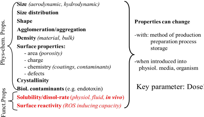
Verification
Claims the paper makes
The claims and statistics of this paper haven’t been checked yet.
Run the check — free, no account needed. It reads the open-access text and checks the sources it cites.
Claims other papers make about this one
Each is checked against this paper’s own text: does it say what the citing paper says it does?
In the citing paper
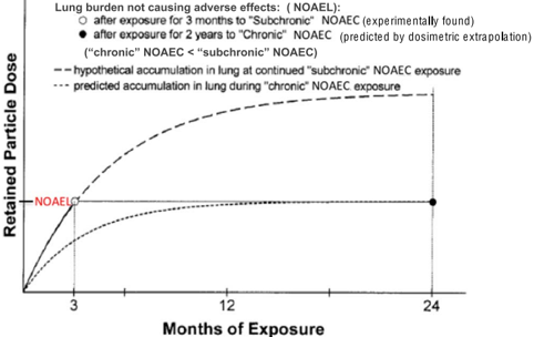
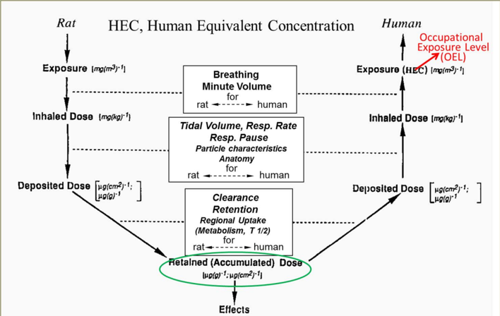
“Results derived from a rodent study can then be the basis for risk extrapolation to human exposure scenarios by deriving a Human Equivalent Concentration (HEC), provided that species differences in respiratory tract dosimetry and retention kinetics are considered [90].”
From Particle toxicology and health - where are we? (2019) · cites this paper as [90]
Self-check
Percentages, sums and reported differences re-computed from the paper’s own numbers.
The self-check needs the paper’s full text.
Citations
Cites
Jiang, J., Oberdörster, G., & Biswas, P. (2009) · Journal of Nanoparticle Research · doi:10.1007/s11051-008-9446-4 · Cited by 1,713
Self-citationTOXICOKINETICS AND EFFECTS OF FIBROUS AND NONFIBROUS PARTICLES
Oberdörster, G. (2002) · Inhalation Toxicology · doi:10.1080/089583701753338622 · Cited by 221
Self-citationApplication of short-term inhalation studies to assess the inhalation toxicity of nanomaterials
Landsiedel, R., Ma-Hock, L., Hofmann, T., Wiemann, M., Strauss, V., Treumann, S., Wohlleben, W., Gröters, S., Wiench, K., & van Ravenzwaay, B. (2014) · Particle and Fibre Toxicology · doi:10.1186/1743-8977-11-16 · Cited by 169
Wiemann, M., Vennemann, A., Sauer, U. G., Wiench, K., Ma-Hock, L., & Landsiedel, R. (2016) · Journal of Nanobiotechnology · doi:10.1186/s12951-016-0164-2 · Cited by 152
Gosens, I., Mathijssen, L. E. A. M., Bokkers, B. G. H., Muijser, H., & Cassee, F. R. (2014) · Nanotoxicology · doi:10.3109/17435390.2013.815814 · Cited by 72
Gillespie, P. A., Kang, G. S., Elder, A., Gelein, R., Chen, L., Moreira, A. L., Koberstein, J., Tchou-Wong, K.-M., Gordon, T., & Chen, L. C. (2010) · Nanotoxicology · doi:10.3109/17435390903470101 · Cited by 70
Research works printed but not linked: 121
- Arts, 2014, A critical appraisal of existing concepts for the grouping of nanomaterials, Regul. Toxicol. Pharmacol., 70, 492, 10.1016/j.yrtph.2014.07.025 · DOI
- Bachler, 2013, A physiologically based pharmacokinetic model for ionic silver and silver nanoparticles, Int. J. Nanomedicine, 8, 3365
- Baisch, 2014, Equivalent titanium dioxide nanoparticle deposition by intratracheal instillation and whole body inhalation: the effect of dose rate on acute respiratory tract inflammation, Part. Fibre Toxicol., 11, 5, 10.1186/1743-8977-11-5 · DOI
- Balasubramanian, 2013, The effect of primary particle size on biodistribution of inhaled gold nano-agglomerates, Biomaterials, 34, 5439, 10.1016/j.biomaterials.2013.03.080 · DOI
- Bernstein, 2005, Comparison of Calidria Chrysotile Asbestos to pure Tremolite: final results of the inhalation biopersistence and histopathology examination following short-term exposure, Inhal. Toxicol., 17, 427, 10.1080/08958370591002012 · DOI · Paper page
- Bello, 2009, Nanomaterials properties vs. biological oxidative damage: implications for toxicity screening and exposure assessment, Nanotoxicology, 3, 249, 10.1080/17435390902989270 · DOI
- Bermudez, 2004, Pulmonary responses of mice, rats, and hamsters to subchronic inhalation of ultrafine titanium dioxide particles, Toxicol. Sci., 77, 347, 10.1093/toxsci/kfh019 · DOI · Paper page
- Braakhuis, 2016, Identification of the appropriate dose metric for pulmonary inflammation of silver nanoparticles in an inhalation toxicity study, Nanotoxicology, 10, 63
- Buesen, 2014, Effects of SiO2, ZrO2, and BaSO4 nanomaterials with or without surface functionalization upon 28-day oral exposure to rats, Arch. Toxicol., 88, 1881, 10.1007/s00204-014-1337-0 · DOI
- Calderon-Garciduenas, 2004, Brain inflammation and Alzheimer's-like pathology in individuals exposed to severe air pollution, Toxicol. Pathol., 32, 650, 10.1080/01926230490520232 · DOI
- Campagnolo, 2017, Silver nanoparticles inhaled during pregnancy reach and affect the placenta and the foetus, Nanotoxicology, 11, 687, 10.1080/17435390.2017.1343875 · DOI
- Charvet, 2014, Characterizing the effective density and primary particle diameter of airborne nanoparticles produced by spark discharge using mobility and mass measurements (tandem DMA/APM), J. Nanopart. Res., 16
- Charvet, 2015, On the importance of density in ELPI data post-treatment, Aerosol Sci. Technol., 49, 1263, 10.1080/02786826.2015.1117568 · DOI
- Cho, 2013, Predictive value of in vitro assays depends on the mechanism of toxicity of metal oxide nanoparticles, Part. Fibre Toxicol., 10, 55, 10.1186/1743-8977-10-55 · DOI
- DeLoid, 2014, Estimating the effective density of engineered nanomaterials for in vitro dosimetry, Nat. Commun., 5, 3514, 10.1038/ncomms4514 · DOI · Paper page
- DeLoid, 2015, Advanced computational modeling for in vitro nanomaterial dosimetry, Part. Fibre Toxicol., 12, 32, 10.1186/s12989-015-0109-1 · DOI
- DeLoid, 2017, Preparation, characterization, and in vitro dosimetry of dispersed, engineered nanomaterials, Nat. Protoc., 12, 355, 10.1038/nprot.2016.172 · DOI
- DeLorme, 2012, Ninety-day inhalation toxicity study with a vapor grown carbon nanofiber in rats, Toxicol. Sci., 128, 449, 10.1093/toxsci/kfs172 · DOI
- Drasler, 2017, In vitro approaches to assess the hazard of nanomaterials, NanoImpact, 10.1016/j.impact.2017.08.002 · DOI
- Drew, 2017, A quantitative framework to group nanoscale and microscale particles by hazard potency to derive occupational exposure limits: proof of concept evaluation, Regul. Toxicol. Pharmacol., 89, 253, 10.1016/j.yrtph.2017.08.003 · DOI
- Driscoll, 2000, Intratracheal instillation as an exposure technique for the evaluation of respiratory tract toxicity: uses and limitations, Toxicol. Sci., 55, 24, 10.1093/toxsci/55.1.24 · DOI · Paper page
- Duffin, 2007, Proinflammogenic effects of low-toxicity and metal nanoparticles in vivo and in vitro: highlighting the role of particle surface area and surface reactivity, Inhal. Toxicol., 19, 849, 10.1080/08958370701479323 · DOI · Paper page
- Eastes, 2000, Estimation of dissolution rate from in vivo studies of synthetic vitreous fibers, Inhal. Toxicol., 12, 1037, 10.1080/08958370050164644 · DOI
- Elder, 2006, Translocation of inhaled ultrafine manganese oxide particles to the central nervous system, Environ. Health Perspect., 114, 1172, 10.1289/ehp.9030 · DOI
- Gangwal, 2011, Informing selection of nanomaterial concentrations for ToxCast in vitro testing based on occupational exposure potential, Environ. Health Perspect., 119, 1539, 10.1289/ehp.1103750 · DOI
- Gao, 2017, Progress towards standardized and validated characterizations for measuring physicochemical properties of manufactured nanomaterials relevant to nano-EHS, NanoImpact
- Geraets, 2014, Tissue distribution and elimination after oral and intravenous administration of different titanium dioxide nanoparticles in rats, Part. Fibre Toxicol., 11, 30, 10.1186/1743-8977-11-30 · DOI
- Godwin, 2015, Nanomaterial categorization for assessing risk potential to facilitate regulatory decision-making, ACS Nano, 9, 3409, 10.1021/acsnano.5b00941 · DOI
- Gomez, 2013, Generation of TiO2 aerosols from liquid suspensions: influence of colloid characteristics, Aerosol Sci. Technol., 47, 1383, 10.1080/02786826.2013.845645 · DOI
- Gosens, 2016, Organ burden and pulmonary toxicity of nano-sized copper (II) oxide particles after short-term inhalation exposure, Nanotoxicology, 1-12
- Graham, 2014, In vivo processing of ceria nanoparticles inside liver: impact on free-radical scavenging activity and oxidative stress, ChemPlusChem, 79, 1083, 10.1002/cplu.201402080 · DOI · Paper page
- Graham, 2017, From dose to response: in vivo nanoparticle processing and potential toxicity, Adv. Exp. Med. Biol., 947, 71, 10.1007/978-3-319-47754-1_4 · DOI · Paper page
- Gregoratto, 2010, Modelling particle retention in the alveolar-interstitial region of the human lungs, J. Radiol. Prot., 30, 491, 10.1088/0952-4746/30/3/005 · DOI
- Guldberg, 1995, Method for determining in-vitro dissolution rates of man-made vitreous fibres, Glastech. Ber. Glass Sci. Technol., 68, 181
- Guldberg, 1998, Measurement of in-vitro fibre dissolution rate at acidic ph, Ann. Occup. Hyg., 42, 233, 10.1016/S0003-4878(98)00026-X · DOI
- He, 2014, Electron spin resonance spectroscopy for the study of nanomaterial-mediated generation of reactive oxygen species, J. Food Drug Anal., 22, 49, 10.1016/j.jfda.2014.01.004 · DOI
- Hellack, 2017, Analytical methods to assess the oxidative potential of nanoparticles: a review, Environ. Sci. Nano, 10.1039/C7EN00346C · DOI
- Henderson, 2014, Inter-laboratory validation of bioaccessibility testing for metals, Regul. Toxicol. Pharmacol., 70, 170, 10.1016/j.yrtph.2014.06.021 · DOI
- Hendrickson, 2016, Size dependent differences in biodistribution of titanium dioxide nanoparticles after sub-acute intragastric administrations to rats, Curr. Nanosci., 12, 00, 10.2174/1573413711666151008013943 · DOI
- Hering, 1995, On-line determination of particle size and density in the nanometer size range, Aerosol Sci. Technol., 23, 155, 10.1080/02786829508965301 · DOI
- Heringa, 2016, Risk assessment of titanium dioxide nanoparticles via oral exposure, including toxicokinetic considerations, Nanotoxicology, 1
- Hinderliter, 2010, ISDD: a computational model of particle sedimentation, diffusion and target cell dosimetry for in vitro toxicity studies, Part. Fibre Toxicol., 7, 36, 10.1186/1743-8977-7-36 · DOI
- Hirn, 2011, Particle size-dependent and surface charge-dependent biodistribution of gold nanoparticles after intravenous administration, Eur. J. Pharm. Biopharm., 77, 407, 10.1016/j.ejpb.2010.12.029 · DOI
- IARC, 2014, Carcinogenicity of fluoro-edenite, silicon carbide fibres and whiskers, and carbon nanotubes, Lancet Oncol., 15, 1427, 10.1016/S1470-2045(14)71109-X · DOI
- ICRP, 1994, 66, 1
- Ivask, 2017, Complete transformation of ZnO and CuO nanoparticles in culture medium and lymphocyte cells during toxicity testing, Nanotoxicology, 10.1080/17435390.2017.1282049 · DOI
- Jang, 2001, Controlled synthesis of titanium dioxide nanoparticles in a modified diffusion flame reactor, Mater. Res. Bull., 36, 627, 10.1016/S0025-5408(01)00552-9 · DOI
- Jiang, 2008, Does nanoparticle activity depend upon size and crystal phase?, Nanotoxicology, 2, 33, 10.1080/17435390701882478 · DOI
- Kasai, 2015, Thirteen-week study of toxicity of fiber-like multi-walled carbon nanotubes with whole-body inhalation exposure in rats, Nanotoxicology, 9, 413, 10.3109/17435390.2014.933903 · DOI
- Kasai, 2016, Lung carcinogenicity of inhaled multi-walled carbon nanotube in rats, Part. Fibre Toxicol., 13, 53, 10.1186/s12989-016-0164-2 · DOI
- Keller, 2014, Time course of lung retention and toxicity of inhaled particles: short-term exposure to nano-ceria, Arch. Toxicol., 88, 2033, 10.1007/s00204-014-1349-9 · DOI · Paper page
- Konduru, 2014, Biokinetics and effects of barium sulfate nanoparticles, Part. Fibre Toxicol., 11, 55, 10.1186/s12989-014-0055-3 · DOI · Paper page
- Konduru, 2015, Silica coating influences the corona and biokinetics of cerium oxide nanoparticles, Part. Fibre Toxicol., 12, 31, 10.1186/s12989-015-0106-4 · DOI
- Kreyling, 1990, Interspecies comparison of lung clearance of “insoluble” particles, J. Aerosol Med., 3, s-93, 10.1089/jam.1990.3.Suppl_1.S-93 · DOI
- Kreyling, 1990, In vitro dissolution of uniform cobalt oxide particles by human and canine alveolar macrophages, Am. J. Respir. Cell Mol. Biol., 2, 413, 10.1165/ajrcmb/2.5.413 · DOI
- Kreyling, 1991, Interspecies comparison of phagolysosomal pH in alveolar macrophages, Inhal. Toxicol., 3, 91, 10.3109/08958379109145276 · DOI
- Kreyling, 2014, Air–blood barrier translocation of tracheally instilled gold nanoparticles inversely depends on particle size, ACS Nano, 8, 222, 10.1021/nn403256v · DOI · Paper page
- Kreyling, 2017, Quantitative biokinetics of titanium dioxide nanoparticles after intravenous injection in rats (Part 1), Nanotoxicology, 1
- Kreyling, 2017, Quantitative biokinetics of titanium dioxide nanoparticles after oral application in rats (Part 2), Nanotoxicology, 1
- Kreyling, 2017, Quantitative biokinetics of titanium dioxide nanoparticles after intratracheal instillation in rats (Part 3), Nanotoxicology, 1
- Kuhlbusch, 2017, Nanomaterial exposures for worker, consumer and the public, NanoImpact
- Lundborg, 1985, Dissolution of metals by human and rabbit alveolar macrophages, Br. J. Ind. Med., 42, 642
- Lundborg, 1995, Phagolysosomal morphology and dissolution of cobalt oxide particles by human and rabbit alveolar macrophages, Exp. Lung Res., 21, 51, 10.3109/01902149509031744 · DOI
- Maher, 2016, Magnetite pollution nanoparticles in the human brain, Proc. Natl. Acad. Sci. U. S. A., 89, 7683
- Ma-Hock, 2009, Inhalation toxicity of multiwall carbon nanotubes in rats exposed for 3 months, Toxicol. Sci., 112, 468, 10.1093/toxsci/kfp146 · DOI · Paper page
- Ma-Hock, 2013, Comparative inhalation toxicity of multi-wall carbon nanotubes, graphene, graphite nanoplatelets and low surface carbon black, Part. Fibre Toxicol., 10, 23, 10.1186/1743-8977-10-23 · DOI
- Malmgren, 1978, Uptake and retrograde axonal transport of various exogenous macromolecules in normal and crushed hypoglossal nerves, Brain Res., 153, 477, 10.1016/0006-8993(78)90333-5 · DOI
- Maricq, 2004, The effective density and fractal dimension of soot particles from premixed flames and motor vehicle exhaust, Aerosol Sci., 35, 1251, 10.1016/j.jaerosci.2004.05.002 · DOI
- Methner, 2010, Nanoparticle Emission Assessment Technique (NEAT) for the identification and measurement of potential inhalation exposure to engineered nanomaterials—part B: results from 12 field studies, J. Occup. Environ. Hyg., 7, 163, 10.1080/15459620903508066 · DOI
- Meuller, 2012, Review of spark discharge generators for production of nanoparticle aerosols, Aerosol Sci. Technol., 46, 1256, 10.1080/02786826.2012.705448 · DOI
- Miller, 2013, Estimating lung burdens based on individual particle density estimated from scanning electron microscopy and cascade impactor samples, Inhal. Toxicol., 25, 813, 10.3109/08958378.2013.850562 · DOI
- Miller, 2016, Improvements and additions to multiple path particle dosimetry model, J. Aerosol Sci., 99, 14, 10.1016/j.jaerosci.2016.01.018 · DOI
- Morrow, 1992, Contemporary issues in toxicology — dust overloading of the lungs: update and appraisal, Toxicol. Appl. Pharmacol., 113, 1, 10.1016/0041-008X(92)90002-A · DOI
- Mortensen, 2008, In vivo skin penetration of quantum dot nanoparticles in the murine model: the effect of UVR, Nano Lett., 8, 2779, 10.1021/nl801323y · DOI
- Nel, 2013, A multi-stakeholder perspective on the use of alternative test strategies for nanomaterial safety assessment, ACS Nano, 7, 6422, 10.1021/nn4037927 · DOI · Paper page
- Nguyen-Legros, 1981, Evolution du lieu de stockage du dextran-fer transporté par le flux axonal retrograde dans le système nerveux central du rat, Acta Neuropathol., 54, 101, 10.1007/BF00689402 · DOI
- Noël, 2013, Generating nano-aerosols from TiO2 (5nm) nanoparticles showing different agglomeration states. application to toxicological studies, J. Occup. Environ. Hyg., 10, 86, 10.1080/15459624.2012.748340 · DOI
- Nurkiewicz, 2009, Pulmonary nanoparticle exposure disrupts systemic microvascular nitric oxide signaling, Toxicol. Sci., 110, 191, 10.1093/toxsci/kfp051 · DOI
- Nyberg, 1989, Intraphagosomal pH in alveolar macrophages studied with fluorescein-labeled amorphous silica particles, Exp. Lung Res., 15, 49, 10.3109/01902148909069608 · DOI
- Nyberg, 1992, Phagolysosomal pH in alveolar macrophages, Environ. Health Perspect., 97, 149, 10.1289/ehp.9297149 · DOI
- Oberdörster, 1989, Dosimetric principles for extrapolating results of rat inhalation studies to humans, using nickel as an example, Health Phys., 57, 213, 10.1097/00004032-198907001-00027 · DOI
- Oberdorster, 1992, Role of the alveolar macrophage in lung injury: studies with ultrafine particles, Environ. Health Perspect., 97, 193
- Oberdörster, 2010, Safety assessment for nanotechnology and nanomedicine: concepts of nanotoxicology, J. Intern. Med., 267, 89, 10.1111/j.1365-2796.2009.02187.x · DOI
- Oberdörster, 2012, Nanotoxicology: in vitro - in vivo dosimetry, EHP Correspondence, 120, A13
- Oberdörster, 2005, Nanotoxicology: an emerging discipline evolving from studies of ultrafine particles, EHP, 113, 823, 10.1289/ehp.7339 · DOI · Paper page
- Oberdörster, 2015, Inhalation exposure to carbon nanotubes (CNT) and carbon nanofibers (CNF): methodology and dosimetry, J. Toxicol. Environ. Health Part B, 18, 121, 10.1080/10937404.2015.1051611 · DOI · Paper page
- Olsson, 1978, A simple histochemical method for double labelling of neurons by retrorade axonal transport, Neurosci. Lett., 8, 265, 10.1016/0304-3940(78)90133-7 · DOI
- Olsson, 1981, Neuronal uptake of iron: somatopetal axonal transport and fate of cationized and native ferritin and iron-dextran after intramuscular injections, Neuropathol. Appl. Neurobiol., 7, 85, 10.1111/j.1365-2990.1981.tb00078.x · DOI
- Pal, 2014, Screening for oxidative damage by engineered nanomaterials: a comparative evaluation of FRAS and DCFH, J. Nanopart. Res., 16, 2167, 10.1007/s11051-013-2167-3 · DOI
- Park, 2003, A closure study of aerosol mass concentration measurements: comparison of values obtained with filters and by direct measurements of mass distributions, Atmos. Environ., 37, 1223, 10.1016/S1352-2310(02)01016-6 · DOI
- Pauluhn, 2010, Multi-walled carbon nanotubes (Baytubes): approach for derivation of occupational exposure limit, Regul. Toxicol. Pharmacol., 57, 78, 10.1016/j.yrtph.2009.12.012 · DOI
- Pekkanen, 2002, Particulate air pollution and risk of ST-segment depression during repeated submaximal exercise tests among subjects with coronary heart disease. The exposure and risk assessment for fine and ultrafine particles in ambient air (ULTRA) study, Circulation, 106, 933, 10.1161/01.CIR.0000027561.41736.3C · DOI
- Potter, 1991, Glass fiber dissolution in a physiological saline solution, Glastech. Ber., 64, 16
- Prow, 2011, Nanoparticles and microparticles for skin drug delivery, Adv. Drug Deliv. Rev., 63, 470, 10.1016/j.addr.2011.01.012 · DOI
- Riebeling, 2016, A redox proteomics approach to investigate the mode of action of nanomaterials, Toxicol. Appl. Pharmacol., 299, 24, 10.1016/j.taap.2016.01.019 · DOI
- Rinderknecht, 2007, Surface functionalization affects the role of nanoparticle disposition, Am. J. Respir. Crit. Care Med., 175, A246
- Rushton, 2010, Concept of assessing nanoparticle hazards considering nanoparticle dosemetric and chemical/biological response-metrics, J. Toxicol. Environ. Health A, 73, 445, 10.1080/15287390903489422 · DOI · Paper page
- Ryman-Rasmussen, 2009, Inhaled carbon nanotubes reach the subpleural tissue in mice, Nat. Nanotechnol., 4, 747, 10.1038/nnano.2009.305 · DOI
- Sayre, 2017, Methods and data for regulatory risk assessment of nanomaterials: questions for an expert consultation, NanoImpact, 8, 20, 10.1016/j.impact.2017.07.001 · DOI
- Schleh, 2013, Biodistribution of inhaled gold nanoparticles in mice and the influence of surfactant protein D, J. Aerosol Med. Pulm. Drug Deliv., 26, 24, 10.1089/jamp.2011.0951 · DOI
- Sebastian, 2002, test guideline: in-vitro acellular dissolution of man-made vitreous silicate fibres, Glas. Sci. Technol., 75, 263
- Shatkin, 2016, Comment on Shvedova et al. (2016), “gender differences in murine pulmonary responses elicited by cellulose nanocrystals”, Part. Fibre Toxicol., 13, 59, 10.1186/s12989-016-0170-4 · DOI
- Shi, 2017, The roles of surface chemistry, dissolution rate, and delivered dose in the cytotoxicity of copper nanoparticles, Nanoscale, 9, 4739, 10.1039/C6NR09102D · DOI
- Shvedova, 2008, Inhalation vs. aspiration of single-walled carbon nanotubes in C57BL/6 mice: inflammation, fibrosis, oxidative stress, and mutagenesis, Am. J. Physiol. Lung Cell. Mol. Physiol., 295, L552, 10.1152/ajplung.90287.2008 · DOI
- Slikker, 2004, Dose-dependent transitions in mechanisms of toxicity, Toxicol. Appl. Pharmacol., 201, 203, 10.1016/j.taap.2004.06.019 · DOI
- Snipes, 1983, Retention of monodisperse or polydisperse aluminosilicate particle inhaled by dogs, rats and mice, Toxicol. Appl. Pharmacol., 69, 345, 10.1016/0041-008X(83)90258-2 · DOI
- Spencer, 2007, Simultaneous measurement of the effective density and chemical composition of ambient aerosol particles, Environ. Sci. Technol., 41, 1303, 10.1021/es061425+ · DOI
- Stapleton, 2012, Impairment of coronary arteriolar endothelium-dependent dilation after multi-walled carbon nanotube inhalation: a time-course study, Int. J. Mol. Sci., 13, 13781, 10.3390/ijms131113781 · DOI
- Stefaniak, 2005, Characterization of phagolysosomal simulant fluid for study of beryllium aerosol particle dissolution, Toxicol. in vitro, 19, 123, 10.1016/j.tiv.2004.08.001 · DOI
- Stone, 2017, Nanomaterials vs ambient ultrafine particles: an opportunity to exchange toxicology knowledge, Environ. Health Perspect., 10.1289/EHP424 · DOI · Paper page
- Sung, 2009, Subchronic inhalation toxicity of silver nanoparticles, Toxicol. Sci., 108, 452, 10.1093/toxsci/kfn246 · DOI
- Suzui, 2016, Multiwalled carbon nanotubes intratracheally instilled into the rat lung induce development of pleural malignant mesothelioma and lung tumors, Cancer Sci., 107, 924, 10.1111/cas.12954 · DOI
- Tsai, 2009, Characterization and evaluation of nanoparticle release during the synthesis of single-walled and multiwalled carbon nanotubes by chemical vapor deposition, Environ. Sci. Technol., 43, 6017, 10.1021/es900486y · DOI
- Utembe, 2015, Dissolution and biodurability: important parameters needed for risk assessment of nanomaterials, Part. Fibre Toxicol., 12, 11, 10.1186/s12989-015-0088-2 · DOI
- Wang, 2015, Characteristics of airborne fractal-like agglomerates of carbon nanotubes, Carbon, 93, 441, 10.1016/j.carbon.2015.05.079 · DOI
- Warheit, 2005, Comparative pulmonary toxicity inhalation and instillation studies with different TiO2 particle formulations: impact of surface treatments on particle toxicity, Toxicol. Sci., 88, 514, 10.1093/toxsci/kfi331 · DOI
- Warheit, 2006, Pulmonary instillation studies with nanoscale TiO2 rods and dots in rats: toxicity is not dependent upon particle size and surface area, Toxicol. Sci., 91, 227, 10.1093/toxsci/kfj140 · DOI
- Weldon, 2016, Occupational exposure limit for silver nanoparticles: considerations on the derivation of a general health-based value, Nanotoxicology, 10, 945, 10.3109/17435390.2016.1148793 · DOI
- Wohlleben, 2013, Nanospecific guidance in REACH: a comparative physical-chemical characterization of 15 materials with methodical correlations, J. Ceram. Sci. Technol., 04, 93
- van der Zande, 2012, Distribution, elimination, and toxicity of silver nanoparticles and silver ions in rats after 28-day oral exposure, ACS Nano, 6, 7427, 10.1021/nn302649p · DOI
- Zareba, 2009, ECG parameters and exposure to carbon ultrafine particles in young healthy subjects, Inhal. Toxicol., 21, 223, 10.1080/08958370802492407 · DOI
Cited by
Sorted by citations
Particle toxicology and health - where are we?
Riediker, M., Zink, D., Kreyling, W., Oberdörster, G., Elder, A., Graham, U., Lynch, I., Duschl, A., Ichihara, G., Ichihara, S., et al. (2019) · Particle and Fibre Toxicology · doi:10.1186/s12989-019-0302-8 · Cited by 237
Self-citation
Metadata
- Authors
- Oberdörster, G., & Kuhlbusch, T. A. J.
- Venue
- NanoImpact
- Publisher
- Elsevier BV
- Published
- 2018-04
- Volume
- 10
- Pages
- 38-60
- ISSN
- 2452-0748
- Cited by
- 88
- References
- 6
- Language
- en
- Licence
- Funders
- EU H2020 (646325)
About these checks
Registry, retraction, Cites and statistics results are computed, not judged, with one exception: a printed reference that no index lists is read once by a model before it counts as a citation error. Claims are extracted by a model and kept only when their quote appears verbatim in the text; each cited source is then checked, with a model’s help, for whether it says what the claim relies on. Those verdicts can be wrong — read the claim’s quote and the cited source before relying on one.
Statistics and arithmetic are recomputed, not judged.
Confidence bands are shown only for fields calibrated on our annotated test set. None is calibrated yet, so no band is shown.
What counts as a claim ›How we measure confidence ›How we check this ›
Are you an author, or do you see a problem with this page? Request a review
