

The DOI matches its Crossref record; 73 of the 73 references it cites were found in a registry or an index.
Muscle insulin sensitivity and glucose metabolism are controlled by the intrinsic muscle clock
Open accessAbstract
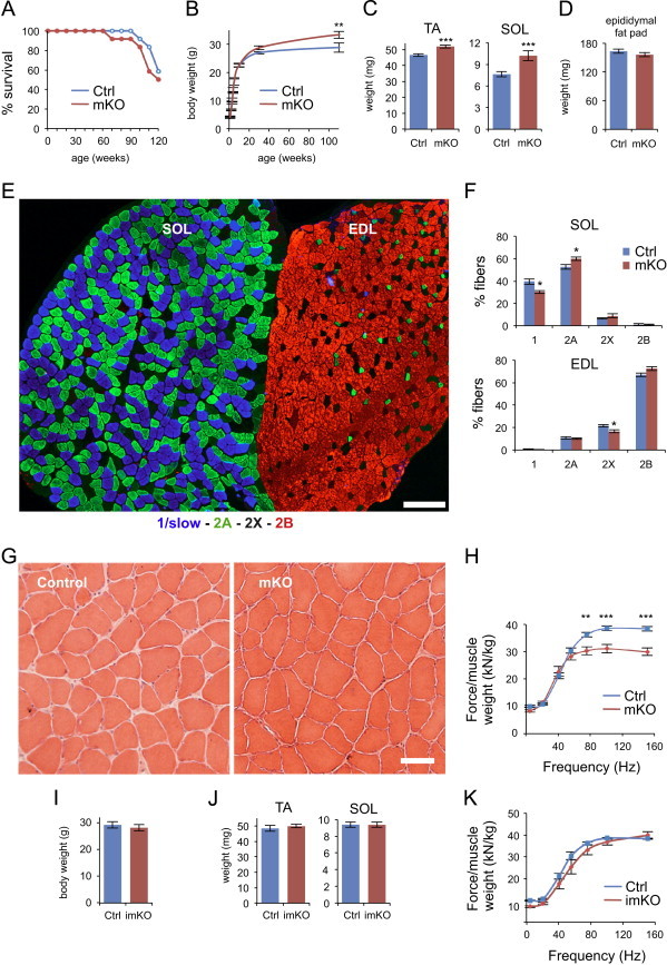
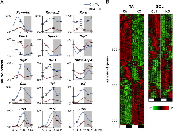
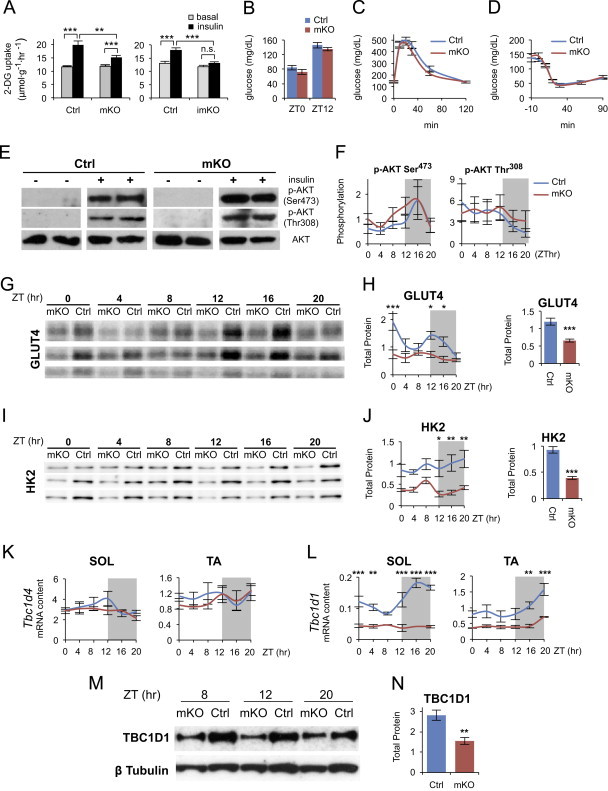
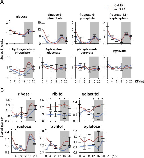
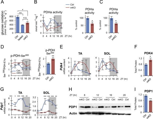
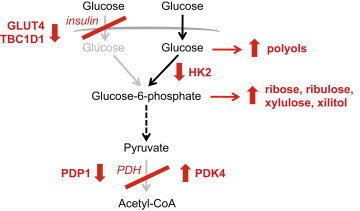
Circadian rhythms control metabolism and energy homeostasis, but the role of the skeletal muscle clock has never been explored. We generated conditional and inducible mouse lines with muscle-specific ablation of the core clock gene Bmal1. Skeletal muscles from these mice showed impaired insulin-stimulated glucose uptake with reduced protein levels of GLUT4, the insulin-dependent glucose transporter, and TBC1D1, a Rab-GTPase involved in GLUT4 translocation. Pyruvate dehydrogenase (PDH) activity was also reduced due to altered expression of circadian genes Pdk4 and Pdp1, coding for PDH kinase and phosphatase, respectively. PDH inhibition leads to reduced glucose oxidation and diversion of glycolytic intermediates to alternative metabolic pathways, as revealed by metabolome analysis. The impaired glucose metabolism induced by muscle-specific Bmal1 knockout suggests that a major physiological role of the muscle clock is to prepare for the transition from the rest/fasting phase to the active/feeding phase, when glucose becomes the predominant fuel for skeletal muscle.
Overview
The claims and statistics of this paper haven’t been checked yet.
Run the check — free, no account needed. It reads the open-access text and checks the sources it cites.
Integrity signals
Registry
Cites
Statistics
Figures
Verification
Claims the paper makes
The claims and statistics of this paper haven’t been checked yet.
Run the check — free, no account needed. It reads the open-access text and checks the sources it cites.
Claims other papers make about this one
Each is checked against this paper’s own text: does it say what the citing paper says it does?
In the citing paper
“Disruption of the muscle clock has also been shown to have significant impact on glucose intolerance and insulin resistance with very small changes in fiber types (Dyar et al., 2014; Schroder et al., 2015; Harfmann et al., 2016).”
From Myosteatosis in the Context of Skeletal Muscle Function Deficit: An Interdisciplinary Workshop at the National Institute on Aging (2020) · cites this paper as Dyar et al., 2014
In the citing paper
“In addition, prolonged loss of Bmal1 function was also associated with increased fibrosis within the muscle.”
From Myosteatosis in the Context of Skeletal Muscle Function Deficit: An Interdisciplinary Workshop at the National Institute on Aging (2020) · cites this paper as Dyar et al., 2014
In the citing paper
“These animals showed reduced protein levels of GLUT4 and an impaired insulin-stimulated glucose uptake in their muscles.”
From Circadian control of glucose metabolism (2014) · cites this paper as [153]
In the citing paper
“For example, the hepatic clock regulates fasting glycemic control and glucose clearance (8), the pancreatic clock regulates insulin secretion and its response to glucose (9-11), the adipose clock regulates lipid storage and mobilization (12,13), and the skeletal muscle clock regulates glucose uptake and metabolism (14).”
From Circadian rhythms, metabolism, and chrononutrition in rodents and humans (2016) · cites this paper as [14]
Self-check
Percentages, sums and reported differences re-computed from the paper’s own numbers.
The self-check needs the paper’s full text.
Citations
Cites
Subramanian, A., Tamayo, P., Mootha, V. K., Mukherjee, S., Ebert, B. L., Gillette, M. A., Paulovich, A., Pomeroy, S. L., Golub, T. R., Lander, E. S., et al. (2005) · Proceedings of the National Academy of Sciences · doi:10.1073/pnas.0506580102 · Cited by 53,625 · cited 5×
“jsp) [27] and gene sets from the KEGG database.”
BackgroundMethodInfluentialExploration, normalization, and summaries of high density oligonucleotide array probe level data
Irizarry, R. A. (2003) · Biostatistics · doi:10.1093/biostatistics/4.2.249 · Cited by 10,603 · cited 2×
“Expression values were generated from fluorescence signals using the robust multi-array average procedure (RMA) [23].”
MethodObesity and Metabolic Syndrome in Circadian Clock Mutant Mice
Turek, F. W., Joshu, C., Kohsaka, A., Lin, E., Ivanova, G., McDearmon, E., Laposky, A., Losee-Olson, S., Easton, A., Jensen, D. R., et al. (2005) · Science · doi:10.1126/science.1108750 · Cited by 2,485 · cited 1×
“Global inactivation of different clock genes leads to altered glucose metabolism: Clock mutant mice display hyperglycemia, obesity and metabolic syndrome [9]; Per1/Per2 double knockout mice and Cry1/Cr2 double knockout mice exhibit glucose”
BackgroundDibner, C., Schibler, U., & Albrecht, U. (2010) · Annual Review of Physiology · doi:10.1146/annurev-physiol-021909-135821 · Cited by 2,421 · cited 2×
“In addition to the core clock genes, all tissues contain a large number of oscillating genes, most of which are tissue-specific and regulated either directly by the core oscillator or by extrinsic circadian signals [7].”
BackgroundEvolving gene/transcript definitions significantly alter the interpretation of GeneChip data
Dai, M. (2005) · Nucleic Acids Research · doi:10.1093/nar/gni179 · Cited by 1,807 · cited 1×
“Speci fi cally, intensity levels have been background adjusted, normalized using quantile normalization, and log 2 expression values calculated using median polish summarization and Entrez custom chip de fi nition fi les for mouse arrays (version 14.1.0) [24].”
MethodInfluentialDeFronzo, R. A., Jacot, E., Jequier, E., Maeder, E., Wahren, J., & Felber, J. P. (1981) · Diabetes · doi:10.2337/diab.30.12.1000 · Cited by 1,770 · cited 1×
“Skeletal muscle is a major site of insulin-stimulated glucose disposal [56] and muscle insulin resistance has been considered to be one of the earliest factors in the pathogenesis of the metabolic syndrome [57].”
BackgroundOccupational and Environmental Medicine
Rosenstock, L., Rest, K. M., BENSON, J. A., Cannella, J. M., Cohen, J. J., Cullen, M. R., Davidoff, F. F., Landrigan, P. J., Reynolds, R. C., Clever, L. H., et al. (1991) · New England Journal of Medicine · doi:10.1056/nejm199109263251305 · Cited by 1,627
Mop3 is an essential component of the master circadian pacemaker in mammals.
Bunger, M. K., Wilsbacher, L. D., Moran, S. M., Clendenin, C., Radcliffe, L. A., Hogenesch, J. B., Simon, M. C., Takahashi, J. S., & Bradfield, C. A. (2000) · Cell · doi:10.1016/s0092-8674(00)00205-1 · Cited by 1,621 · cited 3×
“Global Bmal1 knockout causes not only loss of circadian rhythm of locomotor activity but also reduced total activity levels [42] and rescue experiments in which BMAL1 was constitutively expressed in skeletal muscle of Bmal1 null mice showed that musclespecific rescue restored activity levels, though not circadian rhyt…”
BackgroundDisruption of the clock components CLOCK and BMAL1 leads to hypoinsulinaemia and diabetes
Marcheva, B., Ramsey, K. M., Buhr, E. D., Kobayashi, Y., Su, H., Ko, C. H., Ivanova, G., Omura, C., Mo, S., Vitaterna, M. H., et al. (2010) · Nature · doi:10.1038/nature09253 · Cited by 1,525 · cited 1×
“Selective knockout of Bmal1 in the liver causes hypoglycemia [10], while ablation of the same gene in the endocrine pancreas leads to fasting hyperglycemia and severe glucose intolerance [15].”
BackgroundLipid-induced insulin resistance: unravelling the mechanism
Samuel, V. T., Petersen, K. F., & Shulman, G. I. (2010) · The Lancet · doi:10.1016/s0140-6736(10)60408-4 · Cited by 1,189 · cited 1×
“What is the cause of the altered response to insulin in muscles from Bmal1 mKO mice? The canonical insulin signaling pathway is apparently not responsible for this change, since AKT phosphorylation, which is reduced in obesity and diabetes [60], was unchanged, and was similarly increased by insulin in muscles from con…”
Background
Cited by
Sorted by citations
Items 401–458 of 458
Ghosh, S. (2018) · EngagedScholarship @ Cleveland State University (Cleveland State University)
Sleep, dietary patterns and metabolic health in UK adults
Potter, G. D. (2018) · White Rose eTheses Online (University of Leeds, The University of Sheffield, University of York)
Kim, H.-K., & Shibata, S. (2020) · 体力科學 · doi:10.7600/jspfsm.69.401
Circadian clock and liver energy metabolism
Gao, W., Shu, Y.-Y., Ye, J., & Pan, X. (2020) · World Chinese Journal of Digestology · doi:10.11569/wcjd.v28.i20.1025
Circadian Secretion of Glucagon-like Peptide-1
Martchenko, A. (2020) · TSpace (University of Toronto)
Importance of biological clock epigenetics in the manifestation of polycystic ovary syndrome
Rahman, Z., Bazaz, M. R., Pasam, T., Kumar, M. S., & Dandekar, M. P. (2022) · Biological Sciences · doi:10.55006/biolsciences.2021.1107
Role of the Synchronization of Circadian Clock by Meal-Timing in Obesity and Type 2 Diabetes
Jakubowicz, D. J., Tsameret, S., Landau, Z., & Wainstein, J. (2021) · Advances in biochemistry in health and disease · doi:10.1007/978-3-030-84763-0_18 · Cited by 1
Novel aspects of exercise training to promote human metabolic health
Mancilla, R. (2022) · Research Publications (Maastricht University) · doi:10.26481/dis.20220210rm
Role of CK1ε-regulated PERIOD2 in STZ-induced diabetic myocardial injury
Huang, Q., Jiang, M., Xia, Z., Lei, S., Zhao, B., & Qiu, Z. (2022) · Frontiers in Bioscience-Landmark · doi:10.31083/j.fbl2702058
BMAL1 and MEX3A co-regulate intestinal stem cell successionPreprint
Chang, F., Wang, C., Chou, Y., Chen, T.-J., Wu, Y., Sridharan, S., Hsu, P., & Hwang‐Verslues, W. W. (2022) · bioRxiv (Cold Spring Harbor Laboratory) · doi:10.1101/2022.03.08.483537
Lamia, K. A. (2017) · Faculty Opinions – Post-Publication Peer Review of the Biomedical Literature · doi:10.3410/f.726881060.793537955
Muñoz‐Cánoves, P. (2019) · Faculty Opinions – Post-Publication Peer Review of the Biomedical Literature · doi:10.3410/f.733953661.793554697
CIR-Myo News: Proceedings of the 2014 Spring Padua Muscle Days
Editors, T. (2014) · European Journal of Translational Myology · doi:10.4081/ejtm.2014.3299
Exercício físico como sincronizador do ritmo circadiano no câncer.
Teixeira, A. A. D. S. (2020) · Digital Library of Theses and Dissertations (Universidade de São Paulo) · doi:10.11606/t.42.2020.tde-05052021-134229
Chavanne, A., & Jacobi, D. (2023) · Nutrition Clinique et Métabolisme · doi:10.1016/s0985-0562(24)00008-6
A 24-hour perspective on insulin resistance
Harmsen, J. (2023) · Research Publications (Maastricht University) · doi:10.26481/dis.20231213jh
Circadian Aspects of Skeletal Muscle Biology
Delezie, J., & Lamia, K. A. (2024) · Royal Society of Chemistry eBooks · doi:10.1039/bk9781839167553-00345
Li, Y., Zhang, Y., Hu, Q., Egranov, S. D., Xing, Z., Zhang, Z., Liang, K., Ye, Y., Pan, Y., Chatterjee, S. S., et al. (2021) · Figshare · doi:10.6084/m9.figshare.16529930
Li, Y., Zhang, Y., Hu, Q., Egranov, S. D., Xing, Z., Zhang, Z., Liang, K., Ye, Y., Pan, Y., Chatterjee, S. S., et al. (2021) · Figshare · doi:10.6084/m9.figshare.16529945
Li, Y., Zhang, Y., Hu, Q., Egranov, S. D., Xing, Z., Zhang, Z., Liang, K., Ye, Y., Pan, Y., Chatterjee, S. S., et al. (2021) · Figshare · doi:10.6084/m9.figshare.16529927
Li, Y., Zhang, Y., Hu, Q., Egranov, S. D., Xing, Z., Zhang, Z., Liang, K., Ye, Y., Pan, Y., Chatterjee, S. S., et al. (2021) · Figshare · doi:10.6084/m9.figshare.16529924
Li, Y., Zhang, Y., Hu, Q., Egranov, S. D., Xing, Z., Zhang, Z., Liang, K., Ye, Y., Pan, Y., Chatterjee, S. S., et al. (2021) · Figshare · doi:10.6084/m9.figshare.16529939
Li, Y., Zhang, Y., Hu, Q., Egranov, S. D., Xing, Z., Zhang, Z., Liang, K., Ye, Y., Pan, Y., Chatterjee, S. S., et al. (2021) · Figshare · doi:10.6084/m9.figshare.16529942
Cruz, M. G. M. E., Manetta, I. P., Gozal, D., Romero-Garcia, A., Cebola, P., Torres-Hortelano, J. M., Molla, M. V. L., & Ettlin, D. A. (2024) · Journal of Dental Sleep Medicine · doi:10.15331/jdsm.7328
Zhang, S., Zhang, S., Wang, Z., Adachi, T., Yoshida, Y., & Takahashi, A. (2024) · Life Sciences in Space Research · doi:10.1016/j.lssr.2024.08.004
Enhanced metabolic adaptations following late dark phase wheel running in high-fat diet-fed mice
Ashcroft, S. P., Ehrlich, A. M., Burek, K., Pendergrast, L. A., Yonamine, C. Y., Treebak, J. T., & Zierath, J. R. (2025) · Molecular Metabolism · doi:10.1016/j.molmet.2025.102116
Rajan, A., & Melkani, G. C. (2025) · Elsevier eBooks · doi:10.1016/b978-0-323-95424-2.00047-9
Okazaki, Y., Murray, J., Ehsani, A., Clark, J., Whitson, R. H., Hirose, L., Yanaka, N., & Itakura, K. (2020) · Figshare · doi:10.6084/m9.figshare.13059827
Li, Y., Zhang, Y., Hu, Q., Egranov, S. D., Xing, Z., Zhang, Z., Liang, K., Ye, Y., Pan, Y., Chatterjee, S. S., et al. (2021) · Figshare · doi:10.6084/m9.figshare.16529933
Time of day of skeletal muscle injury is a factor in short- and long-term regeneration outcomes
Scurto, D. L., Ouellette, J. M., Kirincic, T., Crain, N., Marangelli, V., Mozafari, A., Milne, K. J., & Krause, M. P. (2026) · American Journal of Physiology-Cell Physiology · doi:10.1152/ajpcell.00531.2025
Timing of coffee consumption and insulin resistance: evidence from human and animal studies
Liu, P., Yao, G., Wu, Y., & Zhou, Q. (2026) · Frontiers in Immunology · doi:10.3389/fimmu.2026.1775412
Interactions between circadian clocks and feeding behaviour
Sen, S. (2018) · theses.fr (ABES) · doi:10.70675/a9aacc00z1b8ez46fbz9494z07029578de62
The role of the circadian transcription factor KLF10 in liver physiology
Ruberto, A. A. (2019) · theses.fr (ABES) · doi:10.70675/cb3492c2z5778z48e3za7d7zf17279fd718b
Zhang, Y. (2019) · theses.fr (ABES) · doi:10.70675/f9bc8234z71f7z4d14z88f2zf9c8dcdf114f · Cited by 1
Gu, Y., Vidal‐Puig, A., & Xu, Y. (2026) · Reviews in Endocrine and Metabolic Disorders · doi:10.1007/s11154-026-10037-6
“…remains uncertain, but one plausible hypothe - sis is that tighter central coordination became advantageous under the energetic demands of mammalian endothermy, where body temperature regulation, fuel allocation, and activity state must be coordinated with high precision [ 6, 7, 17, 19, 51, 52 ].”
Kahn, R. E., Dayanidhi, S., & Lieber, R. L. (2026) · Physiological Reports · doi:10.14814/phy2.70902
“…residing in skeletal muscle have been shown to regulate isometric force as evidenced by whole-body and muscle-specific molecular clock-knockout (KO) mouse models displaying reduced isometric force production (Andrews et al., 2010; Dyar et al., 2014; Kumar et al., 2024; Schroder et al., 2015).”
Xu, L., Jia, J., Yan, L., Wang, Y., Miao, S., & Zhang, Y. (2026) · Journal of Physiology and Biochemistry · doi:10.1007/s13105-026-01191-1
“Moreover, BMAL1 regulates glucose uptake, catabolism, and glycogen metabolism in skeletal muscle by controlling the transcription of target genes, such as Tbc1d1 , Glut4 , Hk2 , Pfkm , Pdha1 , Pdp1 , and Gbe1 [11– 14 , 48 ].”
Co-expression network analysis reveals repression of Igf2bp2 by REV-ERBβ in skeletal musclePreprint
Vishnu, A. M., Zhang, Q., Srivastava, S., Koronowski, K. B., & Srivastava, A. (2026) · bioRxiv (Cold Spring Harbor Laboratory) · doi:10.64898/2026.05.13.724827
Restoration of circadian rhythm as novel targets against sarcopenia
Wang, T., & Hong, Z. (2026) · Chinese Medical Journal · doi:10.1097/cm9.0000000000004087
“[141,146] The skeletal muscle-specific Bmal1 knockout mice maintain typical circadian behavior under free-running conditions, unlike global Bmal1 knockouts.”
Kim, H., Kawamura, T., Bori, Z., Koch, L. G., Britton, S. L., & Radak, Z. O. (2026) · Frontiers in Physiology · doi:10.3389/fphys.2026.1818866
Morena, F., Viggars, M. R., Bohmke, N. J., Ewachiw, T. E., & Esser, K. A. (2026) · Life Metabolism · doi:10.1093/lifemeta/loag021
Timing the strain: skeletal muscle clock as regulator of physical activity
Kawano, I., & Asher, G. (2026) · npj Metabolic Health and Disease · doi:10.1038/s44324-026-00121-5
“Acentralfunctionofthisanticipatoryprogram,as reviewedabove,isthegatingofskeletalmuscleinsulinsensitivityand substrateutilizationtowardtheactivephase 32,86,104 .”
InfluentialSordi, A. F., Santos, F. H. D., da Silva, B. G., de Moraes, S. M. F., Vieira, E. C., Branco, B. H. M., Pedrosa, M. M. D., & Peres, S. B. (2026) · Chronobiology International · doi:10.1080/07420528.2026.2710867
“⍺ /β-tubulin was used as a loading control for normalization of protein expression, as commonly applied in studies involving adipose tissue and metabolic regulation, and circadian experimental designs (Dyar et al. 2014; Wang et al. 2019; Ribas-Latre et al. 2021).”
Time matters: BMAL1 and cardiovascular-kidney-metabolic health
Reynolds, L. D., Guan, Z., & Pollock, J. S. (2026) · American Journal of Physiology-Regulatory, Integrative and Comparative Physiology · doi:10.1152/ajpregu.00041.2026
Multiorgan and gut microbial alterations in ovariectomized mice: a multiomics analysis
Ma, C., Lei, S., Zhang, G., Hu, X., Qiu, X., Wang, Q., & Kang, X. (2026) · Frontiers in Immunology · doi:10.3389/fimmu.2026.1847257
“Basal autophagy maintains muscle mass and myo fi ber integrity (71), whereas the intrinsic muscle clock regulates insulin sensitivity and glucose metabolism (72).”
Bernstein, R., Bovilla, V. R., Cone, S. J., Sharma, T., Hoyo, C., & Gaddameedhi, S. (2026) · Chronobiology International · doi:10.1080/07420528.2026.2732184
Neto, I. V. D. S., da Rocha, A. L., Pinto, A. P., Ramos, E. L. P., Ropelle, E. R., Pauli, J. R., de Freitas, E. C., & da Silva, A. S. R. (2026) · Journal of Cellular Physiology · doi:10.1002/jcp.70239
“Conversely, exercise during the rest phase, when skeletal muscle Nr1d1 expression is relatively high, has been associated with greater activation of PGC‐1α‐related transcriptional programs and oxidative metabolic responses (Dyar et al. 2014; S. Sato et al. 2019).”
Wang, S., Zhang, Z., Huang, J., Tong, Y., Wu, C., Kobori, H., Seki, Y., Ma, S., & Suzuki, K. (2026) · Nutrients · doi:10.3390/nu18203384
“BMAL1 promotes adipogenesis and lipid storage [24], REV-ERB α / β coordinate hepatic lipid metabolism [25], and the intrinsic skeletal muscle clock regulates muscle insulin sensitivity and glucose metabolism [26].”
Emerging evidence suggests that circadian disruption and muscle wasting form a self-reinforcing loop
Li, F., & Liu, C. (2026) · Chronobiology International · doi:10.1080/07420528.2026.2743668
Li, W., Han, B., Zhu, D., 栾丽霞, & Dang, X. (2026) · Frontiers in Cardiovascular Medicine · doi:10.3389/fcvm.2026.1980726
“…revealed that muscle-specific deletion of the circadian gene Bmal1 impairs insulin-stimulated glucose uptake in skeletal muscle, accompanied by a significant reduction in PDH activity, indicating an intimate functional link between circadian clock components and PDH-mediated glucose oxidation (21).”
Ben-David, R., Sarvin, N., Carmeli, E., Dahan, N., Shlomi, T., & Shemer, S. (2026) · American Journal of Physiology-Cell Physiology · doi:10.1152/ajpcell.00315.2026
Data from: ZeitZeiger: supervised learning for high-dimensional data from an oscillatory system
Hughey, J., Hastie, T. J., & Butte, A. J. (2016) · Data Archiving and Networked Services (DANS) · doi:10.5061/dryad.hn8gp.1
The clock‐output gene DBP regulates muscle insulin sensitivity and its fluctuations
Yodokawa, C., Kajimura, Y., Taguchi, A., Hiroshige, S., Nagao, Y., Akiyama, M., Yodokawa, T., Masuda, K., Mikami, H., Tanizawa, Y., et al. (2026) · Journal of Diabetes Investigation · doi:10.1111/jdi.70481
Metadata
- Authors
- Dyar, K. A., Ciciliot, S., Wright, L. E., Biensø, R. S., Tagliazucchi, G. M., Patel, V. R., Forcato, M., Paz, M. I. P., Gudiksen, A., Solagna, F., Albiero, M., Moretti, I., Eckel-Mahan, K. L., Baldi, P., Sassone-Corsi, P., Rizzuto, R., Bicciato, S., Pilegaard, H., Blaauw, B., & Schiaffino, S.
- Affiliations
- Veneto Institute of Molecular Medicine (IT)
- University of Padua (IT)
- University of Copenhagen (DK)
- University of Modena and Reggio Emilia (IT)
- University of California, Irvine (US)
- Corresponding author
- Dyar, Kenneth A.
- Schiaffino, Stefano
- ORCID
- Venue
- Molecular Metabolism
- Publisher
- Elsevier BV
- Published
- 2014-02
- Volume
- 3
- Issue
- 1
- Pages
- 29-41
- ISSN
- 2212-8778 (ISSN-L)
- PMC
- PMC3929910
- OpenAlex
- W1964634897
- Cited by
- 454
- References
- 73
- Citations by year
- 2026: 34 · 2025: 37 · 2024: 40 · 2023: 46 · 2022: 42 · 2021: 33 · 2020: 53 · 2019: 34 · 2018: 36 · 2017: 28 · 2016: 42 · 2015: 19 · 2014: 9
- FWCI
- 12.78
- Type
- journal article
- Language
- en
- Update notices
- erratum (2014-12-01)
- Open access
- gold
- doi.org
- Licence
- Locations
- Molecular Metabolism (publishedVersion, cc-by-nc-sa)
- Europe PMC (PubMed Central) (publishedVersion, cc-by-nc-sa)
- PubMed Central (publishedVersion, cc-by-nc-sa)
- PubMed (publishedVersion)
- Research at the University of Copenhagen (University of Copenhagen) (publishedVersion, other-oa)
- eScholarship (California Digital Library) (submittedVersion)
- eScholarship (California Digital Library) (submittedVersion, cc-by-nc-sa)
- IRIS UNIMORE (University of Modena and Reggio Emilia) (submittedVersion, cc-by-nc-sa)
- Padua Research Archive (University of Padova) (submittedVersion)
- Funders
- European Commission (223576, 294777)
- Italian Space Agency (ASI, project OSMA)
- Italian Ministries of Health (Ricerca Finalizzata) and of Education, University and Research (PRIN, FIRB), NIA (2P01AG025532-06A1)
- Cariparo Foundation (Padua)
- Italian Association for Cancer Research (AIRC) and Telethon–Italy (GPP1005A, GGP11082)
- Topics
- Life Sciences › Neuroscience › Endocrine and Autonomic Systems › Circadian rhythm and melatonin
- Life Sciences › Biochemistry, Genetics and Molecular Biology › Molecular Biology › Metabolism, Diabetes, and Cancer
- Health Sciences › Medicine › Physiology (medical) › Adipose Tissue and Metabolism
- Keywords
- circadian rhythm, Bmal1, insulin sensitivity, glucose uptake, GLUT4, TBC1D1, pyruvate dehydrogenase, PDK4, PDP1, glucose oxidation, glycolysis, metabolome, conditional knockout mice
About these checks
Registry, retraction, Cites and statistics results are computed, not judged, with one exception: a printed reference that no index lists is read once by a model before it counts as a citation error. Claims are extracted by a model and kept only when their quote appears verbatim in the text; each cited source is then checked, with a model’s help, for whether it says what the claim relies on. Those verdicts can be wrong — read the claim’s quote and the cited source before relying on one.
Statistics and arithmetic are recomputed, not judged.
Confidence bands are shown only for fields calibrated on our annotated test set. None is calibrated yet, so no band is shown.
What counts as a claim ›How we measure confidence ›How we check this ›
Are you an author, or do you see a problem with this page? Request a review
