

The DOI matches its Crossref record; 73 of the 73 references it cites were found in a registry or an index.
Muscle insulin sensitivity and glucose metabolism are controlled by the intrinsic muscle clock
Open accessAbstract
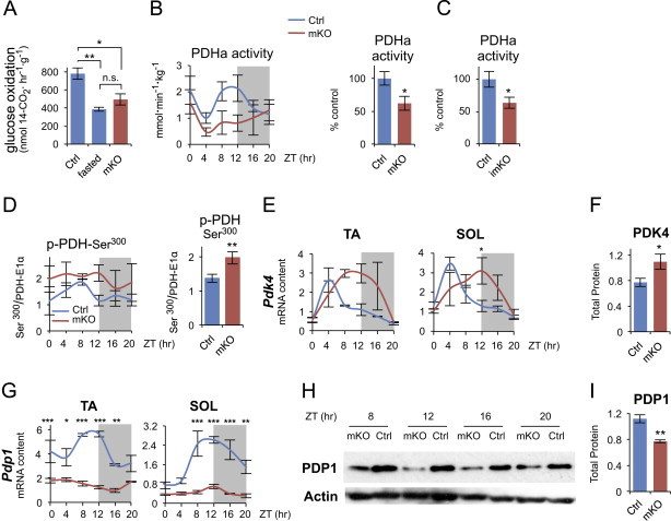
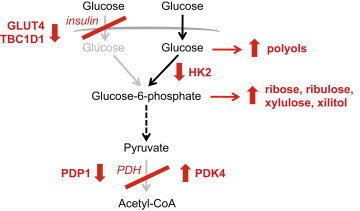
Circadian rhythms control metabolism and energy homeostasis, but the role of the skeletal muscle clock has never been explored. We generated conditional and inducible mouse lines with muscle-specific ablation of the core clock gene Bmal1. Skeletal muscles from these mice showed impaired insulin-stimulated glucose uptake with reduced protein levels of GLUT4, the insulin-dependent glucose transporter, and TBC1D1, a Rab-GTPase involved in GLUT4 translocation. Pyruvate dehydrogenase (PDH) activity was also reduced due to altered expression of circadian genes Pdk4 and Pdp1, coding for PDH kinase and phosphatase, respectively. PDH inhibition leads to reduced glucose oxidation and diversion of glycolytic intermediates to alternative metabolic pathways, as revealed by metabolome analysis. The impaired glucose metabolism induced by muscle-specific Bmal1 knockout suggests that a major physiological role of the muscle clock is to prepare for the transition from the rest/fasting phase to the active/feeding phase, when glucose becomes the predominant fuel for skeletal muscle.
Overview
The claims and statistics of this paper haven’t been checked yet.
Run the check — free, no account needed. It reads the open-access text and checks the sources it cites.
Integrity signals
Registry
Cites
Statistics
Figures
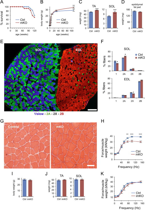
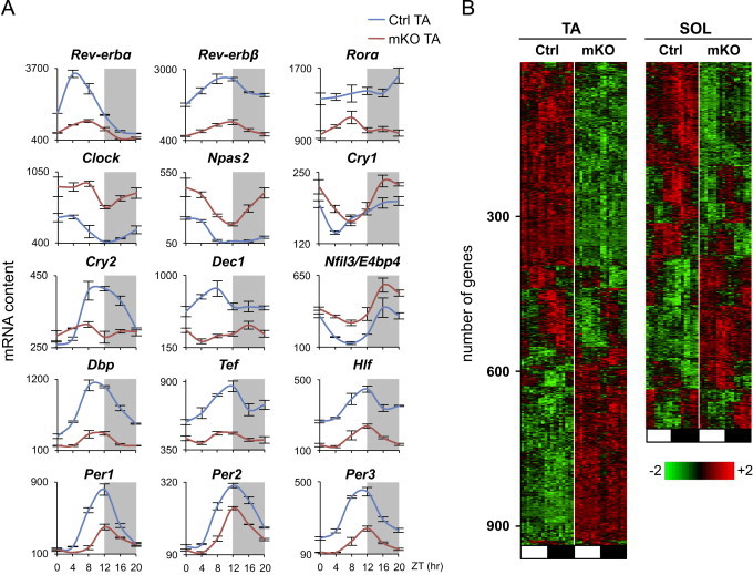
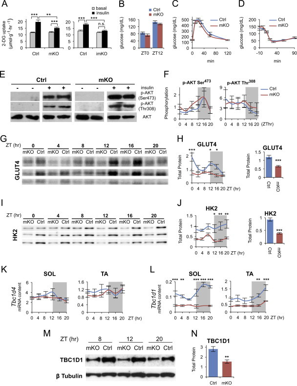
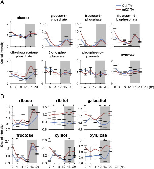
Verification
Claims the paper makes
The claims and statistics of this paper haven’t been checked yet.
Run the check — free, no account needed. It reads the open-access text and checks the sources it cites.
Claims other papers make about this one
Each is checked against this paper’s own text: does it say what the citing paper says it does?
In the citing paper
“Disruption of the muscle clock has also been shown to have significant impact on glucose intolerance and insulin resistance with very small changes in fiber types (Dyar et al., 2014; Schroder et al., 2015; Harfmann et al., 2016).”
From Myosteatosis in the Context of Skeletal Muscle Function Deficit: An Interdisciplinary Workshop at the National Institute on Aging (2020) · cites this paper as Dyar et al., 2014
In the citing paper
“In addition, prolonged loss of Bmal1 function was also associated with increased fibrosis within the muscle.”
From Myosteatosis in the Context of Skeletal Muscle Function Deficit: An Interdisciplinary Workshop at the National Institute on Aging (2020) · cites this paper as Dyar et al., 2014
In the citing paper
“These animals showed reduced protein levels of GLUT4 and an impaired insulin-stimulated glucose uptake in their muscles.”
From Circadian control of glucose metabolism (2014) · cites this paper as [153]
In the citing paper
“For example, the hepatic clock regulates fasting glycemic control and glucose clearance (8), the pancreatic clock regulates insulin secretion and its response to glucose (9-11), the adipose clock regulates lipid storage and mobilization (12,13), and the skeletal muscle clock regulates glucose uptake and metabolism (14).”
From Circadian rhythms, metabolism, and chrononutrition in rodents and humans (2016) · cites this paper as [14]
Self-check
Percentages, sums and reported differences re-computed from the paper’s own numbers.
The self-check needs the paper’s full text.
Citations
Cites
Subramanian, A., Tamayo, P., Mootha, V. K., Mukherjee, S., Ebert, B. L., Gillette, M. A., Paulovich, A., Pomeroy, S. L., Golub, T. R., Lander, E. S., et al. (2005) · Proceedings of the National Academy of Sciences · doi:10.1073/pnas.0506580102 · Cited by 53,625 · cited 5×
“jsp) [27] and gene sets from the KEGG database.”
BackgroundMethodInfluentialExploration, normalization, and summaries of high density oligonucleotide array probe level data
Irizarry, R. A. (2003) · Biostatistics · doi:10.1093/biostatistics/4.2.249 · Cited by 10,603 · cited 2×
“Expression values were generated from fluorescence signals using the robust multi-array average procedure (RMA) [23].”
MethodObesity and Metabolic Syndrome in Circadian Clock Mutant Mice
Turek, F. W., Joshu, C., Kohsaka, A., Lin, E., Ivanova, G., McDearmon, E., Laposky, A., Losee-Olson, S., Easton, A., Jensen, D. R., et al. (2005) · Science · doi:10.1126/science.1108750 · Cited by 2,485 · cited 1×
“Global inactivation of different clock genes leads to altered glucose metabolism: Clock mutant mice display hyperglycemia, obesity and metabolic syndrome [9]; Per1/Per2 double knockout mice and Cry1/Cr2 double knockout mice exhibit glucose”
BackgroundDibner, C., Schibler, U., & Albrecht, U. (2010) · Annual Review of Physiology · doi:10.1146/annurev-physiol-021909-135821 · Cited by 2,421 · cited 2×
“In addition to the core clock genes, all tissues contain a large number of oscillating genes, most of which are tissue-specific and regulated either directly by the core oscillator or by extrinsic circadian signals [7].”
BackgroundEvolving gene/transcript definitions significantly alter the interpretation of GeneChip data
Dai, M. (2005) · Nucleic Acids Research · doi:10.1093/nar/gni179 · Cited by 1,807 · cited 1×
“Speci fi cally, intensity levels have been background adjusted, normalized using quantile normalization, and log 2 expression values calculated using median polish summarization and Entrez custom chip de fi nition fi les for mouse arrays (version 14.1.0) [24].”
MethodInfluentialDeFronzo, R. A., Jacot, E., Jequier, E., Maeder, E., Wahren, J., & Felber, J. P. (1981) · Diabetes · doi:10.2337/diab.30.12.1000 · Cited by 1,770 · cited 1×
“Skeletal muscle is a major site of insulin-stimulated glucose disposal [56] and muscle insulin resistance has been considered to be one of the earliest factors in the pathogenesis of the metabolic syndrome [57].”
BackgroundOccupational and Environmental Medicine
Rosenstock, L., Rest, K. M., BENSON, J. A., Cannella, J. M., Cohen, J. J., Cullen, M. R., Davidoff, F. F., Landrigan, P. J., Reynolds, R. C., Clever, L. H., et al. (1991) · New England Journal of Medicine · doi:10.1056/nejm199109263251305 · Cited by 1,627
Mop3 is an essential component of the master circadian pacemaker in mammals.
Bunger, M. K., Wilsbacher, L. D., Moran, S. M., Clendenin, C., Radcliffe, L. A., Hogenesch, J. B., Simon, M. C., Takahashi, J. S., & Bradfield, C. A. (2000) · Cell · doi:10.1016/s0092-8674(00)00205-1 · Cited by 1,621 · cited 3×
“Global Bmal1 knockout causes not only loss of circadian rhythm of locomotor activity but also reduced total activity levels [42] and rescue experiments in which BMAL1 was constitutively expressed in skeletal muscle of Bmal1 null mice showed that musclespecific rescue restored activity levels, though not circadian rhyt…”
BackgroundDisruption of the clock components CLOCK and BMAL1 leads to hypoinsulinaemia and diabetes
Marcheva, B., Ramsey, K. M., Buhr, E. D., Kobayashi, Y., Su, H., Ko, C. H., Ivanova, G., Omura, C., Mo, S., Vitaterna, M. H., et al. (2010) · Nature · doi:10.1038/nature09253 · Cited by 1,525 · cited 1×
“Selective knockout of Bmal1 in the liver causes hypoglycemia [10], while ablation of the same gene in the endocrine pancreas leads to fasting hyperglycemia and severe glucose intolerance [15].”
BackgroundLipid-induced insulin resistance: unravelling the mechanism
Samuel, V. T., Petersen, K. F., & Shulman, G. I. (2010) · The Lancet · doi:10.1016/s0140-6736(10)60408-4 · Cited by 1,189 · cited 1×
“What is the cause of the altered response to insulin in muscles from Bmal1 mKO mice? The canonical insulin signaling pathway is apparently not responsible for this change, since AKT phosphorylation, which is reduced in obesity and diabetes [60], was unchanged, and was similarly increased by insulin in muscles from con…”
Background
Cited by
Sorted by citations
Items 201–300 of 458
The Tissue Clock Network: Driver and Gatekeeper of Circadian Physiology
Harder, L., & Oster, H. (2020) · BioEssays · doi:10.1002/bies.201900158 · Cited by 24
Nakao, R., Shimba, S., & Oishi, K. (2017) · Scientific Reports · doi:10.1038/s41598-017-03119-8 · Cited by 24
Food deprivation during active phase induces skeletal muscle atrophy via IGF-1 reduction in mice
Abe, T., Kazama, R., Okauchi, H., & Oishi, K. (2019) · Archives of Biochemistry and Biophysics · doi:10.1016/j.abb.2019.108160 · Cited by 25
Cardiolipin Synthesis in Skeletal Muscle Is Rhythmic and Modifiable by Age and Diet
Nohara, K., Kim, E., Wirianto, M., Mileykovskaya, E., Dowhan, W., Chen, Z., & Yoo, S. H. (2020) · Oxidative Medicine and Cellular Longevity · doi:10.1155/2020/5304768 · Cited by 25
“As mentioned above, fuel entry to muscle mitochondria is controlled at the pyruvate dehydrogenase level [20].”
BackgroundLinkage of circadian rhythm disruptions with Alzheimer's disease and therapeutic interventions
Madhamanchi, K., Zhang, J., & Melkani, G. C. (2025) · Acta Pharmaceutica Sinica B · doi:10.1016/j.apsb.2025.04.011 · Cited by 31
“On a molecular level, TRF influences the BMAL1/CLOCK pathway, which governs the expression of CCG involved in glucose metabolism via insulin secretion, glucose uptake 158 , cellular repair, and protein clearance 159 .”
Molecular clocks, satellite cells, and skeletal muscle regeneration
Kahn, R. E., Dayanidhi, S., Lacham‐Kaplan, O., & Hawley, J. A. (2023) · American Journal of Physiology-Cell Physiology · doi:10.1152/ajpcell.00073.2023 · Cited by 26
Aras, E., Ramadori, G., Kinouchi, K., Liu, Y., Ioris, R. M., Brénachot, X., Ljubicic, S., Veyrat‐Durebex, C., Mannucci, S., Galiè, M., et al. (2019) · Cell Reports · doi:10.1016/j.celrep.2019.04.093 · Cited by 23
Petrenko, V., Stolovich-Rain, M., Vandereycken, B., Giovannoni, L., Storch, K.-F., Dor, Y., Chera, S., & Dibner, C. (2020) · Genes & Development · doi:10.1101/gad.343137.120 · Cited by 22
“In addition, loss of functional clocks evoked insulin resistance in skeletal muscles (Dyar et al. 2014; Harfmann et al. 2016) and impaired cell-autonomous adoptative changes to increased insulin needs upon diet-induced obesity (Rakshit et al. 2016).”
BackgroundFrom Chronodisruption to Sarcopenia: The Therapeutic Potential of Melatonin
Fernández‐Martínez, J., Ramírez‐Casas, Y., Yang, Y., Aranda‐Martínez, P., Martínez-Ruiz, L., Escames, G., & Acuña-Castroviejo, D. (2023) · Biomolecules · doi:10.3390/biom13121779 · Cited by 22
Pourabdi, R., Shahidi, F., Tabandeh, M. R., & Salehpour, M. (2024) · Journal of Physiology and Biochemistry · doi:10.1007/s13105-024-01066-3 · Cited by 22
“Deficiency of BMAL1 in skeletal muscle leads to hyper-glycemia, insulin-resistance, and mitochondrial rhythm dysfunction [9, 17 , 26].”
InfluentialCollao, N., Sanders, O., Caminiti, T., Messeiller, L., & Lisio, M. D. (2023) · Journal of Cachexia Sarcopenia and Muscle · doi:10.1002/jcsm.13185 · Cited by 22
Attention to Innate Circadian Rhythm and the Impact of Its Disruption on Diabetes
Lee, D. Y., Jung, I., Park, S. Y., Yu, J. H., Seo, J. A., Kim, K. J., Kim, N. H., Yoo, H. J., Kim, S. G., Choi, K. M., et al. (2024) · Diabetes & Metabolism Journal · doi:10.4093/dmj.2023.0193 · Cited by 21
Astrocyte Clocks and Glucose Homeostasis
Barca‐Mayo, O., & López, M. (2021) · Frontiers in Endocrinology · doi:10.3389/fendo.2021.662017 · Cited by 23
Metabolomics reveal circadian control of cellular metabolism
Ch, R., Chevallier, O. P., & Elliott, C. T. (2020) · TrAC Trends in Analytical Chemistry · doi:10.1016/j.trac.2020.115986 · Cited by 22
Martchenko, S. E., Martchenko, A., Biancolin, A. D., Waller, A. S., & Brubaker, P. L. (2021) · Molecular Metabolism · doi:10.1016/j.molmet.2021.101340 · Cited by 21
Circadian Clock and Sirtuins in Diabetic Lung: A Mechanistic Perspective
Zhou, S., Dai, Y., Zeng, X., & Chen, H. (2020) · Frontiers in Endocrinology · doi:10.3389/fendo.2020.00173 · Cited by 21
“Muscle-specific Bmal1 KO mice showed insulin resistance and impaired glucose metabolism in skeletal muscles (106).”
BackgroundBMAL1/PGC1α4-FNDC5/irisin axis impacts distinct outcomes of time-of-day resistance exercise
Guo, M., Shen, F., Guo, X., Zhang, J., Ma, Y., Wu, X., Zuo, H., Yao, J., Hu, Y., Wang, D., et al. (2024) · Journal of sport and health science/Journal of Sport and Health Science · doi:10.1016/j.jshs.2024.100968 · Cited by 21
Petrenko, V., Saini, C., Perrin, L., & Dibner, C. (2016) · Journal of Visualized Experiments · doi:10.3791/54673 · Cited by 20
Procopio, S. B., & Esser, K. A. (2024) · Journal of Molecular and Cellular Cardiology · doi:10.1016/j.yjmcc.2024.11.011 · Cited by 19
Kelu, J. J., & Hughes, S. M. (2025) · Proceedings of the National Academy of Sciences · doi:10.1073/pnas.2422446122 · Cited by 20
“…mutant ( Bmal1 knockout) mice suggest the necessity of the muscle clock in the circadian regulation of lipid, glucose, and protein metabolism ( 7 – 9 ); whereas studies utilizing clock - disrupted mice with reinstated Bmal1 expression in muscle indicate a partial sufficiency of the muscle…”
Malik, Z. A., Davies, K. A. B., Hall, E. C. R., Barrett, J., Pullinger, S. A., Erskine, R. M., Shepherd, S. O., Iqbal, Z., Edwards, B., & Burniston, J. G. (2020) · Proteomes · doi:10.3390/proteomes8030022 · Cited by 18
“Muscle-specific disruption of the molecular clock results in impairments to glucose metabolism and insulin homeostasis [15] in mice, and exercise at different times of day is associated with different metabolic responses in muscle [16].”
BackgroundChrono-Nutrition Has Potential in Preventing Age-Related Muscle Loss and Dysfunction
Aoyama, S., Nakahata, Y., & Shinohara, K. (2021) · Frontiers in Neuroscience · doi:10.3389/fnins.2021.659883 · Cited by 20
Circadian rhythm in prostate cancer: time to take notice of the clock
Zhu, W., He, Q., Feng, D., Wei, Q., & Yang, L. (2022) · Asian Journal of Andrology · doi:10.4103/aja202255 · Cited by 18
Time-of-day effects on motor unit firing and muscle contractile properties in humans
Hirono, T., Igawa, K., Okudaira, M., Takeda, R., Nishikawa, T., & Watanabe, K. (2024) · Journal of Neurophysiology · doi:10.1152/jn.00368.2023 · Cited by 18
Kiriyama, K., Yamamoto, M., Kim, D., Sun, S., Yamamoto, H., & Oda, H. (2022) · British Journal Of Nutrition · doi:10.1017/s0007114522000356 · Cited by 17
Circadian Rhythm Effects on the Molecular Regulation of Physiological Systems
Crislip, G. R., Johnston, J. G., Douma, L. G., Costello, H. M., Juffre, A., Boyd, K., Li, W. W., Maugans, C. C., Gutierrez‐Monreal, M. A., Esser, K. A., et al. (2021) · Comprehensive physiology · doi:10.1002/cphy.c210011 · Cited by 36
“The next set of circadian transcriptome studies defined the circadian genes across different muscles as well as changes that occur with the muscle-specific loss of the core clock gene, Bmal1 (74, 103, 172, 217).”
InfluentialChrononutrition: Potential, Challenges, and Application in Managing Obesity
Fuad, S. A., Ginting, R. P., & Lee, M.-W. (2025) · International Journal of Molecular Sciences · doi:10.3390/ijms26115116 · Cited by 16
“They found no alteration in muscle weight and force between two littermates after 5 months (~20 weeks) of tamoxifen administration [72].”
Sutton, E. R., & Pekovic‐Vaughan, V. (2025) · Antioxidants · doi:10.3390/antiox14091132 · Cited by 20
“Further to impairments in skeletal muscle structure and contractility, a muscle-specific knock-out of Bmal1 resulted in impaired insulin-dependent glucose uptake and reduced glucose oxidation in skeletal muscle as well as impaired systemic glucose homeostasis [76,104].”
Liang, Q., Lu, Y., Yu, L., Zhu, Q., Xie, W., Wang, Y., Ye, L., Li, Q., Liu, S., Liu, Y., et al. (2021) · Journal of Bone and Mineral Metabolism · doi:10.1007/s00774-021-01271-w · Cited by 17
Bilu, C., Einat, H., Zimmet, P., Vishnevskia‐Dai, V., Schwartz, W. J., & Kronfeld‐Schor, N. (2022) · Scientific Reports · doi:10.1038/s41598-022-06408-z · Cited by 18
Jakubowicz, D. J., Rosenblum, R. C., Wainstein, J., & Twito, O. (2023) · International Journal of Molecular Sciences · doi:10.3390/ijms24087154 · Cited by 16
“Notably, the clock genes in skeletal muscle regulate GLUT-4 activity, muscle glucose uptake [60,68], muscle protein synthesis during the postprandial state, and muscle protein breakdown in the postabsorptive state [30–32,41].”
BackgroundInfluentialTrebucq, L. L., Lamberti, M. L., Rota, R. P., Aiello, I., Borio, C. S., Bilen, M. F., Golombek, D. A., Plano, S. A., & Chiesa, J. J. (2023) · Frontiers in Nutrition · doi:10.3389/fnut.2023.1154647 · Cited by 19
“Several circadian controls set increased glucose tolerance and insulin sensitivity at the light–dark transition to anticipate activity, such as islet clocks controlling peak insulin secretion (13), or the increase in glucose transporter type 4 (GLUT4) in muscle (14).”
BackgroundFernández‐Martínez, J., Ramírez‐Casas, Y., Aranda‐Martínez, P., López‐Rodríguez, A., Sayed, R. K. A., Escames, G., & Acuña-Castroviejo, D. (2023) · Journal of Pineal Research · doi:10.1111/jpi.12912 · Cited by 18
“19 Our data also agree with other Bmal1 knockout models in soleus, extensor digitorum longus, and tibialis anterior muscles, 7,23,30 and suggest a shift toward a more oxidative fiber type with age.”
MethodResultResveratrol as a potential protective compound against skeletal muscle insulin resistance
Bahramzadeh, A., Bolandnazar, K., & Meshkani, R. (2023) · Heliyon · doi:10.1016/j.heliyon.2023.e21305 · Cited by 17
“The molecular clock in skeletal muscle has been shown to play a role in insulin sensitivity in this tissue by increasing GLUT4 translocation and also the effecting on histone deacetylase 3 (HDAC3) [69,70].”
BackgroundMore than the clock: distinct regulation of muscle function and metabolism by PER2 and RORα
Mansingh, S., Maier, G., Delezie, J., Westermark, P. O., Ritz, D., Duchemin, W., Santos, G., Karrer‐Cardel, B., Steurer, S. A., Albrecht, U., et al. (2024) · The Journal of Physiology · doi:10.1113/jp285585 · Cited by 18
“The circadian clock within skeletal muscle governs a rhythmic pattern of gene expression that influences critical physiological processes integral to muscle function and metabolism (Dyar et al., 2014; Gutierrez-Monreal et al., 2020; Mansingh & Handschin, 2022; Zambon et al., 2003).”
Impact of heart-specific disruption of the circadian clock on systemic glucose metabolism in mice
Nakao, T., Kohsaka, A., Otsuka, T., Thein, Z. L., Le, H. T., Waki, H., Gouraud, S. S., Ihara, H., Nakanishi, M., Sato, F., et al. (2017) · Chronobiology International · doi:10.1080/07420528.2017.1415922 · Cited by 17
“Although animal models of clock dysfunction have uncovered a tissue-specific role of clock genes in the regulation of glucose metabolism, increasing attention is being focused on only major metabolic tissues, such as the pancreas, liver, and skeletal muscle (Dyar et al. 2014; Lee et al. 2011; Marcheva et al. 2010; Per…”
BackgroundInfluentialAnabolic Heterogeneity Following Resistance Training: A Role for Circadian Rhythm?
CAMERA, D. M. (2018) · Frontiers in Physiology · doi:10.3389/fphys.2018.00569 · Cited by 17
“This oscillation is non-existent, along with the downregulation of specific MyoD controlled genes, in skeletal muscle from both BMAL1−/− and CLOCK119 mice (Andrews et al., 2010) as well as in skeletal muscle specific Bmal1 knock outmodels (Dyar et al., 2014; Hodge et al., 2015).”
BackgroundRhythm is essential: Unraveling the relation between the circadian clock and cancer
Ogunlusi, O., Ghosh, A., Sarkar, M., Carter, K., Davuluri, H., Chakraborty, M., Eckel‐Mahan, K. L., Keene, A. C., Menet, J. S., Bell‐Pedersen, D., et al. (2025) · Critical Reviews in Oncology/Hematology · doi:10.1016/j.critrevonc.2025.104632 · Cited by 17
Gao, K., Luo, Z., Han, S., Li, Z., Choe, H. M., Paek, H. J., Quan, B., Kang, J., & Yin, X. (2022) · Meat Science · doi:10.1016/j.meatsci.2022.108929 · Cited by 17
Muscle Rev-erb controls time-dependent adaptations to chronic exercise in mice
Liu, J., Xiao, F., Choubey, A., Kumar, S. U., Wang, Y., Hong, S., Yang, T., Otlu, H. G., Oturmaz, E. S., Loro, E., et al. (2025) · Nature Communications · doi:10.1038/s41467-025-60520-y · Cited by 20
“(3) Both mouse lines show reduced glucose uptake and downregulated glycolysis gene expression 61,74 .”
Marot, L. P., Lopes, T. D. V. C., Balieiro, L. C. T., Crispim, C. A., & Moreno, C. R. D. C. (2023) · Nutrients · doi:10.3390/nu15112570 · Cited by 19
Smith, J. G., Molendijk, J., Blazev, R., Chen, W. H., Zhang, Q., Litwin, C., Zinna, V. M., Welz, P.-S., Benitah, S. A., Greco, C. M., et al. (2023) · Molecular & Cellular Proteomics · doi:10.1016/j.mcpro.2023.100655 · Cited by 16
“This process is under circadian control; the central clock in the brain drives mice to consume most food during the dark active phase [50], BMAL1 modulates glucose-stimulated insulin release from pancreatic beta cells [8], and muscle BMAL1 times the expression of key enzymes to meet the demand [51].”
Raza, G. S., Kaya, Y., Stenbäck, V., Sharma, R., Sodum, N., Mutt, S. J., Gagnon, D. D., Tulppo, M. P., Järvelin, M., Herzig, K., et al. (2024) · Molecular Nutrition & Food Research · doi:10.1002/mnfr.202300465 · Cited by 16
Zsuga, J., Móré, C. E., Erdei, T., Papp, C., Harsányi, S., & Gesztelyi, R. (2018) · Frontiers in Neurology · doi:10.3389/fneur.2018.00818 · Cited by 16
Small, L., Brandon, A. E., Parker, B. L., Deshpande, V., Samsudeen, A., Kowalski, G. M., Reznick, J., Wilks, D. L., Preston, E., Bruce, C. R., et al. (2019) · Molecular Metabolism · doi:10.1016/j.molmet.2019.04.006 · Cited by 15
“Disrupting clock machinery in muscle through genetic ablation of bmal1 has been reported to reduce both GLUT4 and hexokinase 2 gene expression [33].”
BackgroundMármol‐Sánchez, E., Ramayo‐Caldas, Y., Quintanilla, R., Cardoso, T. F., González-Prendes, R., Tibau, J., & Amills, M. (2020) · Journal of Animal Science and Biotechnology/Journal of animal science and biotechnology · doi:10.1186/s40104-019-0412-z · Cited by 15
Okazaki, Y., Murray, J., Ehsani, A., Clark, J., Whitson, R. H., Hirose, L., Yanaka, N., & Itakura, K. (2020) · Biological Research · doi:10.1186/s40659-020-00313-3 · Cited by 15
Guo, Y., Wang, Q., Zhang, K., Yao, C., Huang, J., Li, Q., Liu, Z., Zhang, Y., Shan, C., Liu, P., et al. (2020) · The FASEB Journal · doi:10.1096/fj.202001265rr · Cited by 16
Grasset, E., Puel, A., Charpentier, J., Klopp, P., Christensen, J. E., Lelouvier, B., Servant, F., Blasco‐Baqué, V., Tercé, F., & Burcelin, R. (2021) · Acta Diabetologica · doi:10.1007/s00592-021-01790-y · Cited by 15
Fujimoto, R., Ohta, Y., Masuda, K., Taguchi, A., Akiyama, M., Yamamoto, K., Nakabayashi, H., Nagao, Y., Matsumura, T., Hiroshige, S., et al. (2022) · Journal of Diabetes Investigation · doi:10.1111/jdi.13810 · Cited by 15
Mechanosensors control skeletal muscle mass, molecular clocks, and metabolism
Vanmunster, M., Rojo‐García, A. V., Pacolet, A., Dalle, S., Koppo, K., Jonkers, I., Lories, R. J. U., & Suhr, F. (2022) · Cellular and Molecular Life Sciences · doi:10.1007/s00018-022-04346-7 · Cited by 14
“Many processes regulate the maintenance of SkM mass and its physiological metabolism [12, 19, 20].”
BackgroundERRα fosters running endurance by driving myofiber aerobic transformation and fuel efficiency
Xia, H., Scholtes, C., Dufour, C. R., Guluzian, C., & Giguère, V. (2023) · Molecular Metabolism · doi:10.1016/j.molmet.2023.101814 · Cited by 13
Metabolic plasticity and obesity-associated changes in diurnal postexercise metabolism in mice
Pendergrast, L. A., Ashcroft, S. P., Ehrlich, A. M., Treebak, J. T., Krook, A., Dollet, L., & Zierath, J. R. (2024) · Metabolism · doi:10.1016/j.metabol.2024.155834 · Cited by 13
Figueroa‐Toledo, A. M., Gutiérrez‐Pino, J., Carriel‐Nesvara, A., Marchese‐Bittencourt, M., Zbinden‐Foncea, H., & Castro‐Sepúlveda, M. (2024) · The Journal of Physiology · doi:10.1113/jp285955 · Cited by 14
“In rodents, SkM-specific Clock/Bmal1 ablations cause SkM insulin resistance, lower mitochondria density and decreased muscle strength (Dyar et al., 2014; Liu et al., 2016).”
Hierarchy or Heterarchy of Mammalian Circadian Timekeepers?
Bechtel, W. (2024) · Journal of Biological Rhythms · doi:10.1177/07487304241286573 · Cited by 14
Ramírez‐Casas, Y., Fernández‐Martínez, J., Martín‐Estebané, M., Aranda‐Martínez, P., López‐Rodríguez, A., Esquivel‐Ruiz, S., Yang, Y., Escames, G., & Acuña-Castroviejo, D. (2025) · Journal of Pineal Research · doi:10.1111/jpi.70049 · Cited by 16
“As expected, we confirmed the compensatory increase in Clock protein levels in the GM of TMX ‐ treated animals, confirming the data elsewhere published in this model [40, 50].”
Time zones of pancreatic islet metabolism
Petrenko, V., Philippé, J., & Dibner, C. (2018) · Diabetes Obesity and Metabolism · doi:10.1111/dom.13383 · Cited by 14
Wright, T. J., Davis, R. W., Holser, R. R., Hückstädt, L. A., Danesi, C. P., Porter, C. C., Widen, S. G., Williams, T. M., da Costa, D. P., & Sheffield‐Moore, M. (2020) · Frontiers in Physiology · doi:10.3389/fphys.2020.564555 · Cited by 15
Chronic Alcohol Consumption Disrupts the Skeletal Muscle Circadian Clock in Female Mice
Tice, A. L., Laudato, J. A., Gordon, B. S., & Steiner, J. L. (2022) · Journal of Biological Rhythms · doi:10.1177/07487304221141464 · Cited by 14
“…in specific tension, grip strength, thick and thin filament organization, fiber-type distribution and cross-sectional area, mitochondria volume and function, and metabolic gene expression have been reported (Hodge et al., 2015; Schroder et al., 2015; Dyar et al., 2014; Andrews et al., 2010).”
BackgroundInfluentialDifferent levels of circadian (de)synchrony – where does it hurt?
Galinde, A. A., Al‐Μughales, F., Oster, H., & Heyde, I. (2022) · F1000Research · doi:10.12688/f1000research.127234.2 · Cited by 13
Brager, A. J., Heemstra, L. A., Bhambra, R., Ehlen, J. C., Esser, K. A., Paul, K. N., & Novak, C. M. (2016) · Biochimie · doi:10.1016/j.biochi.2016.11.014 · Cited by 13
Muscle interleukin-6 and fasting-induced PDH regulation in mouse skeletal muscle
Gudiksen, A., Bertholdt, L., Vingborg, M. B., Hansen, H. W., Ringholm, S., & Pilegaard, H. (2016) · American Journal of Physiology-Endocrinology and Metabolism · doi:10.1152/ajpendo.00291.2016 · Cited by 12
Genome-wide circadian regulation: A unique system for computational biology
Sun, L., Ma, J., Turck, C. W., Xu, P., & Wang, G. (2020) · Computational and Structural Biotechnology Journal · doi:10.1016/j.csbj.2020.07.002 · Cited by 14
“However, when high-resolution time data are processed, JTK_CYCLE performs better than ARSER, with lower false positive rates[119].”
InfluentialLiu, J., Zhang, T., Luo, J., Chen, S., & Zhang, D. (2023) · International Journal of Environmental Research and Public Health · doi:10.3390/ijerph20043416 · Cited by 15
Circadian rhythm effect on military physical fitness and field training: a narrative review
Rad, R. S., Hosseini, H. M., & Shirvani, H. (2020) · Sport Sciences for Health · doi:10.1007/s11332-020-00692-w · Cited by 12
When should I run—the role of exercise timing in metabolic health
Kirchner, H., Weisner, L., & Wilms, B. (2023) · Acta Physiologica · doi:10.1111/apha.13953 · Cited by 12
The sweet tooth of the circadian clock
Fu, M., & Yang, X. (2017) · Biochemical Society Transactions · doi:10.1042/bst20160183 · Cited by 11
Loss of laminin‐a4 results in pre‐ and postsynaptic modifications at the neuromuscular junction
Chand, K. K., Lee, K. M., Lavidis, N. A., & Noakes, P. G. (2016) · The FASEB Journal · doi:10.1096/fj.201600899r · Cited by 12
A Role for Exercise to Counter Skeletal Muscle Clock Disruption
Erickson, M. L., Esser, K. A., Kraus, W. E., Buford, T. W., & Redman, L. M. (2020) · Exercise and Sport Sciences Reviews · doi:10.1249/jes.0000000000000235 · Cited by 13
Resetting Time: The Role of Exercise Timing in Circadian Reprogramming for Metabolic Health
Hesketh, S. J. (2025) · Obesities · doi:10.3390/obesities5030059 · Cited by 13
Shon, J., Han, Y., Song, S., Kwon, S. Y., Na, K., Lindroth, A. M., & Park, Y. J. (2024) · The Journal of Nutritional Biochemistry · doi:10.1016/j.jnutbio.2024.109590 · Cited by 11
Smith, H., Templeman, I., Davis, M., Slater, T., Clayton, D. J., Varley, I., James, L. J., Middleton, B., Johnston, J. D., Karagounis, L. G., et al. (2024) · The Journal of Clinical Endocrinology & Metabolism · doi:10.1210/clinem/dgae350 · Cited by 12
Molecular Pathways Regulating Circadian Rhythm and Associated Diseases
Ding, M., Zhou, H., Li, Y., & Zheng, Y. (2024) · Frontiers in Bioscience-Landmark · doi:10.31083/j.fbl2906206 · Cited by 14
da Silva, F. M., Cabrera, A. R., Jones, R. G., Schrems, E. R., Muhyudin, R., Washington, T. A., Murach, K. A., & Greene, N. P. (2024) · American Journal of Physiology-Cell Physiology · doi:10.1152/ajpcell.00647.2024 · Cited by 13
Jakubowicz, D. J., Matz, Y., Landau, Z., Rosenblum, R. C., Twito, O., Wainstein, J., & Tsameret, S. (2024) · International Journal of Molecular Sciences · doi:10.3390/ijms252212355 · Cited by 12
Muscle Bmal1 is Dispensable for the Progress of Neurogenic Muscle Atrophy in Mice
Nakao, R., Shimba, S., & Oishi, K. (2016) · Journal of Circadian Rhythms · doi:10.5334/jcr.141 · Cited by 11
The Circadian Clock and Obesity
Sebti, Y., Hebras, A., Pourcet, B., Staels, B., & Duez, H. (2022) · Handbook of experimental pharmacology · doi:10.1007/164_2021_579 · Cited by 13
Sex and limb comparisons of neuromuscular function in the morning versus the evening
Augsburger, G., Soloveva, A., & Carr, J. C. (2022) · Physiological Reports · doi:10.14814/phy2.15260 · Cited by 11
Livelo, C., Guo, Y., & Melkani, G. C. (2022) · International Journal of Molecular Sciences · doi:10.3390/ijms24010422 · Cited by 11
“Another study using muscle-specific Bmal1 disruption led to a reduction of force production, skeletal muscle glucose uptake, and glucose oxidation [107].”
BackgroundOnly time will tell: the interplay between circadian clock and metabolism
Gopalakrishnan, S., & Kannan, N. N. (2020) · Chronobiology International · doi:10.1080/07420528.2020.1842436 · Cited by 12
“Muscle-specific ablation of core clock gene Bmal1 in mice impaired insulin- mediated glucose uptake, glucose oxidation, and the protein levels of insulin-dependent glucose transporter GLUT4 (Dyar et al. 2014; Harfmann et al. 2016).”
BackgroundMetabolic Homeostasis: It’s All in the Timing
Brubaker, P. L., & Martchenko, A. (2021) · Endocrinology · doi:10.1210/endocr/bqab199 · Cited by 14
HIF-1α and BMAL1 in bone regeneration: crosstalk between hypoxia response and circadian rhythm
Weng, Y., Xiong, J., Zhao, Q., & Tan, Z. (2026) · Bone Research · doi:10.1038/s41413-026-00506-8 · Cited by 12
Ye, Z., Huang, K., Dai, X.-Q., Gao, D., Gu, Y., Qian, J., Zhang, F., & Zhai, Q. (2024) · Heliyon · doi:10.1016/j.heliyon.2024.e37475 · Cited by 10
“Disrupting the skeletal muscle clock, as seen in Bmal1 knockout models, results in impaired insulin-stimulated glucose uptake [17], muscle fiber remodeling, and premature aging characteristics, including increased bone calcification and reduced joint collagen [18].”
Negri, M., Pivonello, C., Amatrudo, F., Cimmino, F., Trinchese, G., Vetrani, C., Iaccarino, G., Pivonello, R., Mollica, M. P., & Colao, A. (2025) · Nutrition Reviews · doi:10.1093/nutrit/nuaf007 · Cited by 15
The circadian clock regulates metabolic responses to physical exercise
Cheng, Q., Lü, C., & Qian, R. (2022) · Chronobiology International · doi:10.1080/07420528.2022.2050384 · Cited by 10
“In the process of glucose oxidation, rate-limiting enzymes are controlled by circadian clock genes, such as pyruvate dehydrogenase phosphatase catalytic subunit1 (Pdp1) and pyruvate dehydrogenase kinase 4 (Pdk4) (Dyar et al. 2014).”
BackgroundSex Inclusion in Transcriptome Studies of Daily Rhythms
Obodo, D., Outland, E. H., & Hughey, J. (2022) · Journal of Biological Rhythms · doi:10.1177/07487304221134160 · Cited by 11
Begam, M., & Roche, J. A. (2018) · European Journal of Histochemistry · doi:10.4081/ejh.2018.2896 · Cited by 10
Xu, L., Jia, J., Miao, S., Gong, L., Wang, J., He, S., & Zhang, Y. (2023) · Life Sciences · doi:10.1016/j.lfs.2023.121696 · Cited by 9
Putranata, H., Hengky, A., & Hartoko, B. (2025) · Acta Diabetologica · doi:10.1007/s00592-024-02440-9 · Cited by 9
DNA Methylation-Related circRNA_0116449 Is Involved in Lipid Peroxidation in Traumatic Brain Injury
Zheng, P., Ren, D.-B., Yu, C., Zhang, X., & Zhang, Y.-S. (2022) · Frontiers in Molecular Neuroscience · doi:10.3389/fnmol.2022.904913 · Cited by 8
Dial, M. B., Malek, E. M., Cooper, A. R., Neblina, G. A., Vasileva, N. I., Hines, D. J., & McGinnis, G. R. (2024) · Journal of Applied Physiology · doi:10.1152/japplphysiol.00632.2023 · Cited by 10
Circadian Mechanisms in Bioenergetics and Cell Metabolism
Bass, J. T. (2016) · Research and perspectives in endocrine interactions · doi:10.1007/978-3-319-27069-2_3 · Cited by 9
“Though skeletal muscle ablation of the clock has been achieved in our group and others, the biochemical pathways through which clock abrogation impairs oxidative capacity remain largely unknown (Dyar et al. 2014).”
BackgroundHe, S., Lu, Y., Zhu, R., Wei, H., Wang, J., Zheng, L. X., & Zhang, Y. (2022) · Life · doi:10.3390/life12081233 · Cited by 9
Dynamic modulation of glucose utilisation by glucocorticoid rhythms in health and disease
Zavala, E., Gil-Gómez, C. A., Wedgwood, K. C. A., Burgess, R., Tsaneva-Atanasova, K., & Herrera-Valdez, M. A. (2020) · bioRxiv (Cold Spring Harbor Laboratory) · doi:10.1101/2020.02.27.968354 · Cited by 8
Malik, M. Z., DASHTI, M. H., Fatima, Y., Channanath, A. M., John, S. E., Singh, R. K. B., Al‐Mulla, F., & Thanaraj, T. A. (2023) · Frontiers in Molecular Neuroscience · doi:10.3389/fnmol.2023.1217992 · Cited by 8
Gil, C. I., Löser, A., Lossow, K., Schwarz, M., Weber, D., Grune, T., Kipp, A. P., Klaus, S., & Ost, M. (2023) · Frontiers in Endocrinology · doi:10.3389/fendo.2023.1277866 · Cited by 11
Yan, B., & Li, L. (2025) · Scientific Reports · doi:10.1038/s41598-025-89821-4 · Cited by 8
Cancer-induced FOXP1 disrupts and reprograms skeletal-muscle circadian transcription in cachexia
Ducharme, J. B., Neyroud, D., Schonk, M. M., Gutierrez‐Monreal, M. A., Huo, Z., Tucker, H. O., Esser, K. A., Judge, S. M., & Judge, A. R. (2025) · Cell Reports · doi:10.1016/j.celrep.2025.115689 · Cited by 9
“18 26-28 In this regard, downregulated genes were commonly enriched for pathways associated with carbohydrate metabolism, including insulin-stimulated glucose uptake (e.g., Prkaa2 and Pik3r1) and glucose oxidation (e.g., Pdp1 and Pdp2), while upregulated genes were commonly enriched for pathways…”
Apparent Absence of BMAL1-Dependent Skeletal Muscle–Kidney Cross Talk in Mice
Crislip, G. R., Wohlgemuth, S. E., Wolff, C. A., Gutierrez‐Monreal, M. A., Douglas, C. M., Ebrahimi, E., Cheng, K., Masten, S., Barral, D., Bryant, A. J., et al. (2022) · Biomolecules · doi:10.3390/biom12020261 · Cited by 8
Zhang, S., Zuo, X., Luan, J., Bai, H., Fu, Z., Sun, M., Zhao, X. J., & Feng, X. (2023) · Journal of Environmental Sciences · doi:10.1016/j.jes.2023.09.014 · Cited by 8
Metadata
- Authors
- Dyar, K. A., Ciciliot, S., Wright, L. E., Biensø, R. S., Tagliazucchi, G. M., Patel, V. R., Forcato, M., Paz, M. I. P., Gudiksen, A., Solagna, F., Albiero, M., Moretti, I., Eckel-Mahan, K. L., Baldi, P., Sassone-Corsi, P., Rizzuto, R., Bicciato, S., Pilegaard, H., Blaauw, B., & Schiaffino, S.
- Affiliations
- Veneto Institute of Molecular Medicine (IT)
- University of Padua (IT)
- University of Copenhagen (DK)
- University of Modena and Reggio Emilia (IT)
- University of California, Irvine (US)
- Corresponding author
- Dyar, Kenneth A.
- Schiaffino, Stefano
- ORCID
- Venue
- Molecular Metabolism
- Publisher
- Elsevier BV
- Published
- 2014-02
- Volume
- 3
- Issue
- 1
- Pages
- 29-41
- ISSN
- 2212-8778 (ISSN-L)
- PMC
- PMC3929910
- OpenAlex
- W1964634897
- Cited by
- 454
- References
- 73
- Citations by year
- 2026: 34 · 2025: 37 · 2024: 40 · 2023: 46 · 2022: 42 · 2021: 33 · 2020: 53 · 2019: 34 · 2018: 36 · 2017: 28 · 2016: 42 · 2015: 19 · 2014: 9
- FWCI
- 12.78
- Type
- journal article
- Language
- en
- Update notices
- erratum (2014-12-01)
- Open access
- gold
- doi.org
- Licence
- Locations
- Molecular Metabolism (publishedVersion, cc-by-nc-sa)
- Europe PMC (PubMed Central) (publishedVersion, cc-by-nc-sa)
- PubMed Central (publishedVersion, cc-by-nc-sa)
- PubMed (publishedVersion)
- Research at the University of Copenhagen (University of Copenhagen) (publishedVersion, other-oa)
- eScholarship (California Digital Library) (submittedVersion)
- eScholarship (California Digital Library) (submittedVersion, cc-by-nc-sa)
- IRIS UNIMORE (University of Modena and Reggio Emilia) (submittedVersion, cc-by-nc-sa)
- Padua Research Archive (University of Padova) (submittedVersion)
- Funders
- European Commission (223576, 294777)
- Italian Space Agency (ASI, project OSMA)
- Italian Ministries of Health (Ricerca Finalizzata) and of Education, University and Research (PRIN, FIRB), NIA (2P01AG025532-06A1)
- Cariparo Foundation (Padua)
- Italian Association for Cancer Research (AIRC) and Telethon–Italy (GPP1005A, GGP11082)
- Topics
- Life Sciences › Neuroscience › Endocrine and Autonomic Systems › Circadian rhythm and melatonin
- Life Sciences › Biochemistry, Genetics and Molecular Biology › Molecular Biology › Metabolism, Diabetes, and Cancer
- Health Sciences › Medicine › Physiology (medical) › Adipose Tissue and Metabolism
- Keywords
- circadian rhythm, Bmal1, insulin sensitivity, glucose uptake, GLUT4, TBC1D1, pyruvate dehydrogenase, PDK4, PDP1, glucose oxidation, glycolysis, metabolome, conditional knockout mice
About these checks
Registry, retraction, Cites and statistics results are computed, not judged, with one exception: a printed reference that no index lists is read once by a model before it counts as a citation error. Claims are extracted by a model and kept only when their quote appears verbatim in the text; each cited source is then checked, with a model’s help, for whether it says what the claim relies on. Those verdicts can be wrong — read the claim’s quote and the cited source before relying on one.
Statistics and arithmetic are recomputed, not judged.
Confidence bands are shown only for fields calibrated on our annotated test set. None is calibrated yet, so no band is shown.
What counts as a claim ›How we measure confidence ›How we check this ›
Are you an author, or do you see a problem with this page? Request a review
