

The DOI matches its Crossref record; 73 of the 73 references it cites were found in a registry or an index.
Muscle insulin sensitivity and glucose metabolism are controlled by the intrinsic muscle clock
Open accessAbstract
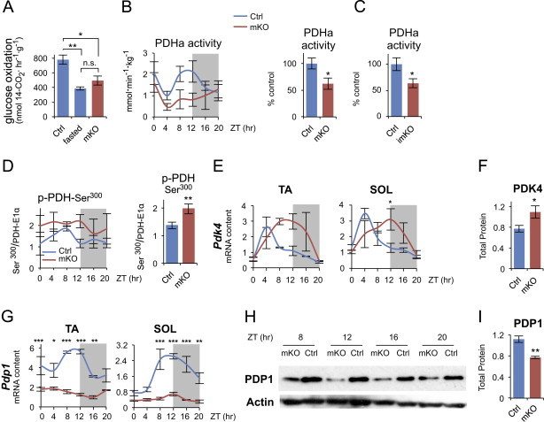
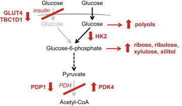
Circadian rhythms control metabolism and energy homeostasis, but the role of the skeletal muscle clock has never been explored. We generated conditional and inducible mouse lines with muscle-specific ablation of the core clock gene Bmal1. Skeletal muscles from these mice showed impaired insulin-stimulated glucose uptake with reduced protein levels of GLUT4, the insulin-dependent glucose transporter, and TBC1D1, a Rab-GTPase involved in GLUT4 translocation. Pyruvate dehydrogenase (PDH) activity was also reduced due to altered expression of circadian genes Pdk4 and Pdp1, coding for PDH kinase and phosphatase, respectively. PDH inhibition leads to reduced glucose oxidation and diversion of glycolytic intermediates to alternative metabolic pathways, as revealed by metabolome analysis. The impaired glucose metabolism induced by muscle-specific Bmal1 knockout suggests that a major physiological role of the muscle clock is to prepare for the transition from the rest/fasting phase to the active/feeding phase, when glucose becomes the predominant fuel for skeletal muscle.
Overview
The claims and statistics of this paper haven’t been checked yet.
Run the check — free, no account needed. It reads the open-access text and checks the sources it cites.
Integrity signals
Registry
Cites
Statistics
Figures
Verification
Claims the paper makes
The claims and statistics of this paper haven’t been checked yet.
Run the check — free, no account needed. It reads the open-access text and checks the sources it cites.
Claims other papers make about this one
Each is checked against this paper’s own text: does it say what the citing paper says it does?
In the citing paper
“Disruption of the muscle clock has also been shown to have significant impact on glucose intolerance and insulin resistance with very small changes in fiber types (Dyar et al., 2014; Schroder et al., 2015; Harfmann et al., 2016).”
From Myosteatosis in the Context of Skeletal Muscle Function Deficit: An Interdisciplinary Workshop at the National Institute on Aging (2020) · cites this paper as Dyar et al., 2014
In the citing paper
“In addition, prolonged loss of Bmal1 function was also associated with increased fibrosis within the muscle.”
From Myosteatosis in the Context of Skeletal Muscle Function Deficit: An Interdisciplinary Workshop at the National Institute on Aging (2020) · cites this paper as Dyar et al., 2014
In the citing paper
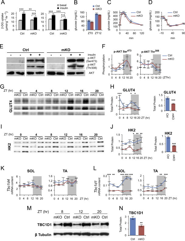
“These animals showed reduced protein levels of GLUT4 and an impaired insulin-stimulated glucose uptake in their muscles.”
From Circadian control of glucose metabolism (2014) · cites this paper as [153]
In the citing paper
“For example, the hepatic clock regulates fasting glycemic control and glucose clearance (8), the pancreatic clock regulates insulin secretion and its response to glucose (9-11), the adipose clock regulates lipid storage and mobilization (12,13), and the skeletal muscle clock regulates glucose uptake and metabolism (14).”
From Circadian rhythms, metabolism, and chrononutrition in rodents and humans (2016) · cites this paper as [14]
Self-check
Percentages, sums and reported differences re-computed from the paper’s own numbers.
The self-check needs the paper’s full text.
Citations
Cites
Subramanian, A., Tamayo, P., Mootha, V. K., Mukherjee, S., Ebert, B. L., Gillette, M. A., Paulovich, A., Pomeroy, S. L., Golub, T. R., Lander, E. S., et al. (2005) · Proceedings of the National Academy of Sciences · doi:10.1073/pnas.0506580102 · Cited by 53,625 · cited 5×
“jsp) [27] and gene sets from the KEGG database.”
BackgroundMethodInfluentialExploration, normalization, and summaries of high density oligonucleotide array probe level data
Irizarry, R. A. (2003) · Biostatistics · doi:10.1093/biostatistics/4.2.249 · Cited by 10,603 · cited 2×
“Expression values were generated from fluorescence signals using the robust multi-array average procedure (RMA) [23].”
MethodObesity and Metabolic Syndrome in Circadian Clock Mutant Mice
Turek, F. W., Joshu, C., Kohsaka, A., Lin, E., Ivanova, G., McDearmon, E., Laposky, A., Losee-Olson, S., Easton, A., Jensen, D. R., et al. (2005) · Science · doi:10.1126/science.1108750 · Cited by 2,485 · cited 1×
“Global inactivation of different clock genes leads to altered glucose metabolism: Clock mutant mice display hyperglycemia, obesity and metabolic syndrome [9]; Per1/Per2 double knockout mice and Cry1/Cr2 double knockout mice exhibit glucose”
BackgroundDibner, C., Schibler, U., & Albrecht, U. (2010) · Annual Review of Physiology · doi:10.1146/annurev-physiol-021909-135821 · Cited by 2,421 · cited 2×
“In addition to the core clock genes, all tissues contain a large number of oscillating genes, most of which are tissue-specific and regulated either directly by the core oscillator or by extrinsic circadian signals [7].”
BackgroundEvolving gene/transcript definitions significantly alter the interpretation of GeneChip data
Dai, M. (2005) · Nucleic Acids Research · doi:10.1093/nar/gni179 · Cited by 1,807 · cited 1×
“Speci fi cally, intensity levels have been background adjusted, normalized using quantile normalization, and log 2 expression values calculated using median polish summarization and Entrez custom chip de fi nition fi les for mouse arrays (version 14.1.0) [24].”
MethodInfluentialDeFronzo, R. A., Jacot, E., Jequier, E., Maeder, E., Wahren, J., & Felber, J. P. (1981) · Diabetes · doi:10.2337/diab.30.12.1000 · Cited by 1,770 · cited 1×
“Skeletal muscle is a major site of insulin-stimulated glucose disposal [56] and muscle insulin resistance has been considered to be one of the earliest factors in the pathogenesis of the metabolic syndrome [57].”
BackgroundOccupational and Environmental Medicine
Rosenstock, L., Rest, K. M., BENSON, J. A., Cannella, J. M., Cohen, J. J., Cullen, M. R., Davidoff, F. F., Landrigan, P. J., Reynolds, R. C., Clever, L. H., et al. (1991) · New England Journal of Medicine · doi:10.1056/nejm199109263251305 · Cited by 1,627
Mop3 is an essential component of the master circadian pacemaker in mammals.
Bunger, M. K., Wilsbacher, L. D., Moran, S. M., Clendenin, C., Radcliffe, L. A., Hogenesch, J. B., Simon, M. C., Takahashi, J. S., & Bradfield, C. A. (2000) · Cell · doi:10.1016/s0092-8674(00)00205-1 · Cited by 1,621 · cited 3×
“Global Bmal1 knockout causes not only loss of circadian rhythm of locomotor activity but also reduced total activity levels [42] and rescue experiments in which BMAL1 was constitutively expressed in skeletal muscle of Bmal1 null mice showed that musclespecific rescue restored activity levels, though not circadian rhyt…”
BackgroundDisruption of the clock components CLOCK and BMAL1 leads to hypoinsulinaemia and diabetes
Marcheva, B., Ramsey, K. M., Buhr, E. D., Kobayashi, Y., Su, H., Ko, C. H., Ivanova, G., Omura, C., Mo, S., Vitaterna, M. H., et al. (2010) · Nature · doi:10.1038/nature09253 · Cited by 1,525 · cited 1×
“Selective knockout of Bmal1 in the liver causes hypoglycemia [10], while ablation of the same gene in the endocrine pancreas leads to fasting hyperglycemia and severe glucose intolerance [15].”
BackgroundLipid-induced insulin resistance: unravelling the mechanism
Samuel, V. T., Petersen, K. F., & Shulman, G. I. (2010) · The Lancet · doi:10.1016/s0140-6736(10)60408-4 · Cited by 1,189 · cited 1×
“What is the cause of the altered response to insulin in muscles from Bmal1 mKO mice? The canonical insulin signaling pathway is apparently not responsible for this change, since AKT phosphorylation, which is reduced in obesity and diabetes [60], was unchanged, and was similarly increased by insulin in muscles from con…”
Background
Cited by
Sorted by citations
Circadian clocks and insulin resistance
Stenvers, D. J., Scheer, F. A. J. L., Schrauwen, P., la Fleur, S. E., & Kalsbeek, A. (2019) · Nature Reviews Endocrinology · doi:10.1038/s41574-018-0122-1 · Cited by 733
Circadian Rhythm and Sleep Disruption: Causes, Metabolic Consequences, and Countermeasures
Potter, G. D., Skene, D. J., Arendt, J., Cade, J. E., Grant, P. J., & Hardie, L. J. (2016) · Endocrine Reviews · doi:10.1210/er.2016-1083 · Cited by 781
Crosstalk between metabolism and circadian clocks
Reinke, H., & Asher, G. (2019) · Nature Reviews Molecular Cell Biology · doi:10.1038/s41580-018-0096-9 · Cited by 598
Meal timing regulates the human circadian system
Wehrens, S. M. T., Christou, S., Isherwood, C., Middleton, B., Gibbs, M. A., Archer, S. N., Skene, D. J., & Johnston, J. D. (2017) · Current Biology · doi:10.1016/j.cub.2017.04.059 · Cited by 596
Widespread seasonal gene expression reveals annual differences in human immunity and physiology
Dopico, X. C., Evangelou, M., Ferreira, R. C., Guo, H., Pekalski, M. L., Smyth, D. J., Cooper, N., Burren, O. S., Fulford, A. J., Hennig, B. J., et al. (2015) · Nature Communications · doi:10.1038/ncomms8000 · Cited by 472
Circadian clock genes and the transcriptional architecture of the clock mechanism
Cox, K. H., & Takahashi, J. S. (2019) · Journal of Molecular Endocrinology · doi:10.1530/jme-19-0153 · Cited by 514
Correa-de-Araujo, R., Addison, O., Miljkovic, I., Goodpaster, B. H., Bergman, B. C., Clark, R. V., Elena, J. W., Esser, K. A., Ferrucci, L., Harris-Love, M. O., et al. (2020) · Frontiers in Physiology · doi:10.3389/fphys.2020.00963 · Cited by 468
Peek, C. B., Levine, D. C., Cedernaes, J., Taguchi, A., Kobayashi, Y., Tsai, S. J., Bonar, N. A., McNulty, M., Ramsey, K. M., & Bass, J. T. (2016) · Cell Metabolism · doi:10.1016/j.cmet.2016.09.010 · Cited by 413
Circadian rhythms and exercise — re-setting the clock in metabolic disease
Gabriel, B. M., & Zierath, J. R. (2019) · Nature Reviews Endocrinology · doi:10.1038/s41574-018-0150-x · Cited by 382
Circadian System and Glucose Metabolism: Implications for Physiology and Disease
Qian, J., & Scheer, F. A. J. L. (2016) · Trends in Endocrinology and Metabolism · doi:10.1016/j.tem.2016.03.005 · Cited by 370
“In muscle-specific Bmal1 knockout mice, neither glucose tolerance nor insulin tolerance tests show any significant differences under chow-diet [39].”
BackgroundAtlas of Circadian Metabolism Reveals System-wide Coordination and Communication between Clocks
Dyar, K. A., Lutter, D., Artati, A., Ceglia, N., Liu, Y., Armenta, D., Jastroch, M., Schneider, S., de Mateo, S., Cervantes, M., et al. (2018) · Cell · doi:10.1016/j.cell.2018.08.042 · Cited by 345
Circadian control of glucose metabolism
Kalsbeek, A., la Fleur, S., & Fliers, E. (2014) · Molecular Metabolism · doi:10.1016/j.molmet.2014.03.002 · Cited by 338
“After submission of our manuscript we became aware of a recent study in which glucose metabolism was studied in a muscle-specific Bmal1 KO [153].”
MethodExercise Metabolism and Adaptation in Skeletal Muscle
Smith, J. A. B., Murach, K. A., Dyar, K. A., & Zierath, J. R. (2023) · Nature Reviews Molecular Cell Biology · doi:10.1038/s41580-023-00606-x · Cited by 342
Time of exercise specifies the impact on muscle metabolic pathways and systemic energy homeostasis
Sato, S., Basse, A. L., Schönke, M., Chen, S., Samad, M., Altıntaş, A., Laker, R. C., Dalbram, E., Barrès, R., Baldi, P., et al. (2019) · Cell Metabolism · doi:10.1016/j.cmet.2019.03.013 · Cited by 298
Endocrine Crosstalk Between Skeletal Muscle and the Brain
Delezie, J., & Handschin, C. (2018) · Frontiers in Neurology · doi:10.3389/fneur.2018.00698 · Cited by 277
“Second, these data imply a muscle-to-brain signaling involved in sleep regulation, either humoral, or, based on the observation that mice lacking Bmal1 in their muscles exhibit potent metabolic disruptions (152, 153), via metabolites.”
BackgroundExercise sustains the hallmarks of health
Qiu, Y., Fernández-García, B., Lehmann, H. I., Li, G., Kroemer, G., López-Otín, C., & Xiao, J. (2023) · Journal of Sport and Health Science · doi:10.1016/j.jshs.2022.10.003 · Cited by 270
Circadian rhythms, metabolism, and chrononutrition in rodents and humans
Johnston, J. D., Ordovás, J. M., Scheer, F. A., & Turek, F. W. (2016) · Advances in Nutrition · doi:10.3945/an.115.010777 · Cited by 258
“For example, the hepatic clock regulates fasting glycemic control and glucose clearance (8), the pancreatic clock regulates insulin secretion and its response to glucose (9–11), the adipose clock regulates lipid storage and mobilization (12, 13), and the skeletal muscle clock regulates glucose uptake and metabolism (1…”
BackgroundHarfmann, B. D., Schroder, E. A., Kachman, M. T., Hodge, B. A., Zhang, X., & Esser, K. A. (2016) · Skeletal Muscle · doi:10.1186/s13395-016-0082-x · Cited by 231
The molecular athlete: exercise physiology from mechanisms to medals
Furrer, R., Hawley, J. A., & Handschin, C. (2023) · Physiological Reviews · doi:10.1152/physrev.00017.2022 · Cited by 253
Demonstration of a day-night rhythm in human skeletal muscle oxidative capacity
van Moorsel, D., Hansen, J., Havekes, B., Scheer, F. A. J. L., Jörgensen, J. A., Hoeks, J., Schrauwen-Hinderling, V. B., Duez, H., Lefebvre, P., Schaper, N. C., et al. (2016) · Molecular Metabolism · doi:10.1016/j.molmet.2016.06.012 · Cited by 218
Clock Regulation of Metabolites Reveals Coupling between Transcription and Metabolism
Krishnaiah, S., Wu, G., Altman, B. J., Growe, J., Rhoades, S. D., Coldren, F., Venkataraman, A., Olarerin-George, A. O., Francey, L. J., Mukherjee, S., et al. (2017) · Cell Metabolism · doi:10.1016/j.cmet.2017.03.019 · Cited by 211
Circadian rhythms in mitochondrial respiration
de Goede, P., Wefers, J., Brombacher, E. C., Schrauwen, P., & Kalsbeek, A. (2018) · Journal of Molecular Endocrinology · doi:10.1530/jme-17-0196 · Cited by 211
“Disrupting the circadian clock by muscle-specific Bmal1 ablation resulted in a substantial decrease in 14C-glucose oxidation in isolated mouse diaphragms (Dyar et al. 2014).”
BackgroundCircadian Rhythms, the Molecular Clock, and Skeletal Muscle
Harfmann, B. D., Schroder, E. A., & Esser, K. A. (2015) · Journal of Biological Rhythms · doi:10.1177/0748730414561638 · Cited by 211
“Loss of the core clock gene Bmal1 results in severe muscle pathology, including fiber-type shifts, decreased mitochondria, impaired mitochondrial respiration, altered sarcomeric structure, and debilitated function (Andrews et al., 2010; Dyar et al., 2014).”
Circadian regulation of metabolism
Bailey, S. M., Udoh, U. S., & Young, M. E. (2014) · Journal of Endocrinology · doi:10.1530/joe-14-0200 · Cited by 219
Single muscle fiber proteomics reveals unexpected mitochondrial specialization
Murgia, M., Nagaraj, N., Deshmukh, A. S., Zeiler, M., Cancellara, P., Moretti, I., Reggiani, C., Schiaffino, S., & Mann, M. (2015) · EMBO Reports · doi:10.15252/embr.201439757 · Cited by 208
“Immunofluorescence with Myh isoform-specific antibodies was performed as described in [44].”
Circadian Etiology of Type 2 Diabetes Mellitus
Javeed, N., & Matveyenko, A. V. (2018) · Physiology · doi:10.1152/physiol.00003.2018 · Cited by 219
Wefers, J., van Moorsel, D., Hansen, J., Connell, N. J., Havekes, B., Hoeks, J., Lichtenbelt, W. D. V. M., Duez, H., Phielix, E., Kalsbeek, A., et al. (2018) · Proceedings of the National Academy of Sciences · doi:10.1073/pnas.1722295115 · Cited by 202
“In the control condition, the core clock genes BMAL1, CRY1, and PER2 displayed diurnal differences in mRNA expression levels, concordant with the results from our previous study (7) (Fig.”
BackgroundInfluentialCedernaes, J., Osler, M. E., Voisin, S., Broman, J.-E., Vogel, H., Dickson, S. L., Zierath, J. R., Schiöth, H. B., & Benedict, C. (2015) · The Journal of Clinical Endocrinology & Metabolism · doi:10.1210/jc.2015-2284 · Cited by 188
Nohara, K., Mallampalli, V. K. P. S., Nemkov, T. G., Wirianto, M., Yang, J., Ye, Y., Sun, Y., Han, L., Esser, K. A., Mileykovskaya, E., et al. (2019) · Nature Communications · doi:10.1038/s41467-019-11926-y · Cited by 185
Circadian regulation of lipid metabolism
Gooley, J. J. (2016) · Proceedings of The Nutrition Society · doi:10.1017/s0029665116000288 · Cited by 194
Circadian metabolism in the light of evolution
Gerhart-Hines, Z., & Lazar, M. A. (2015) · Endocrine Reviews · doi:10.1210/er.2015-1007 · Cited by 178
Hodge, B. A., Wen, Y., Riley, L. A., Zhang, X., England, J. H., Harfmann, B. D., Schroder, E. A., & Esser, K. A. (2015) · Skeletal Muscle · doi:10.1186/s13395-015-0039-5 · Cited by 182
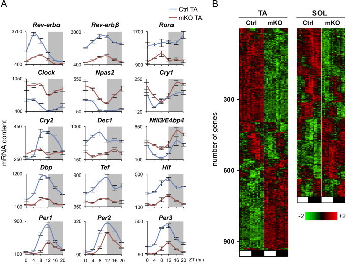
“observed similar expression patterns of Pdk4 and Pdp1 in skeletal muscle and were first to report a shift to carbohydrate utilization at the beginning of the active phase [43].”
InfluentialMechanisms of Communication in the Mammalian Circadian Timing System
Astiz, M., Heyde, I., & Oster, H. (2019) · International Journal of Molecular Sciences · doi:10.3390/ijms20020343 · Cited by 180
Misalignment with the external light environment drives metabolic and cardiac dysfunction
West, A. C., Smith, L. C., Ray, D. W., Loudon, A. S. I., Brown, T. M., & Bechtold, D. A. (2017) · Nature Communications · doi:10.1038/s41467-017-00462-2 · Cited by 173
Interconnections between circadian clocks and metabolism
Guan, D., & Lazar, M. A. (2021) · Journal of Clinical Investigation · doi:10.1172/jci148278 · Cited by 173
Chrono-nutrition for the prevention and treatment of obesity and type 2 diabetes: from mice to men
Hawley, J. A., Sassone‐Corsi, P., & Zierath, J. R. (2020) · Diabetologia · doi:10.1007/s00125-020-05238-w · Cited by 159
Dyar, K. A., Hubert, M. J., Mir, A. A., Ciciliot, S., Lutter, D., Greulich, F., Quagliarini, F., Kleinert, M., Fischer, K., Eichmann, T. O., et al. (2018) · PLoS Biology · doi:10.1371/journal.pbio.2005886 · Cited by 159
Jakubowicz, D., Wainstein, J., Ahrén, B., Bar-Dayan, Y., Landau, Z., Rabinovitz, H. R., & Froy, O. (2015) · Diabetologia · doi:10.1007/s00125-015-3524-9 · Cited by 139
Intrinsic muscle clock is necessary for musculoskeletal health
Schroder, E. A., Harfmann, B. D., Zhang, X., Srikuea, R., England, J. H., Hodge, B. A., Wen, Y., Riley, L. A., Yu, Q., Christie, A., et al. (2015) · The Journal of Physiology · doi:10.1113/jp271436 · Cited by 145
“The most recent research utilizing skeletal muscle-specific models of molecular clock disruption have focused on metabolic effects (Dyar et al. 2014; Hodge et al. 2015).”
BackgroundResultLananna, B. V., & Musiek, E. S. (2020) · Neurobiology of Disease · doi:10.1016/j.nbd.2020.104832 · Cited by 147
Off the Clock: From Circadian Disruption to Metabolic Disease
Maury, E. (2019) · International Journal of Molecular Sciences · doi:10.3390/ijms20071597 · Cited by 148
“Abrogation of the molecular clock restricted to adipose-tissue [116,117], pancreas [22,111], liver [113,118,119], skeletal muscle [120,121], smooth-muscle [122], myeloid cells [123,124] or even arcuate nucleus AgRP neurons [125] further confirmed this link (Figure 3).”
BackgroundTranscriptomic analyses reveal rhythmic and CLOCK-driven pathways in human skeletal muscle
Perrin, L., Loizides-Mangold, U., Chanon, S., Gobet, C., Hulo, N., Isenegger, L., Weger, B. D., Migliavacca, E., Charpagne, A., Betts, J. A., et al. (2018) · eLife · doi:10.7554/elife.34114 · Cited by 139
“DOI: https://doi.org/10.7554/eLife.34114 16 of 30 shift in substrate utilization in skeletal muscle from carbohydrates to lipids with impact on muscle metabolism and glucose homeostasis (Dyar et al., 2014; Dyar et al., 2015; Hodge et al., 2015; Harfmann et al., 2016).”
BackgroundResultInfluentialFasting Imparts a Switch to Alternative Daily Pathways in Liver and Muscle
Kinouchi, K., Magnan, C., Ceglia, N., Liu, Y., Cervantes, M., Pastore, N., Huynh, T., Ballabio, A., Baldi, P., Masri, S., et al. (2018) · Cell Reports · doi:10.1016/j.celrep.2018.11.077 · Cited by 135
Cedernaes, J., Schönke, M., Westholm, J. O., Mi, J., Chibalin, A., Voisin, S., Osler, M., Vogel, H., Hörnaeus, K., Dickson, S. L., et al. (2018) · Science Advances · doi:10.1126/sciadv.aar8590 · Cited by 137
Ribas‐Latre, A., & Eckel‐Mahan, K. L. (2016) · Molecular Metabolism · doi:10.1016/j.molmet.2015.12.006 · Cited by 139
“134 MOLECULAR METABOLISM 5 (2016) 133e152 Published by Elsevier G that many metabolites involved in amino acid, carbohydrate, lipid, nucleotide and xenobiotic metabolic pathways, oscillate in liver [26], muscle [27] and plasma [28], whereas 15%e70% of the metabolome in humans exhibits circadian variation depending on…”
BackgroundBaHammam, A. S., & Pirzada, R. (2023) · Clocks & Sleep · doi:10.3390/clockssleep5030034 · Cited by 144
Metabolic rivalry: circadian homeostasis and tumorigenesis
Kinouchi, K., & Sassone‐Corsi, P. (2020) · Nature reviews. Cancer · doi:10.1038/s41568-020-0291-9 · Cited by 128
Greco, C. M., Koronowski, K. B., Smith, J. G., Shi, J., Kunderfranco, P., Carriero, R., Chen, S., Samad, M., Welz, P.-S., Zinna, V. M., et al. (2021) · Science Advances · doi:10.1126/sciadv.abi7828 · Cited by 128
Gabriel, B. M., Altıntaş, A., Smith, J. A. B., Puig, L. S., Zhang, X., Basse, A. L., Laker, R. C., Gao, H., Liu, Z., Dollet, L., et al. (2021) · Science Advances · doi:10.1126/sciadv.abi9654 · Cited by 121
Circadian Metabolism: From Mechanisms to Metabolomics and Medicine
Brown, S. A. (2016) · Trends in Endocrinology and Metabolism · doi:10.1016/j.tem.2016.03.015 · Cited by 122
Melatonin Signaling a Key Regulator of Glucose Homeostasis and Energy Metabolism
Owino, S., Buonfiglio, D. D. C., Tchio, C. I. M., & Tosini, G. (2019) · Frontiers in Endocrinology · doi:10.3389/fendo.2019.00488 · Cited by 124
Opperhuizen, A., Stenvers, D. J., Jansen, R., Foppen, E., Fliers, E., & Kalsbeek, A. (2017) · Diabetologia · doi:10.1007/s00125-017-4262-y · Cited by 114
CRY1/2 Selectively Repress PPARδ and Limit Exercise Capacity
Jordan, S. D., Kriebs, A., Vaughan, M. E., Duglan, D., Fan, W., Henriksson, E., Huber, A., Papp, S. J., Nguyen, M., Afetian, M. E., et al. (2017) · Cell Metabolism · doi:10.1016/j.cmet.2017.06.002 · Cited by 115
What time is it? Deep learning approaches for circadian rhythms
Agostinelli, F., Ceglia, N., Shahbaba, B., Sassone‐Corsi, P., & Baldi, P. (2016) · Bioinformatics · doi:10.1093/bioinformatics/btw243 · Cited by 106
Perrin, L., Loizides‐Mangold, U., Skarupelova, S., Pulimeno, P., Chanon, S., Robert, M., Bouzakri, K., Modoux, C., Roux‐Lombard, P., Vidal, H., et al. (2015) · Molecular Metabolism · doi:10.1016/j.molmet.2015.07.009 · Cited by 107
Stenvers, D. J., Jongejan, A., Atiqi, S., Vreijling, J. P., Limonard, E. J., Endert, E., Baas, F., Moerland, P. D., Fliers, E., Kalsbeek, A., et al. (2019) · Diabetologia · doi:10.1007/s00125-019-4813-5 · Cited by 105
Jouffe, C., Weger, B. D., Martín, E., Atger, F., Weger, M., Gobet, C., Ramnath, D., Charpagne, A., Morin‐Rivron, D., Powell, E. E., et al. (2022) · Proceedings of the National Academy of Sciences · doi:10.1073/pnas.2200083119 · Cited by 112
Loizides‐Mangold, U., Perrin, L., Vandereycken, B., Betts, J. A., Walhin, J., Templeman, I., Chanon, S., Weger, B. D., Durand, C., Robert, M., et al. (2017) · Proceedings of the National Academy of Sciences · doi:10.1073/pnas.1705821114 · Cited by 105
“Recent data support a fundamental role for the circadian muscle clock in the regulation of glucose uptake, with a significant reduction in Glut4 mRNA and protein levels in two muscle specific Bmal1 knockout models (10, 11).”
BackgroundAoyama, S., & Shibata, S. (2017) · Frontiers in Neuroscience · doi:10.3389/fnins.2017.00063 · Cited by 108
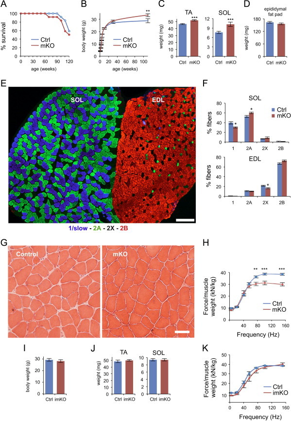
“In addition, Dyar et al. showed that reductions in muscle weight and strength were not observed in inducible muscle-specific Bmal1 knockout mice once Bmal1 knockout was induced after development (Dyar et al., 2014).”
BackgroundMethodInfluentialZeitZeiger: supervised learning for high-dimensional data from an oscillatory system
Hughey, J., Hastie, T. J., & Butte, A. J. (2016) · Nucleic Acids Research · doi:10.1093/nar/gkw030 · Cited by 103
Evidence for widespread dysregulation of circadian clock progression in human cancer
Shilts, J., Chen, G., & Hughey, J. (2018) · PeerJ · doi:10.7717/peerj.4327 · Cited by 101
Debattisti, V., Horn, A. J., Singh, R. P., Seifert, E. L., Hogarth, M. W., Mázala, D. A. G., Huang, K. T., Horváth, R., Jaiswal, J. K., & Hajnóczky, G. (2019) · Cell Reports · doi:10.1016/j.celrep.2019.09.063 · Cited by 102
“Histology—Histology and fiber type staining were performed on TA sections as previously described (Dyar et al., 2013).”
MethodZhang, L., Xin, C., Wang, S., Zhuo, S., Zhu, J., Li, Z., Liu, Y., Yang, L., & Chen, Y. (2024) · Science Advances · doi:10.1126/sciadv.adn4508 · Cited by 103
“Fiber typing was performed with Development Studies Hybridoma Bank (DSHB) mouse monoclonal antibodies for MyHC I (BA-D5), MyHC IIA (SC71), and MyHC IIB (BF-F3), as previously described ( 47 ).”
MethodAt the Interface of Lifestyle, Behavior, and Circadian Rhythms: Metabolic Implications
Bae, S.-A., Fang, M., Rustgi, V. K., Zarbl, H., & Androulakis, I. P. (2019) · Frontiers in Nutrition · doi:10.3389/fnut.2019.00132 · Cited by 103
“During the active phase, glucose is taken up by liver and muscle cells via insulin dependent transporters GLUT4 and GLUT2, which are partly regulated by the clock (88).”
BackgroundMuscle Directs Diurnal Energy Homeostasis through a Myokine-Dependent Hormone Module in Drosophila
Zhao, X. L., & Karpac, J. (2017) · Current Biology · doi:10.1016/j.cub.2017.06.004 · Cited by 93
“Lipid absorption, synthesis, and mobilization are also diurnally regulated in order to temporally coordinate energy homeostasis with changes in energy demands during rest-activity and feedingfasting cycles [8, 9], and muscle lipid consumption is thus under diurnal control [10, 11].”
Coupled network of the circadian clocks: a driving force of rhythmic physiology
Finger, A., Dibner, C., & Kramer, A. (2020) · FEBS Letters · doi:10.1002/1873-3468.13898 · Cited by 101
Metabolism and exercise: the skeletal muscle clock takes centre stage
Martin, R. A., Viggars, M. R., & Esser, K. A. (2023) · Nature Reviews Endocrinology · doi:10.1038/s41574-023-00805-8 · Cited by 109
Qaisar, R., Bhaskaran, S., Premkumar, P., Ranjit, R., Natarajan, K. S., Ahn, B., Riddle, K., Claflin, D. R., Richardson, A., Brooks, S. V., et al. (2018) · Journal of Cachexia, Sarcopenia and Muscle · doi:10.1002/jcsm.12339 · Cited by 95
“For MHC isoforms composition, cryosections of gastrocnemius muscles were stained with monoclonal anti-MHC antibodies as described elsewhere.(35) All images were taken with Nikon Eclipse Ni confocal microscope and analysed with Nikon Elements C image acquisition software.”
MethodCircadian clock network desynchrony promotes weight gain and alters glucose homeostasis in mice
Kolbe, I., Leinweber, B., Brandenburger, M., & Oster, H. (2019) · Molecular Metabolism · doi:10.1016/j.molmet.2019.09.012 · Cited by 91
Time-of-Day-Dependent Physiological Responses to Meal and Exercise
Aoyama, S., & Shibata, S. (2020) · Frontiers in Nutrition · doi:10.3389/fnut.2020.00018 · Cited by 99
“or pancreatic βcell-specific clock gene mutant mice (30, 32, 33, 45, 46).”
BackgroundEffects of Intermittent Fasting on the Circulating Levels and Circadian Rhythms of Hormones
Kim, B. H., Joo, Y., Kim, M., Choe, H. K., Tong, Q., & Kwon, O. (2021) · Endocrinology and Metabolism · doi:10.3803/enm.2021.405 · Cited by 106
“For example, skeletal muscle-specific Bmal1 knockout mice showed impaired insulin-stimulated glucose uptake in skeletal muscle [37].”
BackgroundCLOCK and BMAL1 Regulate Muscle Insulin Sensitivity via SIRT1 in Male Mice
Liu, J., Zhou, B. Z., Yan, M., Huang, R., Wang, Y., He, Z., Yang, Y., Dai, C., Wang, Y., Zhang, F., et al. (2016) · Endocrinology · doi:10.1210/en.2015-2027 · Cited by 90
The functional significance of the skeletal muscle clock: lessons from Bmal1 knockout models
Schiaffino, S., Blaauw, B., & Dyar, K. A. (2016) · Skeletal Muscle · doi:10.1186/s13395-016-0107-5 · Cited by 92
“Muscle histology and ultrastructure are normal, and muscle weight is even increased with a slight decrease in normalized muscle force [27].”
BackgroundResultInfluentialSleep, circadian rhythms, and type 2 diabetes mellitus
Parameswaran, G., & Ray, D. W. (2022) · Clinical Endocrinology · doi:10.1111/cen.14607 · Cited by 85
Endocrine regulation of circadian physiology
Tsang, A. H. K., Astiz, M., Friedrichs, M., & Oster, H. (2016) · Journal of Endocrinology · doi:10.1530/joe-16-0051 · Cited by 86
Coupling the Circadian Clock to Homeostasis: The Role of Period in Timing Physiology
Kim, P., Oster, H., Lehnert, H., Schmid, S. M., Salamat, N., Barclay, J. L., Maronde, E., Inder, W. J., & Rawashdeh, O. (2018) · Endocrine Reviews · doi:10.1210/er.2018-00049 · Cited by 87
Time-of-Day Effects on Short-Duration Maximal Exercise Performance
Mirizio, G. G., Nunes, R. S. M., Vargas, D. A., Foster, C., & Vieira, E. C. (2020) · Scientific Reports · doi:10.1038/s41598-020-66342-w · Cited by 81
Time of Day and Muscle Strength: A Circadian Output?
Douglas, C. M., Hesketh, S. J., & Esser, K. A. (2020) · Physiology · doi:10.1152/physiol.00030.2020 · Cited by 80
Circadian regulation of metabolic homeostasis: causes and consequences
Young, M. E., & McGinnis, G. R. (2016) · Nature and Science of Sleep · doi:10.2147/nss.s78946 · Cited by 82
“133 In fact, muscle-specific Bmal1 −/− mice displayed reduced in vivo contractile force, while inducible Bmal1 −/− mice were similar to controls.”
A role for circadian clock in metabolic disease
Shimizu, I., Yoshida, Y., & Minamino, T. (2016) · Hypertension Research · doi:10.1038/hr.2016.12 · Cited by 81
Potential Roles of Dec and Bmal1 Genes in Interconnecting Circadian Clock and Energy Metabolism
Sato, F., Kohsaka, A., BHAWAL, U. K., & Muragaki, Y. (2018) · International Journal of Molecular Sciences · doi:10.3390/ijms19030781 · Cited by 85
“Insulin-dependent glucose uptake in skeletal muscle was also found to be associated with the function of the Bmal1 gene [53].”
BackgroundMcGinnis, G. R., Tang, Y., Brewer, R. A., Brahma, M. K., Stanley, H. L., Shanmugam, G., Rajasekaran, N. S., Rowe, G. C., Frank, S. J., Wende, A. R., et al. (2017) · Journal of Molecular and Cellular Cardiology · doi:10.1016/j.yjmcc.2017.07.005 · Cited by 77
“(46) In addition to glucose catabolism, insulin promotes the channeling of glucose into glycogen, in part through Akt-dependent GSK3β phosphorylation/inactivation.”
BackgroundJakubowicz, D. J., Landau, Z., Tsameret, S., Wainstein, J., Raz, I., Åhrén, B., Chapnik, N., Barnea, M., Ganz, T., Menaged, M., et al. (2019) · Diabetes Care · doi:10.2337/dc19-1142 · Cited by 78
Dyar, K. A., Ciciliot, S., Tagliazucchi, G. M., Pallafacchina, G., Tothova, J., Argentini, C., Agatea, L., Abraham, R., Ahdesmaki, M. J., Forcato, M., et al. (2015) · Molecular Metabolism · doi:10.1016/j.molmet.2015.09.004 · Cited by 80
Yoshino, J., Almeda-Valdes, P., Patterson, B. W., Okunade, A. L., Imai, S.-I., Mittendorfer, B., & Klein, S. (2014) · The Journal of Clinical Endocrinology & Metabolism · doi:10.1210/jc.2014-1579 · Cited by 75
“However, the morning-to-evening direction of the variation in muscle clock gene expression in people is opposite from the direction observed in nocturnal mice (17, 19, 20).”
BackgroundSterling, P. (2020) · The MIT Press eBooks · doi:10.7551/mitpress/11472.001.0001 · Cited by 118
Abbondante, S., Eckel‐Mahan, K. L., Ceglia, N., Baldi, P., & Sassone‐Corsi, P. (2015) · Journal of Biological Chemistry · doi:10.1074/jbc.m115.681130 · Cited by 73
Brain-muscle communication prevents muscle aging by maintaining daily physiology
Kumar, A., Vaca-Dempere, M., Mortimer, T., Deryagin, O., Smith, J. G., Petrus, P., Koronowski, K. B., Greco, C. M., Segalés, J., Andrés, E., et al. (2024) · Science · doi:10.1126/science.adj8533 · Cited by 72
Saini, C., Brown, S. A., & Dibner, C. (2015) · Frontiers in Neurology · doi:10.3389/fneur.2015.00095 · Cited by 72
Immunometabolism around the Clock
Carroll, R. G., Timmons, G. A., Cervantes‐Silva, M. P., Kennedy, O. D., & Curtis, A. M. (2019) · Trends in Molecular Medicine · doi:10.1016/j.molmed.2019.04.013 · Cited by 72
The pyruvate dehydrogenase complex: Life’s essential, vulnerable and druggable energy homeostat
Stacpoole, P. W., & McCall, C. E. (2023) · Mitochondrion · doi:10.1016/j.mito.2023.02.007 · Cited by 70
Haupt, S., Eckstein, M. L., Wolf, A., Zimmer, R. T., Wachsmuth, N. B., & Moser, O. (2021) · Biomolecules · doi:10.3390/biom11040516 · Cited by 75
Owino, S., Contreras‐Alcantara, S., Baba, K., & Tosini, G. (2016) · PLoS ONE · doi:10.1371/journal.pone.0148214 · Cited by 73
“are insulin resistant and have reduced protein levels of GLUT4[30], while liver specific deletion of Bmal1 leads to a stark increase in glucose tolerance[31].”
BackgroundInfluentialCircadian clock regulation of skeletal muscle growth and repair
Chatterjee, S., & Ma, K. (2016) · F1000Research · doi:10.12688/f1000research.9076.1 · Cited by 67
“The role of the endogenous skeletal muscle molecular clock in regulating muscle metabolic functions and whole body metabolic homeostasis has emerged recently 17,69,70 .”
BackgroundMitochondria and immunity in chronic fatigue syndrome
Anderson, G., & Maes, M. H. J. (2020) · Progress in Neuro-Psychopharmacology and Biological Psychiatry · doi:10.1016/j.pnpbp.2020.109976 · Cited by 65
Circadian Clocks Make Metabolism Run
Sinturel, F., Petrenko, V., & Dibner, C. (2020) · Journal of Molecular Biology · doi:10.1016/j.jmb.2020.01.018 · Cited by 66
Metronidazole Causes Skeletal Muscle Atrophy and Modulates Muscle Chronometabolism
Manickam, R., Oh, H. Y. P., Tan, C. K., Paramalingam, E., & Wahli, W. (2018) · International Journal of Molecular Sciences · doi:10.3390/ijms19082418 · Cited by 66
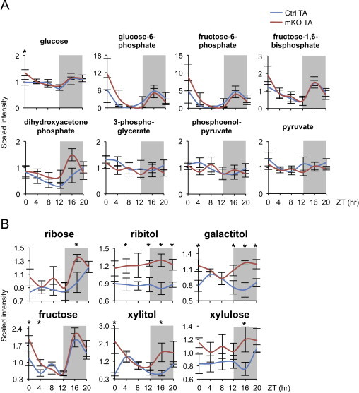
“Skeletal muscle–specific Bmal1 ablation in mice results in decreased glucose uptake and oxidation, with increased Pdk4 expression causing a switch toward increased lipid metabolism in the muscles of these mice [41].”
BackgroundOpperhuizen, A., Wang, D., Foppen, E., Jansen, R., Surovtseva, O. V., de Vries, J., Fliers, E., & Kalsbeek, A. (2016) · European Journal of Neuroscience · doi:10.1111/ejn.13377 · Cited by 64
Glucose Homeostasis: Regulation by Peripheral Circadian Clocks in Rodents and Humans
Gachon, F., Loizides‐Mangold, U., Petrenko, V., & Dibner, C. (2017) · Endocrinology · doi:10.1210/en.2017-00218 · Cited by 63
Valenzuela, F. J., Vera, J., Venegas, C., Muñoz, S., Oyarce, S., Muñoz, K., & Lagunas, C. (2016) · International Journal of Endocrinology · doi:10.1155/2016/2746909 · Cited by 62
“Cellular proliferation is a critical event for the survival and restitution of tissues of all living things and the circadian system is capable of delivering temporary information to the cell cycle [102–104].”
Background
Metadata
- Authors
- Dyar, K. A., Ciciliot, S., Wright, L. E., Biensø, R. S., Tagliazucchi, G. M., Patel, V. R., Forcato, M., Paz, M. I. P., Gudiksen, A., Solagna, F., Albiero, M., Moretti, I., Eckel-Mahan, K. L., Baldi, P., Sassone-Corsi, P., Rizzuto, R., Bicciato, S., Pilegaard, H., Blaauw, B., & Schiaffino, S.
- Affiliations
- Veneto Institute of Molecular Medicine (IT)
- University of Padua (IT)
- University of Copenhagen (DK)
- University of Modena and Reggio Emilia (IT)
- University of California, Irvine (US)
- Corresponding author
- Dyar, Kenneth A.
- Schiaffino, Stefano
- ORCID
- Venue
- Molecular Metabolism
- Publisher
- Elsevier BV
- Published
- 2014-02
- Volume
- 3
- Issue
- 1
- Pages
- 29-41
- ISSN
- 2212-8778 (ISSN-L)
- PMC
- PMC3929910
- OpenAlex
- W1964634897
- Cited by
- 454
- References
- 73
- Citations by year
- 2026: 34 · 2025: 37 · 2024: 40 · 2023: 46 · 2022: 42 · 2021: 33 · 2020: 53 · 2019: 34 · 2018: 36 · 2017: 28 · 2016: 42 · 2015: 19 · 2014: 9
- FWCI
- 12.78
- Type
- journal article
- Language
- en
- Update notices
- erratum (2014-12-01)
- Open access
- gold
- doi.org
- Licence
- Locations
- Molecular Metabolism (publishedVersion, cc-by-nc-sa)
- Europe PMC (PubMed Central) (publishedVersion, cc-by-nc-sa)
- PubMed Central (publishedVersion, cc-by-nc-sa)
- PubMed (publishedVersion)
- Research at the University of Copenhagen (University of Copenhagen) (publishedVersion, other-oa)
- eScholarship (California Digital Library) (submittedVersion)
- eScholarship (California Digital Library) (submittedVersion, cc-by-nc-sa)
- IRIS UNIMORE (University of Modena and Reggio Emilia) (submittedVersion, cc-by-nc-sa)
- Padua Research Archive (University of Padova) (submittedVersion)
- Funders
- European Commission (223576, 294777)
- Italian Space Agency (ASI, project OSMA)
- Italian Ministries of Health (Ricerca Finalizzata) and of Education, University and Research (PRIN, FIRB), NIA (2P01AG025532-06A1)
- Cariparo Foundation (Padua)
- Italian Association for Cancer Research (AIRC) and Telethon–Italy (GPP1005A, GGP11082)
- Topics
- Life Sciences › Neuroscience › Endocrine and Autonomic Systems › Circadian rhythm and melatonin
- Life Sciences › Biochemistry, Genetics and Molecular Biology › Molecular Biology › Metabolism, Diabetes, and Cancer
- Health Sciences › Medicine › Physiology (medical) › Adipose Tissue and Metabolism
- Keywords
- circadian rhythm, Bmal1, insulin sensitivity, glucose uptake, GLUT4, TBC1D1, pyruvate dehydrogenase, PDK4, PDP1, glucose oxidation, glycolysis, metabolome, conditional knockout mice
About these checks
Registry, retraction, Cites and statistics results are computed, not judged, with one exception: a printed reference that no index lists is read once by a model before it counts as a citation error. Claims are extracted by a model and kept only when their quote appears verbatim in the text; each cited source is then checked, with a model’s help, for whether it says what the claim relies on. Those verdicts can be wrong — read the claim’s quote and the cited source before relying on one.
Statistics and arithmetic are recomputed, not judged.
Confidence bands are shown only for fields calibrated on our annotated test set. None is calibrated yet, so no band is shown.
What counts as a claim ›How we measure confidence ›How we check this ›
Are you an author, or do you see a problem with this page? Request a review
