

The DOI matches its Crossref record; 81 of the 91 references it cites were found in a registry or an index.
Investigating white matter fibre density and morphology using fixel-based analysis.
Open accessAbstract
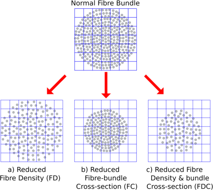
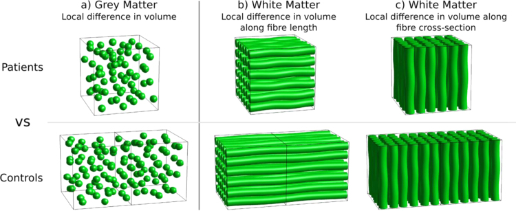
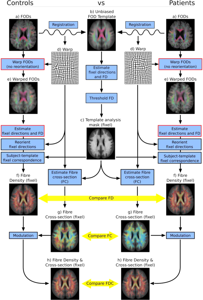
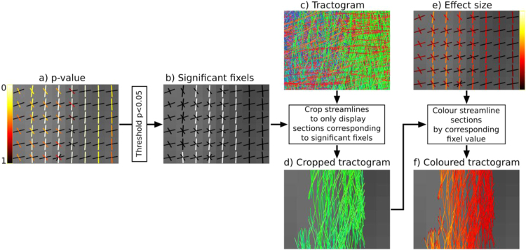
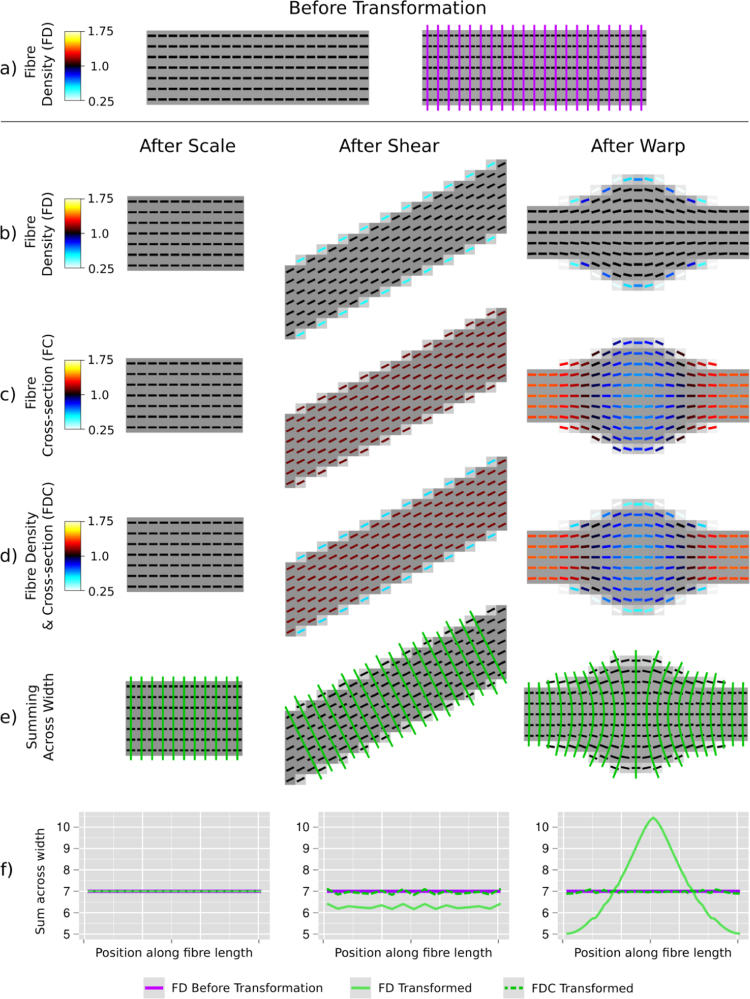
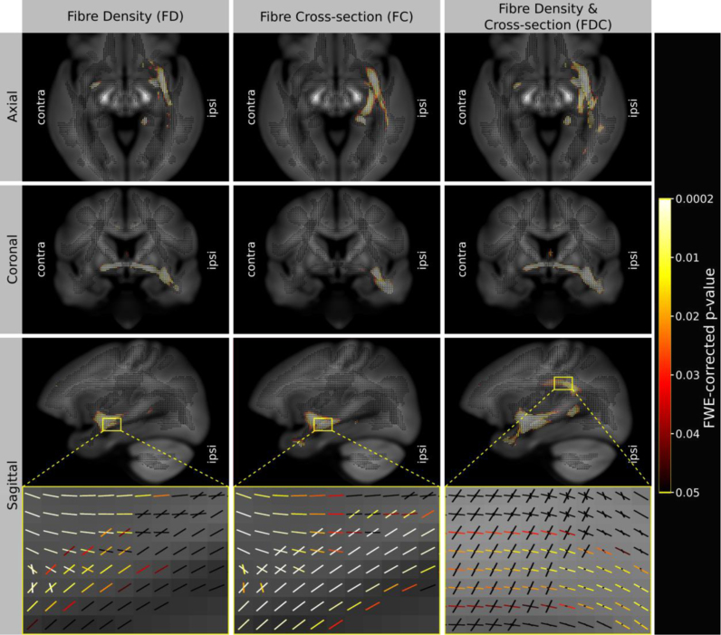
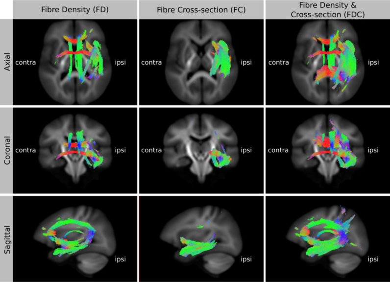
Voxel-based analysis of diffusion MRI data is increasingly popular. However, most white matter voxels contain contributions from multiple fibre populations (often referred to as crossing fibres), and therefore voxel-averaged quantitative measures (e.g. fractional anisotropy) are not fibre-specific and have poor interpretability. Using higher-order diffusion models, parameters related to fibre density can be extracted for individual fibre populations within each voxel ('fixels'), and recent advances in statistics enable the multi-subject analysis of such data. However, investigating within-voxel microscopic fibre density alone does not account for macroscopic differences in the white matter morphology (e.g. the calibre of a fibre bundle). In this work, we introduce a novel method to investigate the latter, which we call fixel-based morphometry (FBM). To obtain a more complete measure related to the total number of white matter axons, information from both within-voxel microscopic fibre density and macroscopic morphology must be combined. We therefore present the FBM method as an integral piece within a comprehensive fixel-based analysis framework to investigate measures of fibre density, fibre-bundle morphology (cross-section), and a combined measure of fibre density and cross-section. We performed simulations to demonstrate the proposed measures using various transformations of a numerical fibre bundle phantom. Finally, we provide an example of such an analysis by comparing a clinical patient group to a healthy control group, which demonstrates that all three measures provide distinct and complementary information. By capturing information from both sources, the combined fibre density and cross-section measure is likely to be more sensitive to certain pathologies and more directly interpretable.
Overview
The claims and statistics of this paper haven’t been checked yet.
Run the check — free, no account needed. It reads the open-access text and checks the sources it cites.
Integrity signals
Registry
Cites
Statistics
Figures
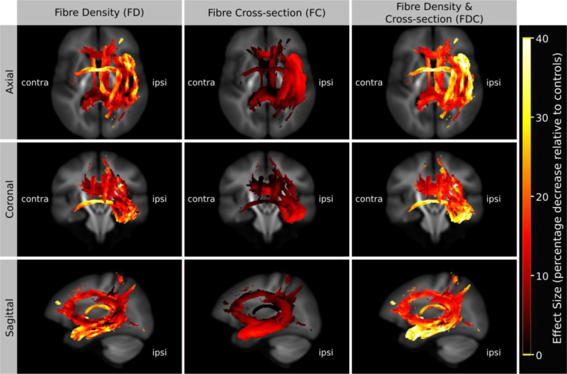
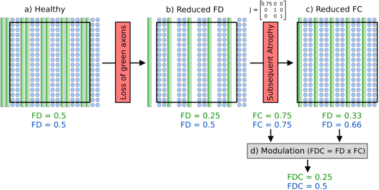
Verification
Claims the paper makes
The claims and statistics of this paper haven’t been checked yet.
Run the check — free, no account needed. It reads the open-access text and checks the sources it cites.
Claims other papers make about this one
Each is checked against this paper’s own text: does it say what the citing paper says it does?
The cited source says what the claim attributes to it.
In the citing paper
“recent fixel-based analysis approaches have been proposed to characterize a specific fiber population within a voxel (i.e., a “fixel”) (Raffelt et al., 2017).”
From Potential Pitfalls of Using Fractional Anisotropy, Axial Diffusivity, and Radial Diffusivity as Biomarkers of Cerebral White Matter Microstructure (2022) · cites this paper as Raffelt et al., 2017
Self-check
Percentages, sums and reported differences re-computed from the paper’s own numbers.
The self-check needs the paper’s full text.
Citations
Cites
Cortical Surface-Based Analysis
Dale, A. M., Fischl, B., & Sereno, M. I. (1999) · NeuroImage · doi:10.1006/nimg.1998.0395 · Cited by 10,910 · cited 1×
“For the FC and FDC analysis, we included intracranial volume (as computed using FreeSurfer from T1-weighted images (Dale et al., 1999)) as a nuisance covariate.”
MethodVoxel-Based Morphometry—The Methods
Ashburner, J., & Friston, K. J. (2000) · NeuroImage · doi:10.1006/nimg.2000.0582 · Cited by 8,676
Tract-based spatial statistics: Voxelwise analysis of multi-subject diffusion data
Smith, S. M., Jenkinson, M., Johansen‐Berg, H., Rueckert, D., Nichols, T. E., Mackay, C. E., Watkins, K. E., Ciccarelli, O., Cader, M. Z., Matthews, P. M., et al. (2006) · NeuroImage · doi:10.1016/j.neuroimage.2006.02.024 · Cited by 6,598 · cited 2×
“Furthermore, as in TBSS (Smith et al., 2006), this approach is likely to suffer from inaccurate FA-based projection in regions of crossing fibres (Bach et al., 2014; De Groot et al., 2013; Schwarz et al., 2014), and lack of fibre-specific inference when investigating voxel-wise measures such as FA.”
MethodBasser, P. J., & Pierpaoli, C. (1996) · Journal of Magnetic Resonance, Series B · doi:10.1006/jmrb.1996.0086 · Cited by 4,306 · cited 2×
“By far the most popular approach to VBA of diffusion MRI is the analysis of diffusion tensor-derived fractional anisotropy (FA) (Basser and Pierpaoli, 1996), with voxel- or clusterlevel statistical inference using packages such as SPM (http://www. fil.ion.ucl.ac.uk/spm/) or FSL…”
MethodPermutation inference for the general linear model
Winkler, A. M., Ridgway, G. R., Webster, M. A., Smith, S. M., & Nichols, T. E. (2014) · NeuroImage · doi:10.1016/j.neuroimage.2014.01.060 · Cited by 3,563 · cited 1×
“Family-wise error corrected p-values were assigned to each fixel using non-parametric permutation testing (Holmes et al., 1996; Winkler et al., 2014) with 5000 permutations.”
MethodSelf-citationProbabilistic diffusion tractography with multiple fibre orientations: What can we gain?
Behrens, T. E. J., Berg, H. J., Jbabdi, S., Rushworth, M. F. S., & Woolrich, M. W. (2007) · NeuroImage · doi:10.1016/j.neuroimage.2006.09.018 · Cited by 3,471
NODDI: Practical in vivo neurite orientation dispersion and density imaging of the human brain
Zhang, H., Schneider, T., Wheeler-Kingshott, C. A., & Alexander, D. C. (2012) · NeuroImage · doi:10.1016/j.neuroimage.2012.03.072 · Cited by 3,288 · cited 6×
“In the context of voxel-averaged DWI measures (e.g. Alexander et al., 2010; Zhang et al., 2012), the ‘density’ of a voxel will also be influenced by partial volume with cerebral spinal fluid.”
BackgroundMethodInfluentialTraining induces changes in white-matter architecture
Scholz, J., Klein, M. C., Behrens, T. E. J., & Johansen-Berg, H. (2009) · Nature Neuroscience · doi:10.1038/nn.2412 · Cited by 1,508 · cited 1×
“, 2012), individual differences and brain-behaviour correlations (Johansen-Berg, 2010), genetics (Meyer-Lindenberg, 2009), structural plasticity (Scholz et al., 2009), treatment response (Szeszko et al.”
BackgroundJeurissen, B., Tournier, J.-D., Dhollander, T., Connelly, A., & Sijbers, J. (2014) · NeuroImage · doi:10.1016/j.neuroimage.2014.07.061 · Cited by 1,484 · cited 1×
“(Jeurissen et al., 2014)) instead of tissue segmentations from T1-weighted images.”
BackgroundSelf-citationDiffusion tensor imaging of white matter tract evolution over the lifespan
Lebel, C., Gee, M., Camicioli, R., Wieler, M., Martin, W., & Beaulieu, C. (2012) · NeuroImage · doi:10.1016/j.neuroimage.2011.11.094 · Cited by 1,100 · cited 1×
“However, most white matter voxels contain contributions from multiple fibre populations (often referred to as crossing fibres), and therefore voxel-averaged quantitative measures (e.g. fractional anisotropy) are not fibre-specific and have poor interpretability.”
Background
Research works printed but not linked: 3
- Jackson, 1993, Optimizing the diagnosis of hippocampal sclerosis using MR imaging, Am. J. Neuroradiol., 14, 753
- Raffelt, D., Crozier, S., Connelly, A., Salvado, O., Tournier, J.-D., 2010. Apparent fibre density: a new measure for high angular resolution diffusion-weighted image analysis. In: Proceedings of the International Society for Magnetic Resonance in Medicine. Presented at the International Society for Magnetic Resonance in Medicine, Stockholm. Sweden, p. 575.
- Tournier, J.-D., Calamante, F., Connelly, A., 2013. A robust spherical deconvolution method for the analysis of low SNR or low angular resolution diffusion data. In: Proceedings of the International Society for Magnetic Resonance in Medicine. Salt Lake City, Utah, USA.
Cited by
Sorted by citations
MRtrix3: A fast, flexible and open software framework for medical image processing and visualisation
Tournier, J.-D., Smith, R., Raffelt, D., Tabbara, R., Dhollander, T., Pietsch, M., Christiaens, D., Jeurissen, B., Yeh, C.-H., & Connelly, A. (2019) · bioRxiv · doi:10.1101/551739 · Cited by 3,043
Self-citationMRtrix3: A fast, flexible and open software framework for medical image processing and visualisation
Tournier, J.-D., Smith, R., Raffelt, D., Tabbara, R., Dhollander, T., Pietsch, M., Christiaens, D., Jeurissen, B., Yeh, C.-H., & Connelly, A. (2019) · NeuroImage · doi:10.1016/j.neuroimage.2019.116137 · Cited by 2,974
Self-citationGorgolewski, K. J., Alfaro-Almagro, F., Auer, T., Bellec, P., Capotă, M., Chakravarty, M. M., Churchill, N. W., Cohen, A. L., Craddock, R. C., Devenyi, G. A., et al. (2017) · PLOS Computational Biology · doi:10.1371/journal.pcbi.1005209 · Cited by 311
Self-citationFibre-specific white matter reductions in Alzheimer’s disease and mild cognitive impairment
Mito, R., Raffelt, D., Dhollander, T., Vaughan, D. N., Tournier, J.-D., Salvado, O., Brodtmann, A., Rowe, C. C., Villemagne, V. L., & Connelly, A. (2018) · Brain · doi:10.1093/brain/awx355 · Cited by 309
“…for both microstructural changes in density (with the fibre density metric), and macrostructural, morphometric changes (with the fibre bundle cross-section metric) enables a more comprehensive analysis of total intra-axonal volume changes within a fibre pathway (FDC metric) (Raffelt et al., 2017).”
BackgroundMethodInfluentialSelf-citationFixel-based Analysis of Diffusion MRI: Methods, Applications, Challenges and Opportunities
Dhollander, T., Clemente, A., Singh, M., Boonstra, F., Civier, O., Duque, J. D., Egorova, N., Enticott, P., Fuelscher, I., Gajamange, S., et al. (2020) · NeuroImage · doi:10.31219/osf.io/zu8fv · Cited by 271
Self-citationFigley, C. R., Uddin, M. N., Wong, K., Kornelsen, J., Puig, J., & Figley, T. D. (2022) · Frontiers in Neuroscience · doi:10.3389/fnins.2021.799576 · Cited by 253
Modelling white matter with spherical deconvolution: How and why?
Dell’Acqua, F., & Tournier, J. (2018) · NMR in Biomedicine · doi:10.1002/nbm.3945 · Cited by 223
“This is equivalent to comparing the partial volume fractions of fibre populations, allowing for the identification of white matter differences along specific tracts across subjects.(90,91) An equivalent index based on a slightly different interpretation of the fODF is the hindrance modulated orientational anisotropy (…”
MethodSelf-citationCousineau, M., Jodoin, P.-M., Garyfallidis, E., Côté, M.-A., Morency, F. C., Rozanski, V. E., Grand'maison, M., Bedell, B. J., & Descoteaux, M. (2017) · NeuroImage: Clinical · doi:10.1016/j.nicl.2017.07.020 · Cited by 158
“Raffelt et al. (2012, 2017) NuFO Number of fiber orientations Number of local maxima of the fODF.”
BackgroundChen, Y., Wang, Y., Song, Z., Fan, Y., Gao, T., & Tang, X. (2023) · Ageing Research Reviews · doi:10.1016/j.arr.2023.101911 · Cited by 152
Differential Tractography as a Track-Based Biomarker for Neuronal InjuryPreprint
Yeh, F.-C. F., Zaydan, I. M., Suski, V. R., Lacomis, D., Richardson, R. M., Maroon, J. C., & Barrios-Martinez, J. (2019) · bioRxiv (Cold Spring Harbor Laboratory) · doi:10.1101/576025 · Cited by 3
Autosomal dominant cerebellar ataxias: Imaging biomarkers with high effect sizes
Adanyeguh, I. M., Perlbarg, V., Henry, P., Rinaldi, D., Petit, E., Valabrègue, R., Brice, A., Dürr, A., & Mochel, F. (2018) · NeuroImage Clinical · doi:10.1016/j.nicl.2018.06.011 · Cited by 103
“The processing steps have previously been fully detailed (Raffelt et al., 2015; 2017) and were performed using the MRtrix software version 0.3.15.”
MethodInfluentialSchilling, K. G., Tax, C. M. W., Rheault, F., Landman, B. A., Anderson, A. W., Descoteaux, M., & Petit, L. (2021) · Human Brain Mapping · doi:10.1002/hbm.25697 · Cited by 102
“referred to as fixels [29], and are parameterized by the mean”
What’s new and what’s next in diffusion MRI preprocessing
Tax, C. M. W., Bastiani, M., Veraart, J., Garyfallidis, E., & Irfanoglu, M. O. (2021) · NeuroImage · doi:10.1016/j.neuroimage.2021.118830 · Cited by 121
“…matrices originating from the tensor-based registration techniques have been extensively used to analyze different pathologies and those originating from FOD based registration led to analysis approaches for fiber bundle morphology with fiber bundle elements (FIXEL) ( Raffelt et al., 2017 ).”
BackgroundSydnor, V. J., Cieslak, M., Duprat, R., Deluisi, J. A., Flounders, M. W., Long, H., Scully, M., Balderston, N. L., Sheline, Y. I., Bassett, D. S., et al. (2022) · Science Advances · doi:10.1126/sciadv.abn5803 · Cited by 103
Babaeeghazvini, P., Rueda‐Delgado, L. M., Gooijers, J., Swinnen, S. P., & Daffertshofer, A. (2021) · Frontiers in Human Neuroscience · doi:10.3389/fnhum.2021.721206 · Cited by 125
Determinants and Biomarkers of Progression Independent of Relapses in Multiple Sclerosis
Calabrese, M., Preziosa, P., Scalfari, A., Colato, E., Marastoni, D., Absinta, M., Battaglini, M., Stefano, N. D., Filippo, M. D., Hametner, S., et al. (2024) · Annals of Neurology · doi:10.1002/ana.26913 · Cited by 115
Impact of b‐value on estimates of apparent fibre density
Genc, S., Tax, C., Raven, E. P., Chamberland, M., Parker, G., & Jones, D. (2020) · bioRxiv · doi:10.1002/hbm.24964 · Cited by 98
Macro- and microstructural changes in cosmonauts’ brains after long-duration spaceflight
Jillings, S., van Ombergen, A., Tomilovskaya, E., Rumshiskaya, A., Litvinova, L., Nosikova, I., Pechenkova, E., Rukavishnikov, I., Kozlovskaya, I., Manko, O., et al. (2020) · Science Advances · doi:10.1126/sciadv.aaz9488 · Cited by 92
Quantitative streamlines tractography: methods and inter-subject normalisation
Smith, R., Raffelt, D., Tournier, J.-D., & Connelly, A. (2020) · doi:10.31219/osf.io/c67kn · Cited by 92
Self-citationBundle-specific tractography with incorporated anatomical and orientational priors
Rheault, F., St‐Onge, E., Sidhu, J., Maier-Hein, K. H., Tzourio‐Mazoyer, N., Petit, L., & Descoteaux, M. (2018) · NeuroImage · doi:10.1016/j.neuroimage.2018.11.018 · Cited by 92
“The E-FOD should not be used to compute FOD metrics such as the AFD (Raffelt et al., 2017) or even NuFO (Tournier et al.”
Diffusion-Weighted Imaging: Recent Advances and Applications
Martínez‐Heras, E., Grussu, F., Prados, F., Solana, E., & Llufriú, S. (2021) · Seminars in Ultrasound CT and MRI · doi:10.1053/j.sult.2021.07.006 · Cited by 117
Effect of Particle Size and Surface Charge on Nanoparticles Diffusion in the Brain White Matter
Tian, Y., Gao, L., Zhan, W., & Dini, D. (2022) · Pharmaceutical Research · doi:10.1007/s11095-022-03222-0 · Cited by 102
“White matter (WM) acts as a relay station and transmits messages between different parts within the central nervous system [19].”
BackgroundGenc, S., Smith, R. E., Malpas, C. B., Anderson, V. A., Nicholson, J. M., Efron, D., Sciberras, E., Seal, M. L., & Silk, T. J. (2018) · NeuroImage · doi:10.1016/j.neuroimage.2018.08.043 · Cited by 85
Self-citationReduced White Matter Fiber Density in Autism Spectrum Disorder
Dimond, D., Schuetze, M., Smith, R. E., Dhollander, T., Cho, I., Vinette, S. A., Eycke, K. T., Lebel, C. A., McCrimmon, A. W., Dewey, D., et al. (2019) · Cerebral Cortex · doi:10.1093/cercor/bhy348 · Cited by 91
Self-citationAction potential propagation and synchronisation in myelinated axons
Schmidt, H., & Knösche, T. (2019) · bioRxiv · doi:10.1371/journal.pcbi.1007004 · Cited by 75
New approaches to studying early brain development in Down syndrome
Baburamani, A. A., Patkee, P. A., Arichi, T., & Rutherford, M. A. (2019) · Developmental Medicine & Child Neurology · doi:10.1111/dmcn.14260 · Cited by 74
“More complex models of voxel-wise diffusion such as a fixel-based analysis (fixel refers to a fibre population in a given voxel) can also provide measures of white matter fibre density, fibre cross-sectional area, and the fibre orientation distribution.(105) Other techniques, such as the neurite orientation distributi…”
MethodSchilling, K. G., Tax, C. M. W., Rheault, F., Hansen, C. B., Yang, Q., Yeh, F.-C. F., Cai, L. Y., Anderson, A. W., & Landman, B. A. (2021) · NeuroImage · doi:10.1016/j.neuroimage.2021.118451 · Cited by 66
The structural connectome in traumatic brain injury: A meta-analysis of graph metrics
Imms, P. E., Clemente, A., Cook, M. J., D'Souza, W. J., Wilson, P. H., Jones, D. K., & Caeyenberghs, K. (2019) · Neuroscience & Biobehavioral Reviews · doi:10.1016/j.neubiorev.2019.01.002 · Cited by 82
“Cognitive deficits (e.g., slow processing speed and poor concentration), motor control deficits (e.g., poor manual dexterity, balance deficits), and behavioural problems (e.g., impulsivity) are particularly common (Rabinowitz & Levin, 2014; Rossi & Sullivan, 1996).”
BackgroundSchroyen, G., Blommaert, J., Weehaeghe, D. V., Sleurs, C., Vandenbulcke, M., Dedoncker, N., Hatse, S., Goris, A., Koole, M. J. B., Smeets, A., et al. (2021) · Cancers · doi:10.3390/cancers13164198 · Cited by 80
“This reduction is likely to reflect impaired ability to transfer information across brain regions, potentially indicating impaired WM integrity [37] and thereby possibly contributing to cognitive impairments seen in chemotherapy-treated patients.”
BackgroundMethodPervasive White Matter Fiber Degeneration in Ischemic Stroke
Egorova, N., Dhollander, T., Khlif, M., Khan, W., Werden, E., & Brodtmann, A. (2020) · Stroke · doi:10.1161/strokeaha.119.028143 · Cited by 69
“These diffusion tensor imaging metrics are nonspecific voxel-averaged measures that do not account for complex microstructural fiber geometry and are known to lead to erroneous interpretations in locations where fibers are crossing.(15) In the current study, we examined white matter degeneration in a cohort of partici…”
BackgroundInfluentialDisentangling the effects of Alzheimer’s and small vessel disease on white matter fibre tracts
Dewenter, A., Jacob, M. A., Cai, M., Gesierich, B. G., Hager, P., Kopczak, A., Biel, D., Ewers, M., Tuladhar, A. M., de Leeuw, F., et al. (2022) · Brain · doi:10.1093/brain/awac265 · Cited by 72
“19 Using this framework, one can simultaneously derive tract-specific measures of fibre density and fibre-bundle cross-section.”
Andrews, D. S., Lee, J. K., Harvey, D. J., Waizbard‐Bartov, E., Solomon, M., Rogers, S. J., Nordahl, C. W., & Amaral, D. G. (2020) · Biological Psychiatry · doi:10.1016/j.biopsych.2020.10.013 · Cited by 76
Fiber-specific white matter reductions in Parkinson hallucinations and visual dysfunction
Zarkali, A., McColgan, P., Leyland, L.-A., Lees, A., Rees, G., & Weil, R. (2020) · Neurology · doi:10.1212/wnl.0000000000009014 · Cited by 67
Early childhood development of white matter fiber density and morphology
Dimond, D., Rohr, C. S., Smith, R. E., Dhollander, T., Cho, I. Y. K., Lebel, C., Dewey, D., Connelly, A., & Bray, S. (2019) · bioRxiv · doi:10.1101/624171 · Cited by 67
Self-citationReview of Diffusion MRI Studies in Chronic White Matter Diseases
Raja, R., Rosenberg, G., & Caprihan, A. (2018) · Neuroscience Letters · doi:10.1016/j.neulet.2018.12.007 · Cited by 67
“Moreover, fixel based analysis [99] which computes metrics on more than one fiber population present within a voxel is potentially another significant method for predictions in SVD.”
MethodWhite matter injury and neurodevelopmental disabilities: A cross-disease (dis)connection
Cainelli, E., Arrigoni, F., & Vedovelli, L. (2020) · Progress in Neurobiology · doi:10.1016/j.pneurobio.2020.101845 · Cited by 74
Histological and MRI markers of white matter damage in focal epilepsy
Deleo, F., Thom, M., Concha, L., Bernasconi, A., Bernhardt, B. C., & Bernasconi, N. (2017) · Epilepsy Research · doi:10.1016/j.eplepsyres.2017.11.010 · Cited by 64
“analysis, a diffusion weighted MRI reconstruction technique which combines the measurements of fiber cross-sectional area, a measure of morphology, with microstructural information of fiber density (Raffelt et al., 2017), thereby providindg a sensitive marker of intra-axonal volume (Raffelt et al.”
A framework for multi-component analysis of diffusion MRI data over the neonatal period
Pietsch, M., Christiaens, D., Hutter, J. M., Cordero‐Grande, L., Price, A. N., Hughes, E., Edwards, A. D., Hajnal, J. V., Counsell, S. J., & Tournier, J. (2018) · NeuroImage · doi:10.1016/j.neuroimage.2018.10.060 · Cited by 68
Self-citationPannek, K., Fripp, J., George, J., Fiori, S., Colditz, P., Boyd, R., & Rose, S. (2018) · NeuroImage: Clinical · doi:10.1016/j.nicl.2018.01.003 · Cited by 60
Diffusion-based tractography atlas of the human acoustic radiation
Maffei, C., Sarubbo, S., & Jovicich, J. (2019) · Scientific Reports · doi:10.1038/s41598-019-40666-8 · Cited by 62
Zarkali, A., McColgan, P., Leyland, L.-A., Lees, A., & Weil, R. (2020) · bioRxiv · doi:10.1002/mds.28477 · Cited by 58
Sex is a defining feature of neuroimaging phenotypes in major brain disorders
Salminen, L. E., Tubi, M. A., Bright, J. K., Thomopoulos, S. I., Wieand, A., & Thompson, P. M. (2021) · Human Brain Mapping · doi:10.1002/hbm.25438 · Cited by 61
Recent advances in diffusion neuroimaging: applications in the developing preterm brain
Pecheva, D., Kelly, C., Kimpton, J., Bonthrone, A., Batalle, D., Zhang, H., & Counsell, S. (2018) · F1000Research · doi:10.12688/f1000research.15073.1 · Cited by 57
A longitudinal fixel-based analysis of white matter alterations in patients with Parkinson's disease
Rau, Y.-A., Wang, S., Tournier, J., Lin, S., Lu, C., Weng, Y.-H., Chen, Y.-L., Ng, S., Yu, S.-W., Wu, Y.-M., et al. (2019) · NeuroImage Clinical · doi:10.1016/j.nicl.2019.102098 · Cited by 52
“The detection by FBA might provide additional information related to white matter changes than conventional voxel-based analysis (Raffelt et al., 2017) and may shed more light to the pathology of PD—based on previous studies in the field.”
InfluentialSelf-citationFibre-specific white matter changes in multiple sclerosis patients with optic neuritis
Gajamange, S., Raffelt, D. A., Dhollander, T., LUI, E. H., van der Walt, A., Kilpatrick, T. J., Fielding, J., Connelly, A., & Kolbe, S. (2017) · NeuroImage Clinical · doi:10.1016/j.nicl.2017.09.027 · Cited by 60
Self-citationZamani, A., Walker, A. K., Rollo, B., Ayers, K. L., Farah, R., O'Brien, T. J., & Wright, D. K. (2022) · Translational Neurodegeneration · doi:10.1186/s40035-022-00291-4 · Cited by 60
“As the name suggests, changes in the combined fibre density and cross-section measure reflect both a macroscopic change to the white matter bundle and a microstructural change due to increased or decreased axon fibre population [15, 38].”
BackgroundMethodMicrostructural Alterations in Hippocampal Subfields Mediate Age-Related Memory Decline in Humans
Radhakrishnan, H., Stark, S. M., & Stark, C. E. L. (2020) · Frontiers in Aging Neuroscience · doi:10.3389/fnagi.2020.00094 · Cited by 49
Genc, S., Malpas, C. B., Ball, G., Silk, T. J., & Seal, M. L. (2018) · Brain Structure and Function · doi:10.1007/s00429-018-1658-5 · Cited by 56
“Higher AFD with older developmental age might therefore reflect bigger axon diameter (Raffelt et al. 2017; Genc et al. 2017b), however this would need to be verified by imaging developmental populations using a high gradient system (McNab et al.”
BackgroundMethodAndica, C., Kamagata, K., Kirino, E., Uchida, W., Irie, R., Murata, S., & Aoki, S. (2021) · Molecular Autism · doi:10.1186/s13229-021-00456-4 · Cited by 53
“Using diffusion kurtosis imaging (DKI) [58] and fixel-based analysis (FBA) [59], reduced axonal density, marked by decreased axonal water fraction, has also been suggested within the CC and long-range association tracts in adults with autism [4, 49].”
MethodCalamuneri, A., Arrigo, A., Mormina, E., Milardi, D., Cacciola, A., Chillemi, G., Marino, S., Gaeta, M., & Quartarone, A. (2018) · Frontiers in Neurology · doi:10.3389/fneur.2018.00716 · Cited by 51
“AFD has been recently successfully applied in normal as well as pathological conditions (27, 28).”
BackgroundMethodChoy, S. W., Bagarinao, E., Watanabe, H., Ho, E. T. W., Maesawa, S., Mori, D., Hara, K., Kawabata, K., Yoneyama, N., Ohdake, R., et al. (2020) · Human Brain Mapping · doi:10.1002/hbm.25008 · Cited by 44
“The acquired DWI data were processed using MRtrix3 (version 3.0, http://www.mrtrix.org) following the recommended FBA (Raffelt et al., 2017) preprocessing pipeline.”
MethodInfluentialCharacteristics of Neural Network Changes in Normal Aging and Early Dementia
Watanabe, H., Bagarinao, E., Maesawa, S., Hara, K., Kawabata, K., Ogura, A., Ohdake, R., Shima, S., Mizutani, Y., Ueda, A., et al. (2021) · Frontiers in Aging Neuroscience · doi:10.3389/fnagi.2021.747359 · Cited by 48
Brain microstructural changes and fatigue after COVID-19
Bispo, D. D. D. C., Brandão, P. R. D. P., Pereira, D. A., Maluf, F. B., Dias, B. A., Paranhos, H. R., von Glehn, F., de Oliveira, A. C. P., Regattieri, N. A. T., Silva, L. S., et al. (2022) · Frontiers in Neurology · doi:10.3389/fneur.2022.1029302 · Cited by 56
Tunç, B., Yankowitz, L., Parker, D., Alappatt, J. A., Pandey, J., Schultz, R., & Verma, R. (2019) · Molecular Autism · doi:10.1186/s13229-019-0301-5 · Cited by 47
Sun, D., Haswell, C. C., Morey, R. A., & Bellis, M. D. D. (2018) · Development and Psychopathology · doi:10.1017/s0954579418000093 · Cited by 44
“, 2008); (c) structural connectivity networks based on diffusion tensor imaging (DTI) of fiber tracts (Hofer & Frahm, 2006; Raffelt et al., 2017); and (d) structural connectivity networks delineated by between subject intraregional correlations in measures of cortical thickness or gray matter volume (Gong, He, Chen, &…”
BackgroundGrazioplene, R. G., Bearden, C., Subotnik, K., Ventura, J., Haut, K. M., Nuechterlein, K., & Cannon, T. D. (2018) · NeuroImage: Clinical · doi:10.1016/j.nicl.2018.02.015 · Cited by 46
“While FA captures information about overall relative diffusion patterns in a voxel, studies in both phantom and human data show that fixel-based analysis can approximate both the density and macrostructural cross-section of distinct fiber bundles traversing a voxel (Raffelt et al., 2015, 2016).”
BackgroundMethodStructural network differences in chronic musculoskeletal pain: Beyond fractional anisotropy
Bishop, J. H., Shpaner, M., Kubicki, A., Clements, S., Watts, R., & Naylor, M. R. (2017) · NeuroImage · doi:10.1016/j.neuroimage.2017.12.021 · Cited by 50
“Fixel-based analysis was then implemented as described in-depth by Raffelt and colleagues (Raffelt et al., 2017).”
Dou, X., Yao, H.-X., Feng, F., Wang, P., Zhou, B., Jin, D., Yang, Z.-Y., Li, J., Zhao, C., Wang, L., et al. (2020) · Cortex; a journal devoted to the study of the nervous system and behavior · doi:10.2139/ssrn.3349212 · Cited by 45
Arun, A. H., Connelly, A., Smith, R., & Calamante, F. (2020) · NeuroImage · doi:10.1016/j.neuroimage.2020.117505 · Cited by 44
Self-citationTract‐specific atrophy in focal epilepsy: Disease, genetics, or seizures?
Vaughan, D., Raffelt, D., Curwood, E., Tsai, M.-H., Tournier, J.-D., Connelly, A., & Jackson, G. (2017) · Annals of Neurology · doi:10.1002/ana.24848 · Cited by 44
“Multiplying FD and FC at each fixel gives a combined measure of the fiber density and cross-sectional area (FDC).(26)”
MethodInfluentialSelf-citationLindsey, H. M., Wilde, E. A., Caeyenberghs, K., & Dennis, E. L. (2019) · Frontiers in Neurology · doi:10.3389/fneur.2019.01296 · Cited by 55
“Fixel-based measures, such as fiber cross-section (FC), a measurement of fixel diameter, can identify tracts that are affected by regions with crossing fibers, overcoming this inherent limitation of the diffusion tensor model (128).”
BackgroundSubtle white matter alterations in schizophrenia identified with a new measure of fiber density
Stämpfli, P., Sommer, S., Manoliu, A., Burrer, A., Schmidt, A., Herdener, M., Seifritz, E., Kaiser, S., & Kirschner, M. (2019) · Scientific Reports · doi:10.1038/s41598-019-40070-2 · Cited by 40
Aging, brain plasticity, and motor learning
Gooijers, J., Pauwels, L., Hehl, M., Seer, C., Cuypers, K., & Swinnen, S. P. (2024) · Ageing Research Reviews · doi:10.1016/j.arr.2024.102569 · Cited by 45
Jünemann, K., Marié, D., Worschech, F., Scholz, D. S., Grouiller, F., Kliegel, M., Ville, D. V. D., James, C. E., Krüger, T. H. C., ALTENMÜLLER, E. O., et al. (2022) · Frontiers in Aging Neuroscience · doi:10.3389/fnagi.2022.817889 · Cited by 46
White matter tract signatures of fiber density and morphology in ADHD.
Fuelscher, I., Hyde, C., Anderson, V., & Silk, T. (2021) · Cortex; a journal devoted to the study of the nervous system and behavior · doi:10.1016/j.cortex.2021.02.015 · Cited by 40
Zhang, J., Zhong, S.-C., Zhou, L., Yu, Y.-M., Tan, X., Wu, M., Sun, P., Zhang, W., Li, J.-B., Cheng, R.-D., et al. (2021) · Neuropsychology Review · doi:10.1007/s11065-021-09482-8 · Cited by 40
White matter alterations at pubertal onset
Genc, S., Seal, M. L., Dhollander, T., Malpas, C. B., Hazell, P., & Silk, T. J. (2017) · NeuroImage · doi:10.1016/j.neuroimage.2017.05.017 · Cited by 46
“Fixel-based analysis Fixel-based analysis (FBA) was performed in accordance with a recommended pipeline (Raffelt et al., 2017).”
Diffusion Imaging Reveals Sex Differences in the White Matter Following Sports-Related Concussion.
Wright, D. K., Symons, G. F., O’Brien, W., McDonald, S., Zamani, A., Major, B., Chen, Z.-B., Costello, D. M., Brady, R., Sun, M.-J., et al. (2021) · Cerebral Cortex · doi:10.1093/cercor/bhab095 · Cited by 39
Lyon, M., Welton, T., Varda, A., Maller, J. J., Broadhouse, K. M., Korgaonkar, M. S., Koslow, S. H., Williams, L. M., Gordon, E., Rush, A. J., et al. (2019) · NeuroImage Clinical · doi:10.1016/j.nicl.2019.101668 · Cited by 39
“In this paper we evaluate fixel-based analysis (FBA) (Raffelt et al., 2017), a new technique for the analysis of diffusion imaging (dMRI), using data from the international Study to Predict Optimized Treatment in Depression (iSPOT-D).”
MethodLiu, T.-T., Gao, F.-S., Zheng, W., You, Y., Zhao, Z.-Y., Lv, Y., Chen, W., Zhang, H.-X., Ji, C., & Wu, D. (2021) · NeuroImage · doi:10.1016/j.neuroimage.2021.118465 · Cited by 37
“Fixel- ased analysis (FBA) is a recently developed model-free approach to dentify crossing fibres with each fibre component in a voxel called a fixel ” ( Raffelt et al., 2017 ; Tournier et al., 2004 ) and provides mi- rostructural measurements including the fibre density (FD) and fibre ross-section…”
Zhang, F., Wu, W., Ning, L., McAnulty, G. B., Waber, D. P., Gagoski, B. A., Sarill, K., Hamoda, H. M., Song, Y., Cai, W., et al. (2018) · NeuroImage · doi:10.1016/j.neuroimage.2018.01.006 · Cited by 35
“Fixel-based analysis (FBA) that takes account of multiple fibers within a voxel has been proposed recently to enable enhanced whole-brain statistics (Raffelt et al., 2015, 2017), showing the potential of incorporating information from multiple fiber populations into WM analyses.”
Autism spectrum disorders: Neuroimaging findings from systematic reviews
Pua, E. P. K., Bowden, S. C., & Seal, M. L. (2017) · Research in Autism Spectrum Disorders · doi:10.1016/j.rasd.2016.11.005 · Cited by 37
Carandini, T., Mancini, M., Bogdan, I., Rae, C. L., Barritt, A. W., Sethi, A., Harrison, N. A., Rashid, W., Scarpini, E. A., Galimberti, D., et al. (2021) · NeuroImage Clinical · doi:10.1016/j.nicl.2021.102587 · Cited by 44
“multiplied by each fixel to obtain FDC values (FDC = FDxFC) (Raffelt et al., 2017).”
BackgroundMethodInfluentialCerebellar structural connectivity and contributions to cognition in frontotemporal dementias
Chen, Y., Landín-Romero, R., Kumfor, F., Irish, M., Hodges, J. R., & Piguet, O. (2020) · Cortex · doi:10.1016/j.cortex.2020.04.013 · Cited by 41
Adab, H. Z., Chalavi, S., Monteiro, T. S., Gooijers, J., Dhollander, T., Mantini, D., & Swinnen, S. P. (2020) · NeuroImage · doi:10.1016/j.neuroimage.2020.116530 · Cited by 38
“The FDC metric was used in these analyses since it is a measure of total intra-axonal volume occupied by white matter fibers and can be denoted as the marker of total capacity of a particular white matter tract for information transmission (Raffelt et al., 2017), i.”
InfluentialFixel-Based Analysis of Visual Pathway White Matter in Primary Open-Angle Glaucoma.
Haykal, S., Ćurčić-Blake, B., Jansonius, N., & Cornelissen, F. (2019) · Investigative Ophthalmology and Visual Science · doi:10.1167/iovs.19-27447 · Cited by 35
“FD can be used to probe WM microstructural changes in a fiber-specific manner, with a decrease in FD indicating a loss of axons.(45,46) FC quantifies gross morphologic (or macrostructural) WM changes by measuring fiber bundle cross-sectional area in a plane perpendicular to fixel orientation.”
BackgroundMethodInfluentialReview and Perspective on Brain Bases of Tinnitus
Husain, F., & Khan, R. A. (2023) · Journal of the Association for Research in Otolaryngology · doi:10.1007/s10162-023-00914-1 · Cited by 34
“DWI also lends itself to more sophisticated analysis techniques, such as fixel-based analysis (FBA), which helps account for multiple fibers within a voxel by reporting metrics such as fiber density and fiber-bundle cross section [30].”
Huang, P., & Zhang, M. (2022) · Neuroscience Bulletin · doi:10.1007/s12264-022-00905-x · Cited by 40
“Furthermore, problems such as fiber crossing [107] and water contamination [108] can lead to substantiate biases.”
BackgroundPark, Y. W., Joers, J. M., Guo, B., Hutter, D., Bushara, K. O., Adanyeguh, I. M., Eberly, L. E., Öz, G., & Lenglet, C. (2020) · Frontiers in Neurology · doi:10.3389/fneur.2020.00411 · Cited by 37
“WM FOD metrics were estimated using MRtrix3 (29) with multi-tissue constrained spherical deconvolution (CSD), accounting for WM and gray matter (GM) compartments (42).”
BackgroundMethodEvaluating spatiotemporal microstructural alterations following diffuse traumatic brain injury
Mohamed, A. Z., Corrigan, F., Collins-Praino, L., Plummer, S. L., Soni, N., & Nasrallah, F. (2019) · NeuroImage: Clinical · doi:10.1016/j.nicl.2019.102136 · Cited by 34
Toschi, N., Gisbert, R. A., Passamonti, L., Canals, S., & Santis, S. D. (2019) · Neurobiology of Aging · doi:10.1016/j.neurobiolaging.2019.11.014 · Cited by 35
“While other similar multi-compartment models are available (Fieremans et al, 2011; Zhang et al, 2012; Raffelt et al., 2017), we used CHARMED as our main hypothesis, informed by previous work (De Santis et al.”
MethodPrinciples of temporal association cortex organisation as revealed by connectivity gradients
Freches, G. B., Haak, K., Bryant, K. L., Schurz, M., Beckmann, C., & Mars, R. (2020) · Brain Structure and Function · doi:10.1007/s00429-020-02047-0 · Cited by 33
Horbrügger, M., Loewe, K., Kaufmann, J., Wagner, M., Schippling, S., Pawlitzki, M., & Schoenfeld, M. A. (2019) · NeuroImage Clinical · doi:10.1016/j.nicl.2019.101740 · Cited by 31
Zarkali, A., McColgan, P., Leyland, L. A., Lees, A. J., & Weil, R. S. (2022) · Journal of Neurology, Neurosurgery & Psychiatry · doi:10.1136/jnnp-2021-326630 · Cited by 32
Applications of advanced diffusion MRI in early brain development: a comprehensive review
DiPiero, M. A., Rodrigues, P. G., Gromala, A., & Dean, D. (2022) · Brain Structure and Function · doi:10.1007/s00429-022-02605-8 · Cited by 34
Mitchell, T., Wilkes, B. J., Archer, D. B., Chu, W. T., Coombes, S. A., Lai, S., Mcfarland, N. R., Okun, M., Black, M. L., Herschel, E., et al. (2022) · NeuroImage: Clinical · doi:10.1016/j.nicl.2022.103022 · Cited by 32
“Fixel-based analysis is a novel diffusion MRI technique that combines voxel-based analysis with tract-based analysis to address the problem of crossing fibers within a voxel, which can result in misleading outcomes in voxels with a high number of crossing fibers (Raffelt et al., 2017).”
InfluentialGenc, S., Malpas, C. B., Gülenç, A., Sciberras, E., Efron, D., Silk, T. J., & Seal, M. L. (2020) · Developmental Cognitive Neuroscience · doi:10.1016/j.dcn.2020.100853 · Cited by 37
Li, Y., Guo, T., Guan, X., Gao, T., Sheng, W., Zhou, C., Wu, J., Xuan, M., Gu, Q., Zhang, M., et al. (2020) · NeuroImage Clinical · doi:10.1016/j.nicl.2020.102355 · Cited by 31
“Moreover, to ensure that the data are centered around zero and normally distributed, we calculated the log-FC instead of the FC for comparison (Raffelt et al., 2017).”
MethodResultInfluentialGirls' pubertal development is associated with white matter microstructure in late adolescence
Chahal, R., Vilgis, V., Grimm, K. J., Hipwell, A. E., Forbes, E. E., Keenan, K. E., & Guyer, A. E. (2018) · NeuroImage · doi:10.1016/j.neuroimage.2018.07.050 · Cited by 30
The developing brain structural and functional connectome fingerprint
Ciarrusta, J., Christiaens, D., Fitzgibbon, S., Dimitrova, R., Hutter, J., Hughes, E., Duff, E., Price, A., Cordero-Grande, L., Tournier, J.-D., et al. (2021) · bioRxiv · doi:10.1016/j.dcn.2022.101117 · Cited by 31
Self-citationRoy, M., Fortier, M., Rheault, F., Edde, M., Croteau, É., Castellano, C., Langlois, F., St-Pierre, V., Cuenoud, B., Bocti, C., et al. (2021) · medRxiv · doi:10.1002/trc2.12217 · Cited by 31
Linking Cortical and Connectional Pathology in Schizophrenia
Biase, M. A. D., Cropley, V., Cocchi, L., Fornito, A., Calamante, F., Ganella, E. P., Pantelis, C., & Zalesky, A. (2018) · Schizophrenia Bulletin · doi:10.1093/schbul/sby121 · Cited by 33
Multi-tissue spherical deconvolution of tensor-valued diffusion MRI
Jeurissen, B., & Szczepankiewicz, F. (2021) · NeuroImage · doi:10.1016/j.neuroimage.2021.118717 · Cited by 30
Kelley, S., Plass, J., Bender, A. R., & Polk, T. (2021) · Cerebral Cortex · doi:10.1093/cercor/bhab056 · Cited by 30
“However, in white matter voxels with crossing fibers one cannot know whether decreases in FA reflect reduced signal from the largest fiber population, increased signal from orthogonal populations, or other combinations of intra-voxel influences (Riffert et al. 2014).”
BackgroundMethodResultInfluentialChahal, R., Kirshenbaum, J., Ho, T., Mastrovito, D., & Gotlib, I. (2020) · Developmental Cognitive Neuroscience · doi:10.1016/j.dcn.2020.100899 · Cited by 30
“Further, FDC has been shown to be "more sensitive to certain pathologies and more directly interpretable" than are voxel-averaged quantitative measures (e.g., fractional anisotropy) (page 58, Raffelt et al., 2017).”
BackgroundMethodInfluentialKelly, C. E., Thompson, D. K., Genc, S., Chen, J., Yang, J., Adamson, C., Beare, R., Seal, M., Doyle, L., Cheong, J., et al. (2020) · medRxiv · doi:10.1101/2020.04.01.20049585 · Cited by 30
“is strongly attenuated, leaving the remaining signal localised to the intra-axonal water compartment, and allowing us to make fibre-specific inferences using FBA.(8,35) The observed lower FD in VP children compared with FT children suggests VP children have lower axonal densities within fibre tracts, which could refle…”
InfluentialMaternal depressive symptoms, neonatal white matter, and toddler social-emotional development
Lautarescu, A., Bonthrone, A. F., Pietsch, M., Batalle, D., Cordero-Grande, L., Tournier, J.-D., Christiaens, D., Hajnal, J. V., Chew, A., Falconer, S., et al. (2022) · Translational Psychiatry · doi:10.1038/s41398-022-02073-y · Cited by 29
“FBA provides information regarding white matter structure for fibre bundles within a voxel through estimates of microstructure via local fibre density (FD), and morphometry via the fibre cross-section (FC) metric; their product, the fibre density and cross section (FDC), serves as a proxy of a fibre bundle’s capacity…”
BackgroundSelf-citationMapping the macrostructure and microstructure of the in vivo human hippocampus using diffusion MRI
Karat, B. G., DeKraker, J., Hussain, U., Köhler, S., & KHAN, A. R. (2023) · Human Brain Mapping · doi:10.1002/hbm.26461 · Cited by 33
Cognitive Training in Young Patients With Traumatic Brain Injury: A Fixel-Based Analysis
Verhelst, H., Giraldo, D. L., Linden, C. V., Vingerhoets, G., Jeurissen, B., & Caeyenberghs, K. (2019) · Neurorehabilitation and neural repair · doi:10.1177/1545968319868720 · Cited by 32
Hyde, C., Fuelscher, I., Enticott, P., Jones, D., Farquharson, S., Silk, T., Williams, J., & Caeyenberghs, K. (2018) · NeuroImage: Clinical · doi:10.1016/j.nicl.2018.101625 · Cited by 29
Metadata
- Authors
- Raffelt, D. A., Tournier, J.-D., Smith, R. E., Vaughan, D. N., Jackson, G., Ridgway, G. R., & Connelly, A.
- Affiliations
- Florey Institute of Neuroscience and Mental Health (AU)
- King's College London (GB)
- The University of Melbourne (AU)
- Wellcome Centre for Human Neuroimaging (GB)
- Corresponding author
- Raffelt, David A.
- ORCID
- Venue
- NeuroImage
- Publisher
- Elsevier BV
- Published
- 2017-01
- Volume
- 144
- Issue
- Pt A
- Pages
- 58-73
- ISSN
- 1053-8119 (ISSN-L)
- PMC
- PMC5182031
- OpenAlex
- W2520744072
- Cited by
- 620
- References
- 91
- Citations by year
- 2026: 74 · 2025: 79 · 2024: 78 · 2023: 76 · 2022: 84 · 2021: 91 · 2020: 71 · 2019: 30 · 2018: 26 · 2017: 9
- FWCI
- 19.39
- Type
- journal article
- Language
- en
- Open access
- hybrid
- doi.org
- Locations
- NeuroImage (publishedVersion, cc-by)
- PubMed Central (publishedVersion, cc-by)
- PubMed (publishedVersion)
- UCL Discovery (University College London) (submittedVersion)
- Research Portal (King's College London) (submittedVersion, cc-by)
- Funders
- National Health and Medical Research Council (NHMRC)
- Victorian Government’s Operational Infrastructure
- UK Medical Research Council (MR/J014257/2)
- Wellcome Trust (091593/Z/10/Z)
- Topics
- Health Sciences › Medicine › Radiology, Nuclear Medicine and Imaging › Advanced Neuroimaging Techniques and Applications
- Life Sciences › Neuroscience › Cognitive Neuroscience › Functional Brain Connectivity Studies
- Health Sciences › Medicine › Radiology, Nuclear Medicine and Imaging › Advanced MRI Techniques and Applications
- Keywords
- fixel-based analysis, crossing fibers, axon number
- MeSH terms
- Humans, Nerve Fibers, Myelinated, Diffusion Magnetic Resonance Imaging, White Matter
About these checks
Registry, retraction, Cites and statistics results are computed, not judged, with one exception: a printed reference that no index lists is read once by a model before it counts as a citation error. Claims are extracted by a model and kept only when their quote appears verbatim in the text; each cited source is then checked, with a model’s help, for whether it says what the claim relies on. Those verdicts can be wrong — read the claim’s quote and the cited source before relying on one.
Statistics and arithmetic are recomputed, not judged.
Confidence bands are shown only for fields calibrated on our annotated test set. None is calibrated yet, so no band is shown.
What counts as a claim ›How we measure confidence ›How we check this ›
Are you an author, or do you see a problem with this page? Request a review
