

The DOI matches its Crossref record; 128 of the 131 references it cites were found in a registry or an index.
Effectiveness of regional DTI measures in distinguishing Alzheimer’s disease, MCI, and normal aging
Open accessAbstract
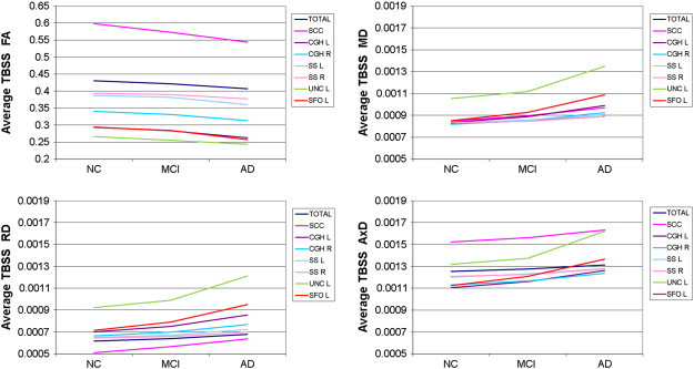
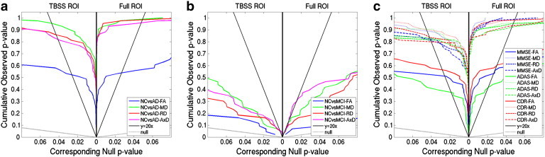
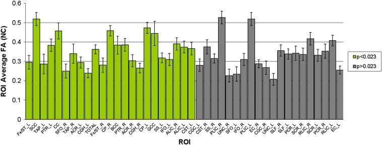
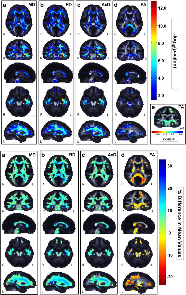
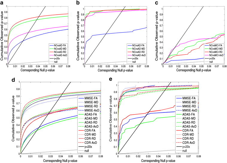
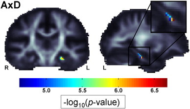
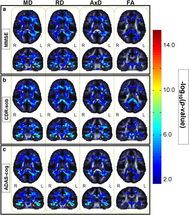
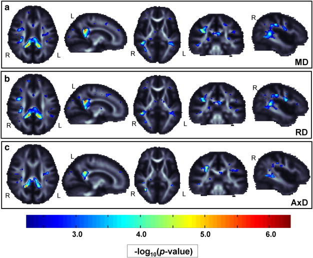
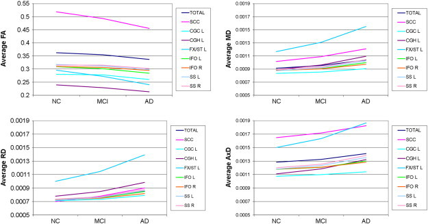
The Alzheimer's Disease Neuroimaging Initiative (ADNI) recently added diffusion tensor imaging (DTI), among several other new imaging modalities, in an effort to identify sensitive biomarkers of Alzheimer's disease (AD). While anatomical MRI is the main structural neuroimaging method used in most AD studies and clinical trials, DTI is sensitive to microscopic white matter (WM) changes not detectable with standard MRI, offering additional markers of neurodegeneration. Prior DTI studies of AD report lower fractional anisotropy (FA), and increased mean, axial, and radial diffusivity (MD, AxD, RD) throughout WM. Here we assessed which DTI measures may best identify differences among AD, mild cognitive impairment (MCI), and cognitively healthy elderly control (NC) groups, in region of interest (ROI) and voxel-based analyses of 155 ADNI participants (mean age: 73.5 ± 7.4; 90 M/65 F; 44 NC, 88 MCI, 23 AD). Both VBA and ROI analyses revealed widespread group differences in FA and all diffusivity measures. DTI maps were strongly correlated with widely-used clinical ratings (MMSE, CDR-sob, and ADAS-cog). When effect sizes were ranked, FA analyses were least sensitive for picking up group differences. Diffusivity measures could detect more subtle MCI differences, where FA could not. ROIs showing strongest group differentiation (lowest p-values) included tracts that pass through the temporal lobe, and posterior brain regions. The left hippocampal component of the cingulum showed consistently high effect sizes for distinguishing groups, across all diffusivity and anisotropy measures, and in correlations with cognitive scores.
Overview
The claims and statistics of this paper haven’t been checked yet.
Run the check — free, no account needed. It reads the open-access text and checks the sources it cites.
Integrity signals
Registry
Cites
Statistics
Figures
Verification
Claims the paper makes
The claims and statistics of this paper haven’t been checked yet.
Run the check — free, no account needed. It reads the open-access text and checks the sources it cites.
Claims other papers make about this one
Each is checked against this paper’s own text: does it say what the citing paper says it does?
The source backs part of the claim, not all of it — or backs it for a different population, condition or comparison.
In the citing paper
“Among the three cognitive assessments, CDR-sob showed the strongest correlations with dMRI indices, in line with prior ADNI brain imaging studies (Hua et al., 2009; Nir et al., 2013).”
From Diffusion MRI Indices and Their Relation to Cognitive Impairment in Brain Aging: The Updated Multi-protocol Approach in ADNI3 (2019) · cites this paper as Nir et al., 2013
Experimental — accuracy not yet measuredThe source backs part of the claim, not all of it — or backs it for a different population, condition or comparison.
In the citing paper
“Similarly, in ADNI2, AxDDTI was the most sensitive to differences between CN and MCI diagnosis (Nir et al., 2013).”
From Diffusion MRI Indices and Their Relation to Cognitive Impairment in Brain Aging: The Updated Multi-protocol Approach in ADNI3 (2019) · cites this paper as Nir et al., 2013
Experimental — accuracy not yet measuredThe cited source says what the claim attributes to it.
In the citing paper
“As in our prior analysis of ADNI2 (Nir et al., 2013), FADTI was the least sensitive DTI measure.”
From Diffusion MRI Indices and Their Relation to Cognitive Impairment in Brain Aging: The Updated Multi-protocol Approach in ADNI3 (2019) · cites this paper as Nir et al., 2013
The cited source says what the claim attributes to it.
In the citing paper
“dMRI has since been used in numerous studies to understand the effects of AD on white matter (WM) microstructure and brain connectivity (Daianu et al., 2013a,b; Nir et al., 2013; Prasad et al., 2013).”
From Diffusion MRI Indices and Their Relation to Cognitive Impairment in Brain Aging: The Updated Multi-protocol Approach in ADNI3 (2019) · cites this paper as Nir et al., 2013
The cited source says what the claim attributes to it.
In the citing paper
“Lower FADTI and higher MDDTI are most frequently reported in studies of AD (Kavcic et al., 2008; Clerx et al., 2012; Nir et al., 2013; Maggipinto et al., 2017; Mayo et al., 2017)”
From Diffusion MRI Indices and Their Relation to Cognitive Impairment in Brain Aging: The Updated Multi-protocol Approach in ADNI3 (2019) · cites this paper as Nir et al., 2013
The cited source says what the claim attributes to it.
In the citing paper
“These are all regions that show early degenerative changes in MCI and AD (Mielke et al., 2009; Nir et al., 2013; Maggipinto et al., 2017; Powell et al., 2018).”
From Diffusion MRI Indices and Their Relation to Cognitive Impairment in Brain Aging: The Updated Multi-protocol Approach in ADNI3 (2019) · cites this paper as Nir et al., 2013
Self-check
Percentages, sums and reported differences re-computed from the paper’s own numbers.
The self-check needs the paper’s full text.
Citations
Cites
Controlling the False Discovery Rate: A Practical and Powerful Approach to Multiple Testing
Benjamini, Y., & Hochberg, Y. (1995) · Journal of the Royal Statistical Society Series B: Statistical Methodology · doi:10.1111/j.2517-6161.1995.tb02031.x · Cited by 109,482
Folstein, M. F., Folstein, S. E., & McHugh, P. R. (1975) · Journal of Psychiatric Research · doi:10.1016/0022-3956(75)90026-6 · Cited by 88,282
Hirono, N., Mori, E., Ikejiri, Y., Imamura, T., Shimomura, T., Hashimoto, M., Yamashita, H., & Ikeda, M. (1997) · PubMed · Cited by 21,012
Kok, J. S., Berg, I. J., & Scherder, E. J. A. (2013) · Dementia and Geriatric Cognitive Disorders Extra · doi:10.1159/000353441 · Cited by 20,589
Older Adults' Reasons for Using Technology while Aging in Place
Peek, S. T. M., Luijkx, K. G., Rijnaard, M. D., Nieboer, M. E., van der Voort, C. S., Aarts, S., van Hoof, J., Vrijhoef, H. J., & Wouters, E. (2015) · Gerontology · doi:10.1159/000430949 · Cited by 19,852
Sensitivity and False Alarm Rate of a Fall Sensor in Long-Term Fall Detection in the Elderly
Kangas, M., Korpelainen, R., Vikman, I., Nyberg, L., & Jämsä, T. J. (2014) · Gerontology · doi:10.1159/000362720 · Cited by 19,007
Neuropathological stageing of Alzheimer-related changes.
Braak, H., & Braak, E. (1991) · Acta Neuropathologica · doi:10.1007/bf00308809 · Cited by 16,123 · cited 3×
“…et al., 2004; Medina et al., 2006; Rose et al., 2006; Xie et al., 2006; Zhang et al., 2007; Kavcic et al., 2008; Stebbins and Murphy, 2009), consistent with regions showing earliest pathological changes (Braak and Braak, 1991; Braak and Braak, 1995; Thompson et al., 2007) and our results.”
Jenkinson, M., Bannister, P., Brady, M., & Smith, S. (2002) · NeuroImage · doi:10.1006/nimg.2002.1132 · Cited by 11,888
Fast robust automated brain extraction
Smith, S. M. (2002) · Human Brain Mapping · doi:10.1002/hbm.10062 · Cited by 10,496
Tract-based spatial statistics: Voxelwise analysis of multi-subject diffusion data
Smith, S. M., Jenkinson, M., Johansen‐Berg, H., Rueckert, D., Nichols, T. E., Mackay, C. E., Watkins, K. E., Ciccarelli, O., Cader, M. Z., Matthews, P. M., et al. (2006) · NeuroImage · doi:10.1016/j.neuroimage.2006.02.024 · Cited by 6,598
Research works printed but not linked: 2
- Daianu, 2012, Analyzing the structural k-core of brain connectivity networks in normal aging and Alzheimer's disease, MICCAI NIBAD, 52
- Gutman, 2012, Creating unbiased minimal deformation templates for brain volume registration, Proceedings of the Organization for Human Brain Mapping Annual Conference
Cited by
Sorted by citations
Items 201–300 of 349
Marizzoni, M., Ferrari, C., Macis, A., Jovicich, J., Albani, D., Babiloni, C., Cavaliere, L., Didic, M., Forloni, G., Galluzzi, S., et al. (2019) · Journal of Alzheimer s Disease · doi:10.3233/jad-181016 · Cited by 9
Chen, Q., Baran, T. M., Rooks, B., O’Banion, M. K., Mapstone, M. E., Zhang, Z., & Lin, F. V. (2020) · NeuroImage Clinical · doi:10.1016/j.nicl.2020.102413 · Cited by 9
Investigating Tissue-Specific Abnormalities in Alzheimer’s Disease with Multi-Shell Diffusion MRI
Giraldo, D. L., Smith, R. E., Struyfs, H., Niemantsverdriet, E., Roeck, E. E. D., Bjerke, M., Engelborghs, S., Romero, E., Sijbers, J., & Jeurissen, B. (2022) · Journal of Alzheimer s Disease · doi:10.3233/jad-220551 · Cited by 10
MVNet: Multi-Variate Multi-View Brain Network Comparison Over Uncertain Data
Shi, L., Hu, J., Tan, Z.-H., Tao, J., Ding, J., Jin, Y., Wu, Y., & Thompson, P. M. (2021) · IEEE Transactions on Visualization and Computer Graphics · doi:10.1109/tvcg.2021.3098123 · Cited by 9
Self-citationAtlas of proteomic signatures of brain structure and its links to brain disorders
Ren, P., Hou, X., Li, Z., You, J., Li, Y., Zhang, W., Gong, W., Zhang, B., Wu, B., Wang, L., et al. (2025) · Nature Communications · doi:10.1038/s41467-025-60185-7 · Cited by 14
Abushakra, S. R., Doraiswamy, P. M., Hey, J. A., Tosun, D., Barkhof, F., Barakos, J. A., Petrella, J. R., Kesslak, J. P., Power, A., Sabbagh, M. N., et al. (2025) · CNS Drugs · doi:10.1007/s40263-025-01251-y · Cited by 11
Pomilio, A. B., Vitale, A. A., & Lazarowski, A. (2022) · Current Pharmaceutical Design · doi:10.2174/1381612828666220413094918 · Cited by 9
Microstructural mapping of dentate gyrus pathology in Alzheimer’s disease: A 16.4 Tesla MRI study
Shih, N., Kurniawan, N. D., Cabeen, R. P., Korobkova, L., Wong, E., Chui, H. C., Clark, K. A., Miller, C. A., Hawes, D., Jones, K. T., et al. (2023) · NeuroImage Clinical · doi:10.1016/j.nicl.2023.103318 · Cited by 8
Zhou, Y., Wei, L., Gao, S., Wang, J., & Hu, Z. (2023) · Frontiers in Neuroscience · doi:10.3389/fnins.2023.1209378 · Cited by 10
Diffusion tensor imaging of the brain in children with sensory processing disorder: A review
Jang, W.-H., & Lee, S. (2024) · Journal of Neuroimaging · doi:10.1111/jon.13186 · Cited by 8
Hu, J., Zhang, X., Yang, H., Xu, Y., Zhang, M., & Niu, X. (2024) · Frontiers in Aging Neuroscience · doi:10.3389/fnagi.2024.1462132 · Cited by 8
Characterization of ten white matter tracts in a representative sample of Cuban population
Góngora, D., Domínguez, M. Á., & Bobes, M. A. (2016) · BMC Medical Imaging · doi:10.1186/s12880-016-0163-7 · Cited by 8
Goltermann, J., Repple, J., Redlich, R., Dohm, K., Flint, C., Grotegerd, D., Waltemate, L., Lemke, H., Fingas, S. M., Meinert, S., et al. (2021) · European Neuropsychopharmacology · doi:10.1016/j.euroneuro.2021.02.006 · Cited by 8
Diffusion spectrum imaging predicts hippocampal sclerosis in mesial temporal lobe epilepsy patients
Wang, Z., Wei, P., Zhang, M., Wu, C., Shan, Y., Yeh, F.-C. F., Shan, Y., & Lu, J. (2022) · Annals of Clinical and Translational Neurology · doi:10.1002/acn3.51503 · Cited by 8
Keong, N. C. H., Lock, C., Soon, S., Hernowo, A. T., Czosnyka, Z. H., Czosnyka, M., Pickard, J. D., & Narayanan, V. (2022) · Frontiers in Neurology · doi:10.3389/fneur.2022.868026 · Cited by 8
Estimation for partial functional partially linear additive model
Tang, Q., Tu, W. Y., & Kong, L. (2022) · Computational Statistics & Data Analysis · doi:10.1016/j.csda.2022.107584 · Cited by 8
Poirier, S. E., Suskin, N., Khaw, A. V., Thiessen, J. D., Shoemaker, J. K., & Anazodo, U. (2023) · Journal of Magnetic Resonance Imaging · doi:10.1002/jmri.28964 · Cited by 10
Skull-stripping with machine learning deformable organisms
Prasad, G., Joshi, A. A., Feng, A., Toga, A. W., Thompson, P. M., & Terzopoulos, D. (2014) · Journal of Neuroscience Methods · doi:10.1016/j.jneumeth.2014.07.023 · Cited by 8
Self-citationAssociation of Olfaction and Microstructural Integrity of Brain Tissue in Community-Dwelling Adults
Shrestha, S., Zhu, X., Sullivan, K., Blackshear, C. T., Deal, J. A., Sharrett, A. R., Kamath, V. J., Schneider, A. L. C., Jack, C. R., Huang, J., et al. (2023) · Neurology · doi:10.1212/wnl.0000000000207636 · Cited by 8
Self-citationIn vivo effect of LATE‐NC on integrity of white matter connections to the hippocampus
Heywood, A. A., Stocks, J. K., Schneider, J. A., Arfanakis, K., Bennett, D. A. A., Beg, M. F., & Wang, L. (2024) · Alzheimer s & Dementia · doi:10.1002/alz.13808 · Cited by 7
Pateria, N., & Kumar, D. (2025) · Expert Systems with Applications · doi:10.1016/j.eswa.2025.127485 · Cited by 7
Aguila, A. L., Lorenzini, L., Janahi, M., Barkhof, F., & Altmann, A. (2025) · Alzheimer s Research & Therapy · doi:10.1186/s13195-025-01753-3 · Cited by 11
Ariaei, A., Ghorbani, A., Habibzadeh, E., Moghaddam, N., Nezhad, N. C., Abdoli, A., Mazinanian, S., Sadeghi, M., & Mayeli, M. (2024) · BMC Neurology · doi:10.1186/s12883-024-03904-9 · Cited by 8
Patch-Based DTI Grading: Application to Alzheimer’s Disease Classification
Initiative, A. D. N., Hett, K., Ta, V., Giraud, R., Mondino, M., Manjón, J. V., & Pierrick Coupé, A. (2016) · Lecture notes in computer science · doi:10.1007/978-3-319-47118-1_10 · Cited by 7
Episodic Past, Future, and counterfactual thinking in Relapsing-Remitting Multiple sclerosis
Ayala, O. D., Banta, D., Hovhannisyan, M., Duarte, L., Lozano, A., García, J. R., Montañés, P., Davis, S. W., & Brigard, F. D. (2022) · NeuroImage Clinical · doi:10.1016/j.nicl.2022.103033 · Cited by 7
Tu, M.-C., Huang, S., Hsu, Y., Yang, J., Lin, C.-Y. E., & Kuo, L. (2024) · Scientific Reports · doi:10.1038/s41598-024-52910-x · Cited by 7
Clinical Features and Brain MRI Findings in Korean Patients with AGel Amyloidosis
Cheong, E., Paik, W., Choi, Y., Lim, Y., Kim, H., Shim, W. H., & Park, H. J. (2021) · Yonsei Medical Journal · doi:10.3349/ymj.2021.62.5.431 · Cited by 7
Yi, F., Wang, J., Lin, M., Li, B., Han, S., Wang, S., Jin, Y., Hu, N., Chen, Y., & Shang, X. (2024) · Frontiers in Aging Neuroscience · doi:10.3389/fnagi.2024.1429098 · Cited by 7
Liu, Q., & Zhang, X. (2023) · Frontiers in Aging Neuroscience · doi:10.3389/fnagi.2023.1073039 · Cited by 6
Temporal Alterations in White Matter in AnAppKnock-In Mouse Model of Alzheimer’s Disease
Morrissey, Z. D., Gao, J., Shetti, A., Li, W., Zhan, L., Li, W., Fortel, I., Saido, T. C., Saito, T., Ajilore, O., et al. (2024) · eNeuro · doi:10.1523/eneuro.0496-23.2024 · Cited by 8
Multi-shell diffusion MRI of the fornix as a biomarker for cognition in Alzheimer's disease
Sakaie, K., Koenig, K. A., Lerner, A. J., Appleby, B. S., Ogrocki, P. K., Pillai, J. A., Rao, S. M., Leverenz, J. B., & Lowe, M. J. (2024) · Magnetic Resonance Imaging · doi:10.1016/j.mri.2024.03.030 · Cited by 6
Chen, Y., Chang, C., Liang, Y., Tseng, H.-Y., Li, S., Chang, C., Wu, Y.-T., Shao, H.-H., Chen, P., Lai, M.-L., et al. (2024) · Journal of Neural Engineering · doi:10.1088/1741-2552/ad7322 · Cited by 7
Mechanically anisotropic phantoms for magnetic resonance elastography
Eckstein, K. N., Yoon, D., Ruding, M., Balouchzadeh, R., Thompson‐Mazzeo, A., Okamoto, R. J., Johnson, C. L., McGarry, M., & Bayly, P. V. (2024) · Magnetic Resonance in Medicine · doi:10.1002/mrm.30394 · Cited by 6
Yu, B., Ding, Z., Wang, L., Feng, Q., Fan, Y., Xu, X., & Liao, Z. (2022) · Current Alzheimer Research · doi:10.2174/1567205019666220718142130 · Cited by 6
Symmetric data-driven fusion of diffusion tensor MRI: Age differences in white matter
Colmenares, A. M., Hefner, M. B., Calhoun, V. D., Salerno, E. A., Fanning, J. T., Gothe, N. P., McAuley, E., Kramer, A. F., & Burzynska, A. Z. (2023) · Frontiers in Neurology · doi:10.3389/fneur.2023.1094313 · Cited by 6
Mendez, M. F., Karve, S. J., Daianu, M., Jimenez, E. E., & Thompson, P. M. (2015) · PLoS ONE · doi:10.1371/journal.pone.0142445 · Cited by 6
Self-citationRamírez‐Toraño, F., Abbas, K., Bruña, R., Pedro, S. M.-D., Gómez‐Ruiz, N., Barabash, A., Pereda, E., Marcos, A., López-Higes, R., Maestú, F., et al. (2021) · Cerebral Cortex Communications · doi:10.1093/texcom/tgab051 · Cited by 7
Li, C., Initiative, A. D. N., Li, Y., Wu, J., Wu, M., Fang, P., & Chao, Q. (2022) · Journal of Alzheimer s Disease · doi:10.3233/jad-220282 · Cited by 6
Heritability Estimation of Reliable Connectomic Features
Xie, L., Amico, E., Salama, P., Wu, Y., Fang, S., Sporns, O., Saykin, A. J., Goñi, J., Yan, J., & Shen, L. (2018) · Lecture notes in computer science · doi:10.1007/978-3-030-00755-3_7 · Cited by 6
Exploration of Alzheimer’s Disease MRI Biomarkers Using APOE4 Carrier Status in the UK BiobankPreprint
Du, J., Liu, Z., Hanford, L. C., Anderson, K. M., Feng, J., Ge, T., & Buckner, R. L. (2021) · medRxiv · doi:10.1101/2021.09.09.21263324 · Cited by 6
Amyloid, Tau, and APOE in Alzheimer’s Disease: Impact on White Matter TractsPreprint
Chandio, B. Q., Villalón‐Reina, J. E., Nir, T. M., Thomopoulos, S. I., Feng, Y., Benavidez, S. M., Jahanshad, N., Harezlak, J., Garyfallidis, E., Thompson, P. M., et al. (2024) · bioRxiv (Cold Spring Harbor Laboratory) · doi:10.1101/2024.08.05.606560 · Cited by 7
Self-citationSaini, F., Ivain, P., Idris, M., Wells, J., Bianchi, L., Tamayo‐Elizalde, M., Dell’Acqua, F., & Strydom, A. (2025) · Alzheimer s & Dementia · doi:10.1002/alz.70382 · Cited by 5
Probabilistic mapping and automated segmentation of human brainstem white matter bundles
Olchanyi, M., Schreier, D. R., Li, J., Maffei, C., Sorby‐Adams, A., Kinney, H. C., Healy, B. C., Freeman, H. J., Shless, J. S., Destrieux, C., et al. (2026) · Proceedings of the National Academy of Sciences · doi:10.1073/pnas.2509321123 · Cited by 7
Matsumoto, N., Sugimoto, T., Yamashita, F., Mori, F., Kuroda, Y., Fujita, K., Uchida, K., Kishino, Y., Sasaki, M., Arai, H., et al. (2024) · Acta Radiologica · doi:10.1177/02841851241295394 · Cited by 5
Association of Cardiovascular Health With Brain Volume and White Matter Integrity
Trifan, G., Stickel, A. M., Cai, J., Daviglus, M. L., Estrella, M. L., Garcia‐Bedoya, O., Gallo, L. C., Isasi, C. R., Kaplan, R. C., Lamar, M., et al. (2025) · Neurology · doi:10.1212/wnl.0000000000213404 · Cited by 5
Aiello, M., Marizzoni, M., Borrelli, P., Cavaliere, C., Ribaldi, F., Garibotto, V., Scheffler, M., Jelescu, I., Jovicich, J., Catani, M., et al. (2025) · Alzheimer s & Dementia · doi:10.1002/alz.70126 · Cited by 5
Understanding scanner upgrade effects on brain integrity & connectivity measures
Zhan, L., Jahanshad, N., Jin, Y., Nir, T. M., Leonardo, C. D., Bernstein, M. A., Borowski, B., Jack, C. R., & Thompson, P. M. (2014) · IEEE International Symposium on Biomedical Imaging (ISBI) · doi:10.1109/isbi.2014.6867852 · Cited by 5
Self-citationKok, C. Y., Lock, C., Ang, T. Y., & Keong, N. C. H. (2022) · Frontiers in Aging Neuroscience · doi:10.3389/fnagi.2022.787516 · Cited by 5
Konwar, S., Manca, R., Marco, M. D., Soininen, H. S., & Venneri, A. (2023) · Frontiers in Aging Neuroscience · doi:10.3389/fnagi.2023.1096798 · Cited by 6
The association of diffusion tensor MRI measures of normal appearing white matter and cognition
Ng, Y. L., Tan, C. S., Egle, M. S., Gyanwali, B., Tozer, D. J., Markus, H. S., Chen, C., & Hilal, S. (2023) · Cerebral Circulation - Cognition and Behavior · doi:10.1016/j.cccb.2023.100174 · Cited by 5
A Geometric Framework for Feature Mappings in Multimodal Fusion of Brain Image Data
Zhang, W., Mi, L., Thompson, P. M., & Wang, Y. (2019) · Lecture notes in computer science · doi:10.1007/978-3-030-20351-1_48 · Cited by 5
Self-citationHe, F., Initiative, A. D. N., Zhang, Y., Wu, X., Li, Y., Zhao, J., Fang, P., Fan, L., Li, C., Liu, T., et al. (2021) · Journal of Alzheimer s Disease · doi:10.3233/jad-210495 · Cited by 5
Xiao, J., Hornburg, K. J., Cofer, G. P., Cook, J. J. H., Pratson, F., Qi, Y. P., & Johnson, G. A. (2021) · NMR in Biomedicine · doi:10.1002/nbm.4611 · Cited by 5
Nishioka, C., Liang, H., Ong, S., & Sun, S. (2022) · Neuroscience · doi:10.1016/j.neuroscience.2022.06.025 · Cited by 5
Haddad, S. M. H., Pieruccini‐Faria, F., Montero‐Odasso, M., & Bartha, R. (2023) · Journal of Alzheimer s Disease · doi:10.3233/jad-220476 · Cited by 7
Jain, P., Yates, M., de Celis, C. R., Petros Drineas, B., Jahanshad, N., Thompson, P. M., & Paschou, P. (2023) · NeuroImage · doi:10.1016/j.neuroimage.2023.120466 · Cited by 7
Self-citationEffects of mixed metal exposures on MRI diffusion features in the medial temporal lobe
Lee, E., Kim, J., Prado‐Rico, J. M., Du, G., Lewis, M. M., Kong, L., Yanosky, J. D., Eslinger, P. J., Kim, B.-G., Hong, Y., et al. (2023) · NeuroToxicology · doi:10.1016/j.neuro.2024.10.005 · Cited by 6
Reynolds, G., Nir, T. M., Jahanshad, N., Prasad, G., & Thompson, P. M. (2014) · IEEE International Symposium on Biomedical Imaging (ISBI) · doi:10.1109/isbi.2014.6868048 · Cited by 5
Self-citationDetection of Alzheimer’s Disease using Structural Brain Network and Convolutional Neural Network
Srivishagan, S., Thanikasalam, K., Nagulan, R., Pinidiyaarachchi, U. A. J., & Jeyasuthan, M. (2021) · International Conference on Information and Automation for Sustainability (ICIAfS) · doi:10.1109/iciafs52090.2021.9606008 · Cited by 5
Zhao, B., Shan, Y., Yang, Y., Li, T., Luo, T., Zhu, Z., Li, Y., & Hongtu Zhu, B. (2019) · bioRxiv (Cold Spring Harbor Laboratory) · doi:10.1101/842872 · Cited by 5
Lim, S., Kim, H., Lee, Y., & Initiative, A. D. N. (2025) · PLoS ONE · doi:10.1371/journal.pone.0329893 · Cited by 5
Lu, Y.-Q., Liu, Y.-S., Zhou, X., Wu, H., Zhou, Y., Hu, L.-Y., Zhang, H., Long, Y., Pang, C., Xi, J., et al. (2025) · Journal of Alzheimer's Disease · doi:10.1177/13872877251372953 · Cited by 3
Fan, W., Cheng, J., Li, C., Yang, J., Wu, R., Zou, J., & Wang, S. (2025) · Lecture notes in computer science · doi:10.1007/978-3-031-86920-4_6 · Cited by 4
BrainSort: a Machine Learning Toolkit for Brain Connectome Data Analysis and Visualization
Liu, M., Liu, T., Wang, Y., Feng, Y., Xie, Y., Yan, T., & Wu, J. (2020) · Journal of Signal Processing Systems · doi:10.1007/s11265-020-01583-6 · Cited by 4
Du, L., Xu, B., Zhao, Z., Han, X., Gao, W., Shi, S., Liu, X., Chen, Y., Wang, Y., Sun, S., et al. (2020) · Frontiers in Neuroscience · doi:10.3389/fnins.2020.00767 · Cited by 4
Zhang, W., Shi, J., Yu, J., Zhan, L., Thompson, P. M., & Wang, Y. (2017) · IEEE International Symposium on Biomedical Imaging (ISBI) · doi:10.1109/isbi.2017.7950574 · Cited by 4
Self-citationRanking diffusion tensor measures of brain aging and Alzheimer’s disease
Borowski, B., Jack, C. R., Weiner, M. W., Thompson, P. M., Zavaliangos‐Petropulu, A., Nir, T. M., Thomopoulos, S. I., Jahanshad, N., Reid, R. I., & Bernstein, M. A. (2018) · International Symposium on Medical Information Processing and Analysis (SIPAIM) · doi:10.1117/12.2506694 · Cited by 3
Self-citationChiyanika, C., Chan, N. Y., Liu, W., Au, L. W. C., Chen, W., Liu, C., Chen, S., Leung, E. Y. L., Ho, C. L., Cai, Y., et al. (2025) · Journal of Alzheimer s Disease Reports · doi:10.1177/25424823251394399 · Cited by 3
Multimodal magnetic resonance scans of patients with mild cognitive impairment
Cui, Y., Liu, C., Wang, Y., & Xie, H. (2023) · Dementia & Neuropsychologia · doi:10.1590/1980-5764-dn-2023-0017 · Cited by 3
París, G., Pieciak, T., Jones, D. K., Aja‐Fernández, S., Tristán‐Vega, A., & Veraart, J. (2025) · Magnetic Resonance in Medicine · doi:10.1002/mrm.70035 · Cited by 5
DTI versus NODDI White Matter Microstructural Biomarkers of Alzheimer’s Disease
Liou, K., Thomopoulos, S. I., Yoo, H., Shuai, Y., Chehrzadeh, S., Arani, A., Borowski, B., Reid, R. I., Jack, C. R., Weiner, M. W., et al. (2025) · International Symposium on Medical Information Processing and Analysis (SIPAIM) · doi:10.1109/sipaim67325.2025.11283294 · Cited by 4
Self-citationDiffusion Tensor Imaging in Alzheimer’s Studies
Ruiz‐Rizzo, A. L., Finke, K., & Archila-Meléndez, M. E. (2024) · Methods in molecular biology · doi:10.1007/978-1-0716-3774-6_8 · Cited by 3
Teng, J., McKenna, M. R., Gbadeyan, O., Prakash, R. S., & Initiative, A. D. N. (2024) · Network Neuroscience · doi:10.1162/netn_a_00373 · Cited by 6
BundleAGE: Predicting White Matter Age using Along-Tract Microstructural Profiles from Diffusion MRI
Feng, Y., Villalón‐Reina, J. E., Nir, T. M., Chandio, B. Q., Jahanshad, N., & Thompson, P. M. (2024) · International Symposium on Medical Information Processing and Analysis (SIPAIM) · doi:10.1109/sipaim62974.2024.10783507 · Cited by 3
Self-citationNir, T. M., Haddad, E., Thompson, P. M., & Jahanshad, N. (2021) · Current topics in behavioral neurosciences · doi:10.1007/7854_2021_221 · Cited by 3
Self-citationNeuroimaging in dementia syndromes
Rahmat, K., Phuah, A. H., Ramli, N. M., Gowdh, N. F. M., Chan, W. Y., & Tan, M. P. (2022) · Neurology Asia · doi:10.54029/2022aym · Cited by 3
New perspectives in the search for reliable biomarkers in Alzheimer disease
Moreno‐Martínez, L., Osta, R., & Calvo, A. C. (2015) · The European Journal of Psychiatry · doi:10.4321/s0213-61632015000100005 · Cited by 3
Zhao, B., Zhang, J., Ibrahim, J. G., Santelli, R., Li, Y., Li, T., Shan, Y., Zhu, Z., Zhou, F., Liao, H., et al. (2018) · bioRxiv (Cold Spring Harbor Laboratory) · doi:10.1101/288555 · Cited by 3
Dávid, S., Mesri, H. Y., Viergever, M. A., & Leemans, A. (2019) · bioRxiv (Cold Spring Harbor Laboratory) · doi:10.1101/755140 · Cited by 3
Parker, C., Weston, P., Zhang, H., Oxtoby, N., & Initiative, T. A. D. N. (2022) · doi:10.1101/2022.07.12.499784 · Cited by 2
Deep Learning Framework using Sparse Diffusion MRI for Diagnosis of Frontotemporal Dementia
Tiwari, A. S., Singhal, A., Shigwan, S. J., & Singh, R. K. (2023) · IEEE International Conference on Computer Vision Workshops · doi:10.1109/iccvw60793.2023.00413 · Cited by 2
Longitudinal diffusion tensor imaging correlates with amyloid burden in Down syndrome
LeMerise, L., Guerrero-Gonzalez, J. M., McVea, A. K., Zammit, M. D., Kecskemeti, S., Hartley, S. L., Head, E., Mapstone, M., Handen, B. L., Alexander, A. L., et al. (2025) · Alzheimer s & Dementia · doi:10.1002/alz.70572 · Cited by 3
Takahashi, T., Thuy, D. H. D., Takenaka, S., Ono, S., Fukui, M., Okada, Y., Asada, T., Niimi, K., Kimura, K., Ikeda, A., et al. (2025) · Frontiers in Aging Neuroscience · doi:10.3389/fnagi.2025.1651596 · Cited by 2
Gupta, N. K., Yadav, N., & Tiwari, V. (2025) · Cerebral Cortex · doi:10.1093/cercor/bhaf302 · Cited by 2
Alzheimer's Disease Clinical Trials: What Have We Learned From Magnetic Resonance Imaging
By, S., Kahl, A., & Cogswell, P. M. (2024) · Journal of Magnetic Resonance Imaging · doi:10.1002/jmri.29462 · Cited by 2
Pang, R., Wang, J., Kanekar, S. S., Karunanayaka, P., Beselia, G. V., Kanekar, S. G., Meadowcroft, M. D., Connor, J. R., Yang, Q. X., & Initiative, A. D. N. (2025) · Journal of Alzheimer s Disease · doi:10.1177/13872877251363211 · Cited by 2
Diffusion tensor imaging analysis of aged dogs with and without canine cognitive dysfunction
Henry, J., Ukai, M., Antonakakis, M., Zhou, T., Moreno, J. A., Burzynska, A. Z., & McGrath, S. (2025) · BMC Veterinary Research · doi:10.1186/s12917-025-04973-9 · Cited by 2
Isaev, D., Nir, T. M., Jahanshad, N., Villalón‐Reina, J. E., Zhan, L., Leow, A., & Thompson, P. M. (2017) · Proceedings of SPIE, the International Society for Optical Engineering/Proceedings of SPIE · doi:10.1117/12.2257281 · Cited by 2
Self-citationDemir, A., & Rosas, H. D. (2023) · Neuroimage Reports · doi:10.1016/j.ynirp.2023.100174 · Cited by 2
White matter differences in Parkinson’s disease mapped using tractometryPreprint
Corbin, C. K., Gupta, V., Villalon-Reina, J. E., Nir, T. M., Rashid, F. M., Thomopoulos, S. I., Jahanshad, N., & Thompson, P. M. (2017) · bioRxiv (Cold Spring Harbor Laboratory) · doi:10.1101/209858 · Cited by 2
Self-citationXiao, J., Hornburg, K. J., Cofer, G. P., Cook, J. J. H., Qi, Y. P., & Johnson, G. A. (2020) · bioRxiv (Cold Spring Harbor Laboratory) · doi:10.1101/2020.12.02.407338 · Cited by 2
Associations between APOE‐TOMM40 ‘523 haplotypes and limbic system white matter microstructure
Mooney, K. E., Archer, D. B., Sathe, A., Hohman, T. J., Kadiri, O., Lamar, M., Arfanakis, K., Yu, L., Barnes, L. L., & Deters, K. (2025) · Alzheimer s & Dementia Diagnosis Assessment & Disease Monitoring · doi:10.1002/dad2.70099 · Cited by 1
Detection of Alzheimer's Disease with an Ensemble Deep Learning Model Using Diffusion Tensor Imaging
Karakış, R., Gürkahraman, K., Ünsal, E., & Çiğdem, B. (2025) · Engineering cyber-physical systems and critical infrastructures · doi:10.1007/978-3-031-92552-8_3 · Cited by 1
Duran, T., Bilgel, M., An, Y., Kandala, S., Davatzikos, C. A., Landman, B. A., Erus, G., Moghekar, A., Ferrucci, L., Walker, K. A., et al. (2026) · Alzheimer s & Dementia · doi:10.1002/alz.71135 · Cited by 1
Strobel, J., Kassubek, J., Thaiss, W. M., Anderl‐Straub, S., Uzelac, Ž., Jesse, S. M., Michelberger, L., Solbach, C., Beer, A. J., Bee, M., et al. (2026) · NeuroImage Clinical · doi:10.1016/j.nicl.2026.103964 · Cited by 1
Tian, Y., & Whitwell, J. L. (2026) · NeuroImage · doi:10.1016/j.neuroimage.2026.121984 · Cited by 2
Diffusion tensor distribution function metrics boost power to detect deficits in Alzheimer's disease
Nir, T. M., Thompson, P. M., Zavaliangos‐Petropulu, A., Jahanshad, N., Villalón‐Reina, J. E., Zhan, L., Leow, A., Bernstein, M. A., Jack, C. R., & Weiner, M. W. (2016) · IEEE International Symposium on Biomedical Imaging (ISBI) · doi:10.1109/isbi.2016.7493455 · Cited by 1
Self-citationUsing Multiple Diffusion MRI Measures to Predict Alzheimer’s Disease with a TV-L1 Prior
Initiative, A. D. N., Villalón‐Reina, J. E., Nir, T. M., Gutman, B. A., Jahanshad, N., Jack, C. R., Weiner, M. W., Pasternak, O., & Thompson, P. M. (2017) · Mathematics and visualization · doi:10.1007/978-3-319-54130-3_13 · Cited by 1
Self-citationAdvanced Neuroimaging for Prevention of Brain Aging
Hobbs, D. A., & Raji, C. A. (2021) · Springer eBooks · doi:10.1007/978-3-030-82367-2_7 · Cited by 1
Castellano, G., Lella, E., Longo, V., Placidi, G., Polsinelli, M., & Vessio, G. (2023) · Proceedings - IEEE Symposium on Computer-Based Medical Systems · doi:10.1109/cbms58004.2023.00271 · Cited by 1
Metadata
- Authors
- Nir, T. M., Jahanshad, N., Villalon-Reina, J. E., Toga, A. W., Jack, C. R., Weiner, M. W., & Thompson, P. M.
- Affiliations
- University of California, Los Angeles (US)
- Mayo Clinic (US)
- University of California, San Francisco (US)
- Corresponding author
- Thompson, Paul M.
- ORCID
- Venue
- NeuroImage: Clinical
- Publisher
- Elsevier BV
- Published
- 2013
- Volume
- 3
- Pages
- 180-195
- ISSN
- 2213-1582 (ISSN-L)
- PMC
- PMC3792746
- OpenAlex
- W2028748733
- Cited by
- 348
- References
- 131
- Citations by year
- 2026: 14 · 2025: 37 · 2024: 28 · 2023: 33 · 2022: 29 · 2021: 31 · 2020: 23 · 2019: 32 · 2018: 22 · 2017: 21 · 2016: 27 · 2015: 30 · 2014: 21
- FWCI
- 18.49
- Type
- journal article
- Language
- en
- Open access
- gold
- doi.org
- Licence
- Locations
- NeuroImage Clinical (publishedVersion, cc-by-nc-nd)
- Europe PMC (PubMed Central) (publishedVersion, cc-by-nc-nd)
- PubMed Central (publishedVersion, cc-by-nc-nd)
- PubMed (publishedVersion)
- eScholarship (California Digital Library) (submittedVersion, cc-by-nc-nd)
- Funders
- ADNI (U01 AG024904)
- National Institute on Aging
- National Institute of Biomedical Imaging and Bioengineering
- Abbott
- Alzheimer's Association
- Alzheimer's Drug Discovery Foundation
- Amorfix Life Sciences Ltd.
- AstraZeneca
- Bayer HealthCare
- BioClinica, Inc.
- Biogen Idec Inc.
- Bristol-Myers Squibb Company
- Eisai Inc.
- Elan Pharmaceuticals Inc.
- Eli Lilly and Company
- F. Hoffmann-La Roche Ltd.
- Genentech, Inc.
- GE Healthcare
- Innogenetics, N.V.
- IXICO Ltd.
- Janssen Alzheimer Immunotherapy Research & Development, LLC.
- Johnson & Johnson Pharmaceutical Research & Development LLC.
- Medpace, Inc.
- Merck & Co., Inc.
- Meso Scale Diagnostics, LLC.
- Novartis Pharmaceuticals Corporation
- Pfizer Inc.
- Servier
- Synarc Inc.
- Takeda Pharmaceutical Company
- Canadian Institutes of Health Research
- NIH (P30 AG010129, K01 AG030514)
- NIA
- NIBIB
- National Library of Medicine
- National Center for Research Resources (AG016570, EB01651, LM05639, RR019771)
- Topics
- Health Sciences › Medicine › Psychiatry and Mental health › Dementia and Cognitive Impairment Research
- Health Sciences › Medicine › Radiology, Nuclear Medicine and Imaging › Advanced Neuroimaging Techniques and Applications
- Life Sciences › Neuroscience › Cellular and Molecular Neuroscience › Alzheimer's disease research and treatments
- Keywords
- Alzheimer's disease, mild cognitive impairment, normal aging, diffusion tensor imaging, fractional anisotropy, white matter diffusivity, white matter microstructure, temporal lobe, neurodegeneration biomarkers, cognitive impairment detection, voxel-based analysis, region of interest analysis
About these checks
Registry, retraction, Cites and statistics results are computed, not judged, with one exception: a printed reference that no index lists is read once by a model before it counts as a citation error. Claims are extracted by a model and kept only when their quote appears verbatim in the text; each cited source is then checked, with a model’s help, for whether it says what the claim relies on. Those verdicts can be wrong — read the claim’s quote and the cited source before relying on one.
Statistics and arithmetic are recomputed, not judged.
Confidence bands are shown only for fields calibrated on our annotated test set. None is calibrated yet, so no band is shown.
What counts as a claim ›How we measure confidence ›How we check this ›
Are you an author, or do you see a problem with this page? Request a review
