

The DOI matches its Crossref record; 128 of the 131 references it cites were found in a registry or an index.
Effectiveness of regional DTI measures in distinguishing Alzheimer’s disease, MCI, and normal aging
Open accessAbstract
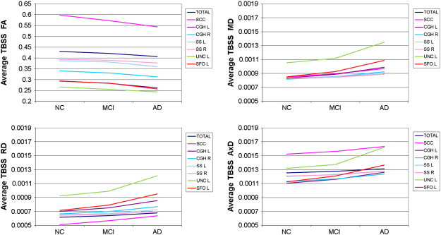
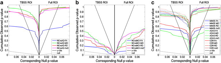
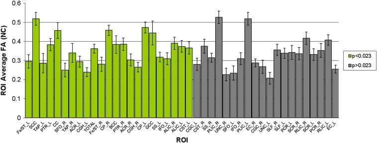
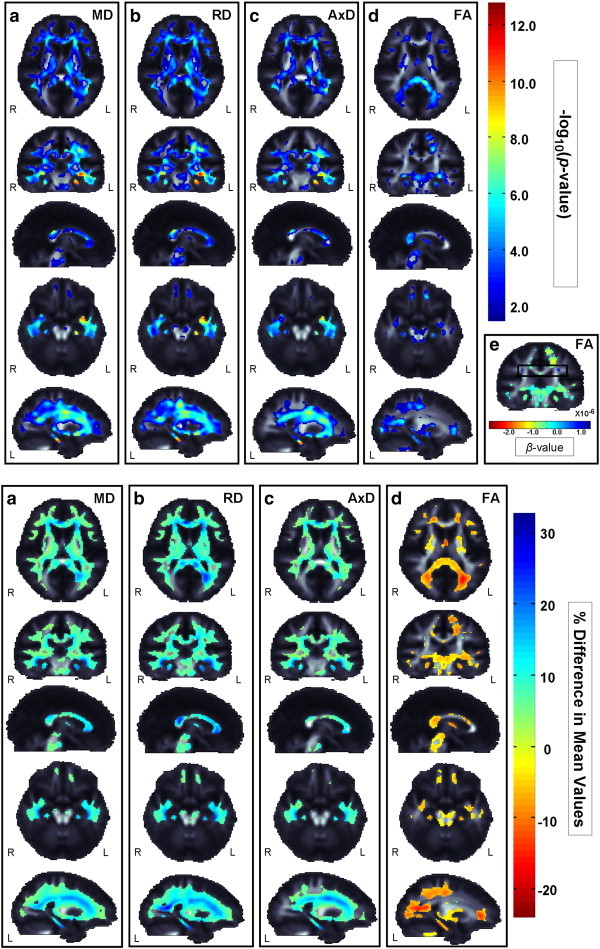
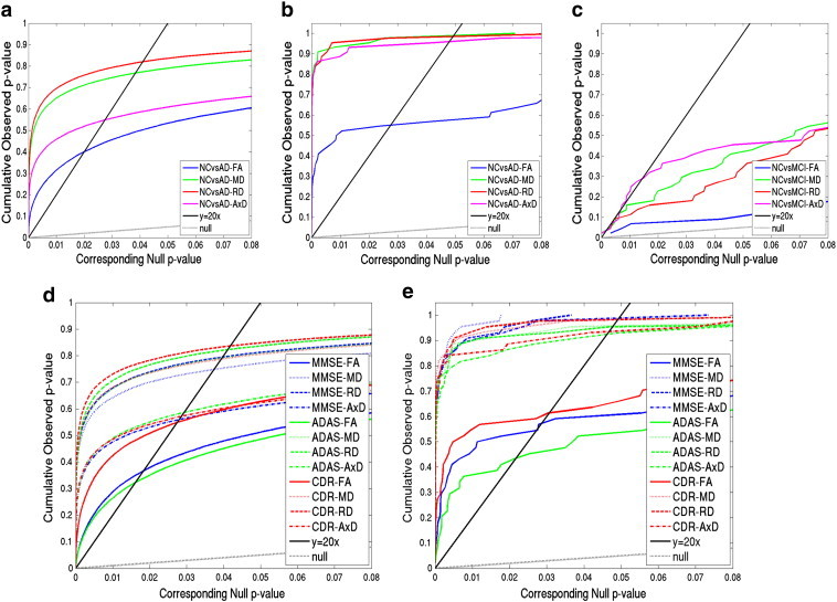
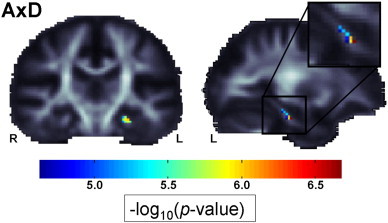
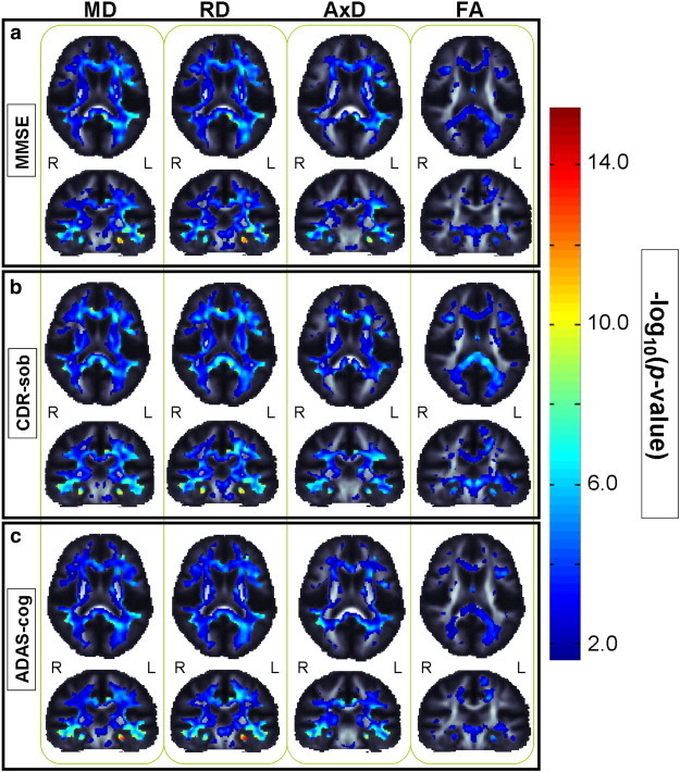
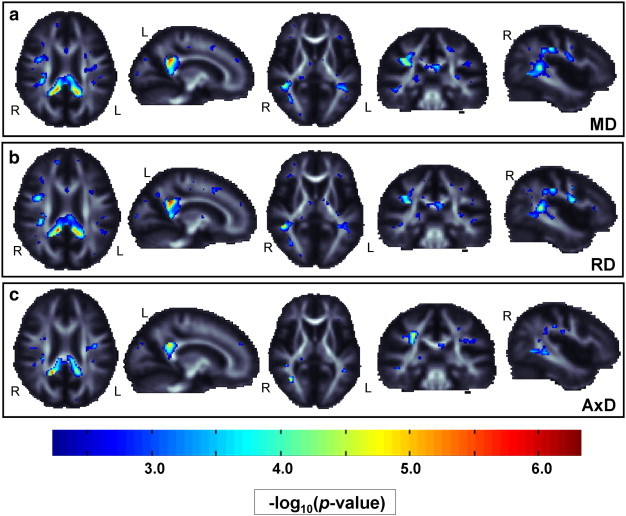
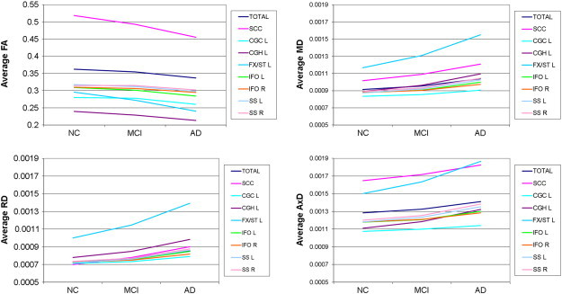
The Alzheimer's Disease Neuroimaging Initiative (ADNI) recently added diffusion tensor imaging (DTI), among several other new imaging modalities, in an effort to identify sensitive biomarkers of Alzheimer's disease (AD). While anatomical MRI is the main structural neuroimaging method used in most AD studies and clinical trials, DTI is sensitive to microscopic white matter (WM) changes not detectable with standard MRI, offering additional markers of neurodegeneration. Prior DTI studies of AD report lower fractional anisotropy (FA), and increased mean, axial, and radial diffusivity (MD, AxD, RD) throughout WM. Here we assessed which DTI measures may best identify differences among AD, mild cognitive impairment (MCI), and cognitively healthy elderly control (NC) groups, in region of interest (ROI) and voxel-based analyses of 155 ADNI participants (mean age: 73.5 ± 7.4; 90 M/65 F; 44 NC, 88 MCI, 23 AD). Both VBA and ROI analyses revealed widespread group differences in FA and all diffusivity measures. DTI maps were strongly correlated with widely-used clinical ratings (MMSE, CDR-sob, and ADAS-cog). When effect sizes were ranked, FA analyses were least sensitive for picking up group differences. Diffusivity measures could detect more subtle MCI differences, where FA could not. ROIs showing strongest group differentiation (lowest p-values) included tracts that pass through the temporal lobe, and posterior brain regions. The left hippocampal component of the cingulum showed consistently high effect sizes for distinguishing groups, across all diffusivity and anisotropy measures, and in correlations with cognitive scores.
Overview
The claims and statistics of this paper haven’t been checked yet.
Run the check — free, no account needed. It reads the open-access text and checks the sources it cites.
Integrity signals
Registry
Cites
Statistics
Figures
Verification
Claims the paper makes
The claims and statistics of this paper haven’t been checked yet.
Run the check — free, no account needed. It reads the open-access text and checks the sources it cites.
Claims other papers make about this one
Each is checked against this paper’s own text: does it say what the citing paper says it does?
The source backs part of the claim, not all of it — or backs it for a different population, condition or comparison.
In the citing paper
“Among the three cognitive assessments, CDR-sob showed the strongest correlations with dMRI indices, in line with prior ADNI brain imaging studies (Hua et al., 2009; Nir et al., 2013).”
From Diffusion MRI Indices and Their Relation to Cognitive Impairment in Brain Aging: The Updated Multi-protocol Approach in ADNI3 (2019) · cites this paper as Nir et al., 2013
Experimental — accuracy not yet measuredThe source backs part of the claim, not all of it — or backs it for a different population, condition or comparison.
In the citing paper
“Similarly, in ADNI2, AxDDTI was the most sensitive to differences between CN and MCI diagnosis (Nir et al., 2013).”
From Diffusion MRI Indices and Their Relation to Cognitive Impairment in Brain Aging: The Updated Multi-protocol Approach in ADNI3 (2019) · cites this paper as Nir et al., 2013
Experimental — accuracy not yet measuredThe cited source says what the claim attributes to it.
In the citing paper
“As in our prior analysis of ADNI2 (Nir et al., 2013), FADTI was the least sensitive DTI measure.”
From Diffusion MRI Indices and Their Relation to Cognitive Impairment in Brain Aging: The Updated Multi-protocol Approach in ADNI3 (2019) · cites this paper as Nir et al., 2013
The cited source says what the claim attributes to it.
In the citing paper
“dMRI has since been used in numerous studies to understand the effects of AD on white matter (WM) microstructure and brain connectivity (Daianu et al., 2013a,b; Nir et al., 2013; Prasad et al., 2013).”
From Diffusion MRI Indices and Their Relation to Cognitive Impairment in Brain Aging: The Updated Multi-protocol Approach in ADNI3 (2019) · cites this paper as Nir et al., 2013
The cited source says what the claim attributes to it.
In the citing paper
“Lower FADTI and higher MDDTI are most frequently reported in studies of AD (Kavcic et al., 2008; Clerx et al., 2012; Nir et al., 2013; Maggipinto et al., 2017; Mayo et al., 2017)”
From Diffusion MRI Indices and Their Relation to Cognitive Impairment in Brain Aging: The Updated Multi-protocol Approach in ADNI3 (2019) · cites this paper as Nir et al., 2013
The cited source says what the claim attributes to it.
In the citing paper
“These are all regions that show early degenerative changes in MCI and AD (Mielke et al., 2009; Nir et al., 2013; Maggipinto et al., 2017; Powell et al., 2018).”
From Diffusion MRI Indices and Their Relation to Cognitive Impairment in Brain Aging: The Updated Multi-protocol Approach in ADNI3 (2019) · cites this paper as Nir et al., 2013
Self-check
Percentages, sums and reported differences re-computed from the paper’s own numbers.
The self-check needs the paper’s full text.
Citations
Cites
Controlling the False Discovery Rate: A Practical and Powerful Approach to Multiple Testing
Benjamini, Y., & Hochberg, Y. (1995) · Journal of the Royal Statistical Society Series B: Statistical Methodology · doi:10.1111/j.2517-6161.1995.tb02031.x · Cited by 109,482
Folstein, M. F., Folstein, S. E., & McHugh, P. R. (1975) · Journal of Psychiatric Research · doi:10.1016/0022-3956(75)90026-6 · Cited by 88,282
Hirono, N., Mori, E., Ikejiri, Y., Imamura, T., Shimomura, T., Hashimoto, M., Yamashita, H., & Ikeda, M. (1997) · PubMed · Cited by 21,012
Kok, J. S., Berg, I. J., & Scherder, E. J. A. (2013) · Dementia and Geriatric Cognitive Disorders Extra · doi:10.1159/000353441 · Cited by 20,589
Older Adults' Reasons for Using Technology while Aging in Place
Peek, S. T. M., Luijkx, K. G., Rijnaard, M. D., Nieboer, M. E., van der Voort, C. S., Aarts, S., van Hoof, J., Vrijhoef, H. J., & Wouters, E. (2015) · Gerontology · doi:10.1159/000430949 · Cited by 19,852
Sensitivity and False Alarm Rate of a Fall Sensor in Long-Term Fall Detection in the Elderly
Kangas, M., Korpelainen, R., Vikman, I., Nyberg, L., & Jämsä, T. J. (2014) · Gerontology · doi:10.1159/000362720 · Cited by 19,007
Neuropathological stageing of Alzheimer-related changes.
Braak, H., & Braak, E. (1991) · Acta Neuropathologica · doi:10.1007/bf00308809 · Cited by 16,123 · cited 3×
“…et al., 2004; Medina et al., 2006; Rose et al., 2006; Xie et al., 2006; Zhang et al., 2007; Kavcic et al., 2008; Stebbins and Murphy, 2009), consistent with regions showing earliest pathological changes (Braak and Braak, 1991; Braak and Braak, 1995; Thompson et al., 2007) and our results.”
Jenkinson, M., Bannister, P., Brady, M., & Smith, S. (2002) · NeuroImage · doi:10.1006/nimg.2002.1132 · Cited by 11,888
Fast robust automated brain extraction
Smith, S. M. (2002) · Human Brain Mapping · doi:10.1002/hbm.10062 · Cited by 10,496
Tract-based spatial statistics: Voxelwise analysis of multi-subject diffusion data
Smith, S. M., Jenkinson, M., Johansen‐Berg, H., Rueckert, D., Nichols, T. E., Mackay, C. E., Watkins, K. E., Ciccarelli, O., Cader, M. Z., Matthews, P. M., et al. (2006) · NeuroImage · doi:10.1016/j.neuroimage.2006.02.024 · Cited by 6,598
Research works printed but not linked: 2
- Daianu, 2012, Analyzing the structural k-core of brain connectivity networks in normal aging and Alzheimer's disease, MICCAI NIBAD, 52
- Gutman, 2012, Creating unbiased minimal deformation templates for brain volume registration, Proceedings of the Organization for Human Brain Mapping Annual Conference
Cited by
Sorted by citations
Cerebral blood flow regulation and neurovascular dysfunction in Alzheimer disease
Kisler, K., Nelson, A. R., Montagne, A., & Zlokovic, B. V. (2017) · Nature Reviews Neuroscience · doi:10.1038/nrn.2017.48 · Cited by 1,243
Brain atrophy in Alzheimer’s Disease and aging
Pini, L., Pievani, M., Bocchetta, M., Altomare, D., Bosco, P., Cavedo, E., Galluzzi, S., Marizzoni, M., & Frisoni, G. B. (2016) · Ageing Research Reviews · doi:10.1016/j.arr.2016.01.002 · Cited by 836
Functional brain connectivity using fMRI in aging and Alzheimer’s disease
Dennis, E. L., & Thompson, P. M. (2014) · Neuropsychology Review · doi:10.1007/s11065-014-9249-6 · Cited by 583
Self-citationWeiner, M. W., Veitch, D. P., Aisen, P. S., Beckett, L. A., Cairns, N. J., Cedarbaum, J., Green, R. C., Harvey, D., Jack, C. R., Jagust, W., et al. (2015) · Alzheimer's & Dementia · doi:10.1016/j.jalz.2014.11.001 · Cited by 348
Self-citationImpact of the Alzheimer's Disease Neuroimaging Initiative, 2004 to 2014
Weiner, M. W., Veitch, D. P., Aisen, P. S. S., Beckett, L. A., Cairns, N. J., Cedarbaum, J. M., Donohue, M., Green, R. C., Harvey, D. J., Jack, C. R., et al. (2015) · Alzheimer s & Dementia · doi:10.1016/j.jalz.2015.04.005 · Cited by 249
Self-citationWhat Happens with the Circuit in Alzheimer's Disease in Mice and Humans?
Zott, B., Busche, M. A., Sperling, R. A., & Konnerth, A. (2018) · Annual Review of Neuroscience · doi:10.1146/annurev-neuro-080317-061725 · Cited by 221
Zhou, T., Liu, M., Thung, K., & Shen, D. G. (2019) · IEEE Transactions on Medical Imaging · doi:10.1109/tmi.2019.2913158 · Cited by 233
Magnetic resonance imaging in Alzheimer’s disease neuroimaging initiative 2
Jack, C. R., Barnes, J., Bernstein, M. A., Borowski, B. J., Brewer, J., Clegg, S., Dale, A. M., Carmichael, O., Ching, C., DeCarli, C., et al. (2015) · Alzheimer's & Dementia · doi:10.1016/j.jalz.2015.05.002 · Cited by 220
Self-citationChen, X., Zhang, H., Zhang, L., Shen, C., Lee, S., & Shen, D. G. (2017) · Human Brain Mapping · doi:10.1002/hbm.23711 · Cited by 190
Calderón‐Garcidueñas, L., Reynoso-Robles, R., Martínez, J. V., Chew, A. G. M., Pérez-Guillé, B., Mukherjee, P. S., Torres‐Jardón, R., Perry, G. L., & González-Maciel, A. (2016) · Environmental Research · doi:10.1016/j.envres.2015.12.031 · Cited by 176
A large-scale multicentre cerebral diffusion tensor imaging study in amyotrophic lateral sclerosis
Müller, H.-P., Turner, M. R., Großkreutz, J., Abrahams, S., Bede, P., Govind, V., Prudlo, J., Ludolph, A. C., Filippi, M., & Kassubek, J. (2016) · Journal of Neurology Neurosurgery & Psychiatry · doi:10.1136/jnnp-2015-311952 · Cited by 176
Schwarz, C. G., Reid, R. I., Gunter, J. L., Senjem, M. L., Przybelski, S. A., Zuk, S. M., Whitwell, J. L., Vemuri, P., Josephs, K. A., Kantarci, K., et al. (2014) · NeuroImage · doi:10.1016/j.neuroimage.2014.03.026 · Cited by 176
Self-citationZhao, B., Zhang, J., Ibrahim, J. G., Luo, T., Santelli, R., Li, Y., Li, T., Shan, Y., Zhu, Z., Zhou, F., et al. (2019) · Molecular Psychiatry · doi:10.1038/s41380-019-0569-z · Cited by 150
Multimodal neuroimaging computing: a review of the applications in neuropsychiatric disorders
Liu, S., Cai, W., Liu, S., Zhang, F., FULHAM, M., Feng, D., Pujol, S., & Kikinis, R. (2015) · Brain Informatics · doi:10.1007/s40708-015-0019-x · Cited by 150
Glucose indices are associated with cognitive and structural brain measures in young adults
Weinstein, G., Maillard, P., Himali, J. J., Beiser, A., Au, R., Wolf, P. A., Seshadri, S. S., & DeCarli, C. S. (2015) · Neurology · doi:10.1212/wnl.0000000000001655 · Cited by 156
Relationship Between DTI Metrics and Cognitive Function in Alzheimer’s Disease
Mayo, C. D., García-Barrera, M. A., Mazerolle, E. L., Ritchie, L. J., Fisk, J. D., Gawryluk, J. R., & Initiative, A. D. N. (2019) · Frontiers in Aging Neuroscience · doi:10.3389/fnagi.2018.00436 · Cited by 154
Daianu, M., Jahanshad, N., Nir, T. M., Jack, C. R., Weiner, M. W., Bernstein, M. A., Thompson, P. M., & Initiative, T. A. D. N. (2015) · Human Brain Mapping · doi:10.1002/hbm.22830 · Cited by 147
Self-citationZhan, L., Zhou, J., Wang, Y., Jin, Y., Jahanshad, N., Prasad, G., Nir, T. M., Leonardo, C. D., Ye, J., Thompson, P. M., et al. (2015) · Frontiers in Aging Neuroscience · doi:10.3389/fnagi.2015.00048 · Cited by 141
Self-citationDeepDTI: High-fidelity six-direction diffusion tensor imaging using deep learning
Tian, Q., Bilgiç, B., Fan, Q., Liao, C., Ngamsombat, C., Hu, Y., Witzel, T., Setsompop, K., Polimeni, J. R., & Huang, S. Y. (2020) · NeuroImage · doi:10.1016/j.neuroimage.2020.117017 · Cited by 137
Racine, A. M., Adluru, N., Alexander, A. L., Christian, B. T., Okonkwo, O. C., Oh, J., Cleary, C. A., Birdsill, A., Hillmer, A. T., Murali, D., et al. (2014) · NeuroImage: Clinical · doi:10.1016/j.nicl.2014.02.001 · Cited by 136
Dementia risk and prevention by targeting modifiable vascular risk factors
Tariq, S., & Barber, P. A. (2017) · Journal of Neurochemistry · doi:10.1111/jnc.14132 · Cited by 140
Tremblay, S., Henry, L. C., Bedetti, C., Larson-Dupuis, C., Gagnon, J., Evans, A. C., Théoret, H., Lassonde, M. C., & Beaumont, L. D. (2014) · Brain · doi:10.1093/brain/awu236 · Cited by 145
Connectome imaging for mapping human brain pathways
Shi, Y., & Toga, A. W. (2017) · Molecular Psychiatry · doi:10.1038/mp.2017.92 · Cited by 135
Self-citationPraet, J., Manyakov, N. V., Muchene, L., Mai, Z., Terzopoulos, V., de Backer, S., Torremans, A., Guns, P.-J., Van De Casteele, T., Bottelbergs, A., et al. (2018) · Alzheimer's Research & Therapy · doi:10.1186/s13195-017-0329-8 · Cited by 133
Cerebrospinal Fluid Biomarkers of Neurovascular Dysfunction in Mild Dementia and Alzheimer'S Disease
Sweeney, M. D., Sagare, A. P., & Zlokovic, B. V. (2015) · Journal of Cerebral Blood Flow & Metabolism · doi:10.1038/jcbfm.2015.76 · Cited by 122
Effects of Arterial Stiffness on Brain Integrity in Young Adults From the Framingham Heart Study
Maillard, P., Mitchell, G. F., Himali, J. J., Beiser, A., Tsao, C. W., Pase, M. P., Satizábal, C. L., Vasan, R. S., Seshadri, S. S., & DeCarli, C. S. (2016) · Stroke · doi:10.1161/strokeaha.116.012949 · Cited by 122
Cerebral white matter free water
Maillard, P., Fletcher, E. M., Singh, B., Martinez, O., Johnson, D. K., Olichney, J., Farias, S. T., & DeCarli, C. S. (2019) · Neurology · doi:10.1212/wnl.0000000000007449 · Cited by 114
Chen, H., Huang, L., Li, H., Qian, Y., Yang, D., Zhao, Q., Luo, C., Li, M., Zhang, B., & Xu, Y. (2020) · CNS Neuroscience & Therapeutics · doi:10.1111/cns.13283 · Cited by 110
Acosta‐Cabronero, J., & Nestor, P. J. (2014) · Frontiers in Aging Neuroscience · doi:10.3389/fnagi.2014.00266 · Cited by 109
Revolutionizing Alzheimer's disease and clinical trials through biomarkers
Mattsson, N., Carrillo, M. C., Dean, R. A., Devous, M. D., Nikolcheva, T., Pesini, P., Salter, H., Potter, W. Z., Sperling, R. A., Bateman, R. J., et al. (2015) · Alzheimer s & Dementia Diagnosis Assessment & Disease Monitoring · doi:10.1016/j.dadm.2015.09.001 · Cited by 102
Zavaliangos-Petropulu, A., Nir, T. M., Thomopoulos, S. I., Reid, R. I., Bernstein, M. A., Borowski, B., Jack Jr., C. R., Weiner, M. W., Jahanshad, N., & Thompson, P. M. (2019) · Frontiers in Neuroinformatics · doi:10.3389/fninf.2019.00002 · Cited by 101
“We hypothesized that the diffusion indices from ADNI2 (Nir et al., 2013, 2017) would still be associated with clinical measures of disease burden in ADNI3—despite the variation in protocols.”
InfluentialSelf-citationHydration water mobility is enhanced around tau amyloid fibers
Fichou, Y., Schirò, G., Gallat, F., Laguri, C., Moulin, M., Combet, J., Zamponi, M., Haertlein, M., Picart, C., Mossou, E., et al. (2015) · Proceedings of the National Academy of Sciences · doi:10.1073/pnas.1422824112 · Cited by 99
White matter alterations in early‐stage Alzheimer's disease: A tract‐specific study
Wen, Q., Mustafi, S. M., Li, J., Risacher, S. L., Tallman, E., Brown, S. A., West, J. D., Harezlak, J., Farlow, M. R., Unverzagt, F. W., et al. (2019) · Alzheimer's & Dementia: Diagnosis, Assessment & Disease Monitoring · doi:10.1016/j.dadm.2019.06.003 · Cited by 96
Abdelaziz, M., Wang, T., & Elazab, A. (2021) · Journal of Biomedical Informatics · doi:10.1016/j.jbi.2021.103863 · Cited by 101
Perivascular space fluid contributes to diffusion tensor imaging changes in white matter
Sepehrband, F., Cabeen, R. P., Choupan, J., Barisano, G., Law, M., Toga, A. W., & Initiative, T. A. D. N. (2018) · NeuroImage · doi:10.1016/j.neuroimage.2019.04.070 · Cited by 88
Self-citationRémy, F., Vayssière, N., Saint‐Aubert, L., Barbeau, E. J., & Pariente, J. (2015) · NeuroImage Clinical · doi:10.1016/j.nicl.2015.01.014 · Cited by 87
Tract-based analysis of white matter degeneration in Alzheimer’s disease
Lee, S., Coutu, J., Wilkens, P. J., Yendiki, A., Rosas, H. D., & Salat, D. H. (2015) · Neuroscience · doi:10.1016/j.neuroscience.2015.05.049 · Cited by 91
Default Mode Network Functional Connectivity in Early and Late Mild Cognitive Impairment
Lee, E., Yoo, K., Lee, Y., Chung, J., Lim, J., Yoon, B., & Jeong, Y. (2016) · Alzheimer Disease & Associated Disorders · doi:10.1097/wad.0000000000000143 · Cited by 86
Dissecting the Fornix in Basic Memory Processes and Neuropsychiatric Disease: A Review
Benear, S. L., Ngo, C. T., & Olson, I. R. (2020) · Brain Connectivity · doi:10.1089/brain.2020.0749 · Cited by 95
Jack, C. R., Arani, A., Borowski, B. J., Cash, D. M., Crawford, K., Das, S. R., DeCarli, C., Fletcher, E., Fox, N. C., Gunter, J. L., et al. (2024) · Alzheimer's & Dementia · doi:10.1002/alz.14166 · Cited by 82
Self-citationSDnDTI: Self-supervised deep learning-based denoising for diffusion tensor MRI
Tian, Q., Li, Z., Fan, Q., Polimeni, J. R., Bilgiç, B., Salat, D. H., & Huang, S. Y. (2022) · NeuroImage · doi:10.1016/j.neuroimage.2022.119033 · Cited by 92
Ezzati, A., Katz, M. J., Lipton, M. L., Zimmerman, M. E., & Lipton, R. B. (2016) · Brain Imaging and Behavior · doi:10.1007/s11682-015-9452-y · Cited by 79
Nir, T. M., Villalón‐Reina, J. E., Prasad, G., Jahanshad, N., Joshi, S. H., Toga, A. W., Bernstein, M. A., Jack, C. R., Weiner, M. W., & Thompson, P. M. (2014) · Neurobiology of Aging · doi:10.1016/j.neurobiolaging.2014.05.037 · Cited by 79
Self-citationNeural correlates of the impulse dyscontrol domain of mild behavioral impairment
Gill, S., Wang, M., Mouchès, P., Rajashekar, D., Sajobi, T. T., MacMaster, F. P., Smith, E. E., Forkert, N. D., Ismail, Z., & Initiative, A. D. N. (2021) · International Journal of Geriatric Psychiatry · doi:10.1002/gps.5540 · Cited by 73
CNN and swin-transformer based efficient model for Alzheimer’s disease diagnosis with sMRI
Xin, J., Wang, A., Guo, R. J., Liu, W., & Tang, X. (2023) · Biomedical Signal Processing and Control · doi:10.1016/j.bspc.2023.105189 · Cited by 72
White matter degeneration in subjective cognitive decline: a diffusion tensor imaging study
Li, X.-Y., Tang, Z.-C., Sun, Y., Tian, J., Liu, Z.-Y., & Han, Y. (2016) · Oncotarget · doi:10.18632/oncotarget.10091 · Cited by 65
Neurofilament relates to white matter microstructure in older adults
Moore, E. E., Hohman, T. J., Badami, F. S., Pechman, K. R., Osborn, K. E., Acosta, L. M. Y., Bell, S. P., Babicz, M. A., Gifford, K. A., Anderson, A. W., et al. (2018) · Neurobiology of Aging · doi:10.1016/j.neurobiolaging.2018.06.023 · Cited by 65
The Worldwide Alzheimer's Disease Neuroimaging Initiative: An update
Hendrix, J. A., Finger, B., Weiner, M. W., Frisoni, G. B., Iwatsubo, T., Rowe, C. C., Kim, S. Y., Guinjoan, S. M., Sevlever, G. E., & Carrillo, M. C. (2015) · Alzheimer s & Dementia · doi:10.1016/j.jalz.2015.05.008 · Cited by 65
Self-citationDaianu, M., Mendez, M. F., Baboyan, V. G., Jin, Y., Melrose, R. J., Jimenez, E. E., & Thompson, P. M. (2016) · Brain Imaging and Behavior · doi:10.1007/s11682-015-9458-5 · Cited by 64
Self-citationTang, X., Qin, Y., Wu, J., Zhang, M., Zhu, W., & Miller, M. I. (2016) · Magnetic Resonance Imaging · doi:10.1016/j.mri.2016.05.001 · Cited by 64
Zhao, B., Shan, Y., Yang, Y., Yu, Z., Li, T., Wang, X., Luo, T., Zhu, Z., Sullivan, P. F., Zhao, H., et al. (2021) · Nature Communications · doi:10.1038/s41467-021-23130-y · Cited by 66
Prediction of Incipient Alzheimer’s Disease Dementia in Patients with Mild Cognitive Impairment
Ardekani, B. A., Initiative, A. D. N., Bermudez, E., Mubeen, A. M., & Bachman, A. H. (2016) · Journal of Alzheimer s Disease · doi:10.3233/jad-160594 · Cited by 63
Jalbrzikowski, M., Villalón‐Reina, J. E., Karlsgodt, K. H., Şentürk, D., Chow, C. C., Thompson, P. M., & Bearden, C. E. (2014) · Frontiers in Behavioral Neuroscience · doi:10.3389/fnbeh.2014.00393 · Cited by 65
Self-citationWilliams, O. A., An, Y., Beason‐Held, L. L., Huo, Y., Ferrucci, L., Landman, B. A., & Resnick, S. M. (2018) · NeuroImage · doi:10.1016/j.neuroimage.2018.12.009 · Cited by 66
Wang, Q., Wang, Y., Liu, J., Sutphen, C. L., Cruchaga, C., Blazey, T., Gordon, B. A., Su, Y., Chen, C., Shimony, J. S., et al. (2019) · NeuroImage: Clinical · doi:10.1016/j.nicl.2019.101767 · Cited by 63
White matter in the older brain is more plastic than in the younger brain
Yotsumoto, Y., Chang, L.-H., Ni, R., Pierce, R. S., Andersen, G. J., Watanabe, T., & Sasaki, Y. (2014) · Nature Communications · doi:10.1038/ncomms6504 · Cited by 62
Anckaerts, C., Blockx, I., Summer, P., Michael, J., Hamaide, J., Kreutzer, C., Boutin, H., Couillard‐Després, S., Verhoye, M., & der Linden, A. V. (2018) · Neurobiology of Disease · doi:10.1016/j.nbd.2018.11.010 · Cited by 61
Diffusion Kurtosis Imaging: A Possible MRI Biomarker for AD Diagnosis?
Struyfs, H., Hecke, W. V., Veraart, J., Sijbers, J., Slaets, S., Belder, M. D., Wuyts, L. L., Peters, B., Sleegers, K., Robberecht, C., et al. (2015) · Journal of Alzheimer s Disease · doi:10.3233/jad-150253 · Cited by 61
Fractional Anisotropy changes in Parahippocampal Cingulum due to Alzheimer’s Disease
da Rocha, J. L. D., Bramati, I. E., Coutinho, G., Tovar-Moll, F. F., & Sitaram, R. (2020) · Scientific Reports · doi:10.1038/s41598-020-59327-2 · Cited by 61
Diffusion MRI and its Role in Neuropsychology
Mueller, B. A., Lim, K. O., Hemmy, L. S., & Camchong, J. (2015) · Neuropsychology Review · doi:10.1007/s11065-015-9291-z · Cited by 65
White matter abnormalities and cognition in aging and Alzheimer disease
Peter, C., Sathe, A., Shashikumar, N., Pechman, K. R., Workmeister, A. W., Jackson, T. B., Huo, Y., Mukherjee, S., Mez, J., Dumitrescu, L. C., et al. (2025) · JAMA Neurology · doi:10.1001/jamaneurol.2025.1601 · Cited by 59
Self-citationZhang, X., Sun, Y., Li, W., Liu, B., Wu, W., Zhao, H., Liu, R., Zhang, Y., Yin, Z., Yu, T., et al. (2019) · NeuroImage Clinical · doi:10.1016/j.nicl.2019.101723 · Cited by 59
Halliday, D., Gawryluk, J. R., García-Barrera, M. A., & MacDonald, S. W. S. (2019) · Frontiers in Human Neuroscience · doi:10.3389/fnhum.2019.00352 · Cited by 60
Haberg, A. K., Olsen, A., Moen, K. G., Schirmer-Mikalsen, K., Visser, E., Finnanger, T. G., Evensen, K. A. I., Skandsen, T., Vik, A., & Eikenes, L. (2014) · Journal of Neuroscience Research · doi:10.1002/jnr.23534 · Cited by 59
Stolp, H. B., Ball, G., So, P.-W., Tournier, J.-D., Jones, M., Thornton, C., & Edwards, A. D. (2018) · Scientific Reports · doi:10.1038/s41598-018-22295-9 · Cited by 59
Functional connectivity of white matter as a biomarker of cognitive decline in Alzheimer’s disease
Gao, Y., Sengupta, A., Li, M., Zu, Z., Rogers, B. P., Anderson, A. W., Ding, Z., Gore, J. C., & Initiative, A. D. N. (2020) · PLoS ONE · doi:10.1371/journal.pone.0240513 · Cited by 58
Wen, M., Heng, H. S. E., Lu, Z., Xu, Z., Chan, L. L., Tan, E. K., & Tan, L. C. S. (2018) · Neurorehabilitation and neural repair · doi:10.1177/1545968317753075 · Cited by 53
Gray matter T1‐w/T2‐w ratios are higher in Alzheimer's disease
Pelkmans, W., Dicks, E., Barkhof, F., Vrenken, H., Scheltens, P., van der Flier, W. M., & Tijms, B. M. (2019) · Human Brain Mapping · doi:10.1002/hbm.24638 · Cited by 53
Liebscher, S., Keller, G. B., Goltstein, P. M., Bonhoeffer, T., & Hübener, M. (2016) · Current Biology · doi:10.1016/j.cub.2016.01.070 · Cited by 52
Lee, P., Ryoo, H., Park, J., Jeong, Y., & Initiative, A. D. N. (2017) · Journal of Clinical Neurology · doi:10.3988/jcn.2017.13.2.144 · Cited by 52
Growing out of attention deficit hyperactivity disorder: Insights from the ‘remitted’ brain
Sudre, G. P., Mangalmurti, A., & Shaw, P. (2018) · Neuroscience & Biobehavioral Reviews · doi:10.1016/j.neubiorev.2018.08.010 · Cited by 55
Multi-modal multiple kernel learning for accurate identification of Tourette syndrome children
Wen, H., Liu, Y., Rekik, I., Wang, S., Chen, Z., Zhang, J., Zhang, Y., Peng, Y., & He, H. (2016) · Pattern Recognition · doi:10.1016/j.patcog.2016.09.039 · Cited by 52
3 D tract‐specific local and global analysis of white matter integrity in A lzheimer's disease
Jin, Y., Huang, C., Daianu, M., Zhan, L., Dennis, E. L., Reid, R. I., Jack, C. R., Hongtu Zhu, B., Thompson, P. M., & Initiative, A. D. N. (2016) · Human Brain Mapping · doi:10.1002/hbm.23448 · Cited by 50
Self-citationMultimodal Hippocampal Subfield Grading For Alzheimer’s Disease Classification
Hett, K., Ta, V.-T., Catheline, G., Tourdias, T., Manjón, J. V., Coupé, P., Initiative, A. D. N., Weiner, M. W., Aisen, P., Petersen, R., et al. (2019) · Scientific Reports · doi:10.1038/s41598-019-49970-9 · Cited by 49
Self-citationWillette, A. A., Calhoun, V. D., Egan, J. M., & Kapogiannis, D. (2014) · Psychiatry Research Neuroimaging · doi:10.1016/j.pscychresns.2014.08.005 · Cited by 52
Kavroulakis, E., Simos, P. G., Kalaitzakis, G., Maris, T. G., Karageorgou, D., Zaganas, I. V., Panagiotakis, S., Basta, M., Vgontzas, A. N., & Papadaki, E. Z. (2017) · Journal of Magnetic Resonance Imaging · doi:10.1002/jmri.25849 · Cited by 53
Arendash, G. W., Cao, C., Abulaban, H., Baranowski, R., Wisniewski, G., Becerra, L. R., Andel, R., Lin, X., Zhang, X., Wittwer, D., et al. (2019) · Journal of Alzheimer s Disease · doi:10.3233/jad-190367 · Cited by 52
Madhavan, A. A., Schwarz, C. G., Duffy, J. R., Strand, E. A., Machulda, M. M., Drubach, D. A., Kantarci, K., Przybelski, S. A., Reid, R. I., Senjem, M. L., et al. (2015) · Journal of Alzheimer s Disease · doi:10.3233/jad-150502 · Cited by 48
Self-citationKumar, R., Waisberg, E., Ong, J., Paladugu, P., Amiri, D., Saintyl, J., Yelamanchi, J., Nahouraii, R., Jagadeesan, R., & Tavakkoli, A. (2024) · Brain Sciences · doi:10.3390/brainsci14121266 · Cited by 52
Discriminative analysis of multivariate features from structural MRI and diffusion tensor images
Li, M., Qin, Y., Gao, F., Zhu, W., & He, X. (2014) · Magnetic Resonance Imaging · doi:10.1016/j.mri.2014.05.008 · Cited by 46
Going round in circles—The Papez circuit in Alzheimer's disease
Forno, G., Lladó, A., & Hornberger, M. (2021) · European Journal of Neuroscience · doi:10.1111/ejn.15494 · Cited by 47
Quantitative T2 mapping of white matter: applications for ageing and cognitive decline
Knight, M. J., McCann, B., Tsivos, D., Dillon, S., Coulthard, E. J., & Kauppinen, R. A. (2016) · Physics in Medicine and Biology · doi:10.1088/0031-9155/61/15/5587 · Cited by 44
Serpa, M. H., Doshi, J. J., Erus, G., Chaim-Avancini, T. M., Cavallet, M., van de Bilt, M. T., Sallet, P. C., Gattaz, W. F., Davatzikos, C. A., Busatto, G. F., et al. (2017) · Psychological Medicine · doi:10.1017/s0033291717001015 · Cited by 42
Central white matter degeneration in bulbar- and limb-onset amyotrophic lateral sclerosis
Cárdenas‐Blanco, A., Machts, J., Acosta‐Cabronero, J., Kaufmann, J., Abdulla, S., Kollewe, K., Petri, S., Heinze, H., Dengler, R., Vielhaber, S., et al. (2014) · Journal of Neurology · doi:10.1007/s00415-014-7434-4 · Cited by 42
Xiao, D., Wang, K., Theriault, L., Charbel, E., & Initiative, A. D. N. (2022) · European Journal of Neuroscience · doi:10.1111/ejn.15815 · Cited by 42
Cerebellar White Matter Disruption in Alzheimer’s Disease Patients: A Diffusion Tensor Imaging Study
Toniolo, S., Serra, L., Olivito, G., Caltagirone, C., Mercuri, N. B., Marra, C., Cercignani, M., & Bozzali, M. (2020) · Journal of Alzheimer s Disease · doi:10.3233/jad-191125 · Cited by 40
Cooccurrence of vascular risk factors and late-life white-matter integrity changes
Maillard, P., Carmichael, O. T., Reed, B., Mungas, D., & DeCarli, C. S. (2015) · Neurobiology of Aging · doi:10.1016/j.neurobiolaging.2015.01.007 · Cited by 41
Yeung, M. K.-C., Chau, A. K.-Y., Chiu, J. Y.-C., Shek, J. T.-L., Leung, J. P.-Y., & Wong, T. C.-H. (2022) · Ageing Research Reviews · doi:10.1016/j.arr.2022.101675 · Cited by 40
Effects of low-level sarin and cyclosarin exposure on white matter integrity in Gulf War Veterans
Chao, L. L., Zhang, Y., & Buckley, S. T. (2015) · NeuroToxicology · doi:10.1016/j.neuro.2015.04.005 · Cited by 40
Protein-based amide proton transfer-weighted MR imaging of amnestic mild cognitive impairment
Zhang, Z., Zhang, W., Yao, J., Chen, X., Gao, F., Jiang, S., Chen, W., Zhou, J., & Wang, G. (2019) · NeuroImage Clinical · doi:10.1016/j.nicl.2019.102153 · Cited by 38
Huang, L., Chen, X., Sun, W., Chen, H., Ye, Q., Yang, D., Li, M., Luo, C., Ma, J., Shao, P., et al. (2021) · Frontiers in Aging Neuroscience · doi:10.3389/fnagi.2020.598242 · Cited by 40
Obesity gene NEGR1 associated with white matter integrity in healthy young adults
Dennis, E. L., Jahanshad, N., Braskie, M. N., Warstadt, N. M., Hibar, D. P., Omid Kohannim, A., Nir, T. M., McMahon, K. L., de Zubicaray, G. I., Montgomery, G. W., et al. (2014) · NeuroImage · doi:10.1016/j.neuroimage.2014.07.041 · Cited by 41
Self-citationDeFord, P., Brown, K., Richards, R. L., King, A., Newburn, K., Westover, K., & Albig, A. R. (2016) · Experimental Cell Research · doi:10.1016/j.yexcr.2016.01.011 · Cited by 39
A Multimodal Structural and Functional Neuroimaging Study of Amnestic Mild Cognitive Impairment
Bharath, S., Joshi, H., John, J. P., Balachandar, R., Sadanand, S., Saini, J., Kumar, K. J., & Varghese, M. (2016) · American Journal of Geriatric Psychiatry · doi:10.1016/j.jagp.2016.05.001 · Cited by 38
Magnetic Resonance Imaging to Detect Early Molecular and Cellular Changes in Alzheimer's Disease
Knight, M. J., McCann, B., Kauppinen, R. A., & Coulthard, E. J. (2016) · Frontiers in Aging Neuroscience · doi:10.3389/fnagi.2016.00139 · Cited by 41
Longitudinal diffusion tensor imaging in dementia with Lewy bodies and Alzheimer's disease
Firbank, M., Watson, R., Mak, E., Aribisala, B. S., Barber, R., Colloby, S. J., He, J., Blamire, A. M., & O'Brien, J. T. (2016) · Parkinsonism & Related Disorders · doi:10.1016/j.parkreldis.2016.01.003 · Cited by 40
Salminen, L. E., Conturo, T. E., Bolzenius, J. D., Cabeen, R. P., Akbudak, E., & Paul, R. H. (2016) · Technology & Innovation · doi:10.21300/18.1.2016.5 · Cited by 37
Lin, S.-Y., Lin, C., Hsieh, T.-J., Lin, C.-F., Chen, S.-H., Chao, Y., Chen, Y., Hsu, C.-C., & Kuo, L. (2019) · NeuroImage Clinical · doi:10.1016/j.nicl.2019.101680 · Cited by 37
White-matter commissures: a clinically focused anatomical review
Lavrador, J. P., Ferreira, V., Lourenço, M., Alexandre, I., Rocha, M., Oliveira, E., Kailaya-Vasan, A., & Neto, L. L. (2019) · Surgical and Radiologic Anatomy · doi:10.1007/s00276-019-02218-7 · Cited by 36
Bigham, B., Zamanpour, S. A., Zemorshidi, F., Boroumand, F., Zare, H., & Initiative, A. D. N. (2020) · Journal of Alzheimer s Disease Reports · doi:10.3233/adr-190149 · Cited by 36
Metadata
- Authors
- Nir, T. M., Jahanshad, N., Villalon-Reina, J. E., Toga, A. W., Jack, C. R., Weiner, M. W., & Thompson, P. M.
- Affiliations
- University of California, Los Angeles (US)
- Mayo Clinic (US)
- University of California, San Francisco (US)
- Corresponding author
- Thompson, Paul M.
- ORCID
- Venue
- NeuroImage: Clinical
- Publisher
- Elsevier BV
- Published
- 2013
- Volume
- 3
- Pages
- 180-195
- ISSN
- 2213-1582 (ISSN-L)
- PMC
- PMC3792746
- OpenAlex
- W2028748733
- Cited by
- 348
- References
- 131
- Citations by year
- 2026: 14 · 2025: 37 · 2024: 28 · 2023: 33 · 2022: 29 · 2021: 31 · 2020: 23 · 2019: 32 · 2018: 22 · 2017: 21 · 2016: 27 · 2015: 30 · 2014: 21
- FWCI
- 18.49
- Type
- journal article
- Language
- en
- Open access
- gold
- doi.org
- Licence
- Locations
- NeuroImage Clinical (publishedVersion, cc-by-nc-nd)
- Europe PMC (PubMed Central) (publishedVersion, cc-by-nc-nd)
- PubMed Central (publishedVersion, cc-by-nc-nd)
- PubMed (publishedVersion)
- eScholarship (California Digital Library) (submittedVersion, cc-by-nc-nd)
- Funders
- ADNI (U01 AG024904)
- National Institute on Aging
- National Institute of Biomedical Imaging and Bioengineering
- Abbott
- Alzheimer's Association
- Alzheimer's Drug Discovery Foundation
- Amorfix Life Sciences Ltd.
- AstraZeneca
- Bayer HealthCare
- BioClinica, Inc.
- Biogen Idec Inc.
- Bristol-Myers Squibb Company
- Eisai Inc.
- Elan Pharmaceuticals Inc.
- Eli Lilly and Company
- F. Hoffmann-La Roche Ltd.
- Genentech, Inc.
- GE Healthcare
- Innogenetics, N.V.
- IXICO Ltd.
- Janssen Alzheimer Immunotherapy Research & Development, LLC.
- Johnson & Johnson Pharmaceutical Research & Development LLC.
- Medpace, Inc.
- Merck & Co., Inc.
- Meso Scale Diagnostics, LLC.
- Novartis Pharmaceuticals Corporation
- Pfizer Inc.
- Servier
- Synarc Inc.
- Takeda Pharmaceutical Company
- Canadian Institutes of Health Research
- NIH (P30 AG010129, K01 AG030514)
- NIA
- NIBIB
- National Library of Medicine
- National Center for Research Resources (AG016570, EB01651, LM05639, RR019771)
- Topics
- Health Sciences › Medicine › Psychiatry and Mental health › Dementia and Cognitive Impairment Research
- Health Sciences › Medicine › Radiology, Nuclear Medicine and Imaging › Advanced Neuroimaging Techniques and Applications
- Life Sciences › Neuroscience › Cellular and Molecular Neuroscience › Alzheimer's disease research and treatments
- Keywords
- Alzheimer's disease, mild cognitive impairment, normal aging, diffusion tensor imaging, fractional anisotropy, white matter diffusivity, white matter microstructure, temporal lobe, neurodegeneration biomarkers, cognitive impairment detection, voxel-based analysis, region of interest analysis
About these checks
Registry, retraction, Cites and statistics results are computed, not judged, with one exception: a printed reference that no index lists is read once by a model before it counts as a citation error. Claims are extracted by a model and kept only when their quote appears verbatim in the text; each cited source is then checked, with a model’s help, for whether it says what the claim relies on. Those verdicts can be wrong — read the claim’s quote and the cited source before relying on one.
Statistics and arithmetic are recomputed, not judged.
Confidence bands are shown only for fields calibrated on our annotated test set. None is calibrated yet, so no band is shown.
What counts as a claim ›How we measure confidence ›How we check this ›
Are you an author, or do you see a problem with this page? Request a review
