

The DOI matches its Crossref record; 4 of the 4 references it cites were found in a registry or an index.
Recent Advances in Studying Toll-like Receptors with the Use of Computational Methods
Open accessPublished version of a preprint
Abstract
Toll-like receptors (TLRs) are transmembrane proteins that recognize various molecular patterns and activate signaling that triggers the immune response. In this review, our goal is to summarize how, in recent years, various computational solutions have contributed to a better understanding of TLRs, regarding both their function and mechanism of action. We update the recent information about small-molecule modulators and expanded the topic toward next-generation vaccine design, as well as studies of the dynamic nature of TLRs. Also, we underline problems that remain unsolved.
Overview
The claims and statistics of this paper haven’t been checked yet.
Run the check — free, no account needed. It reads the open-access text and checks the sources it cites.
Integrity signals
Registry
Cites
Statistics
Figures
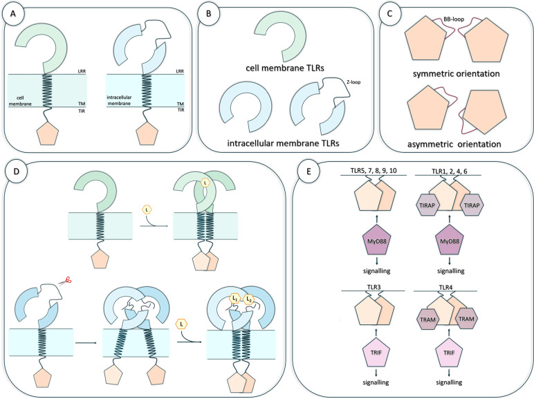
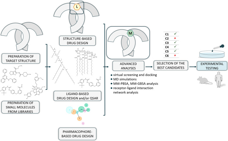
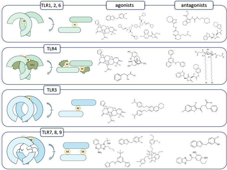
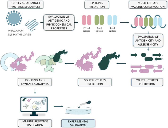
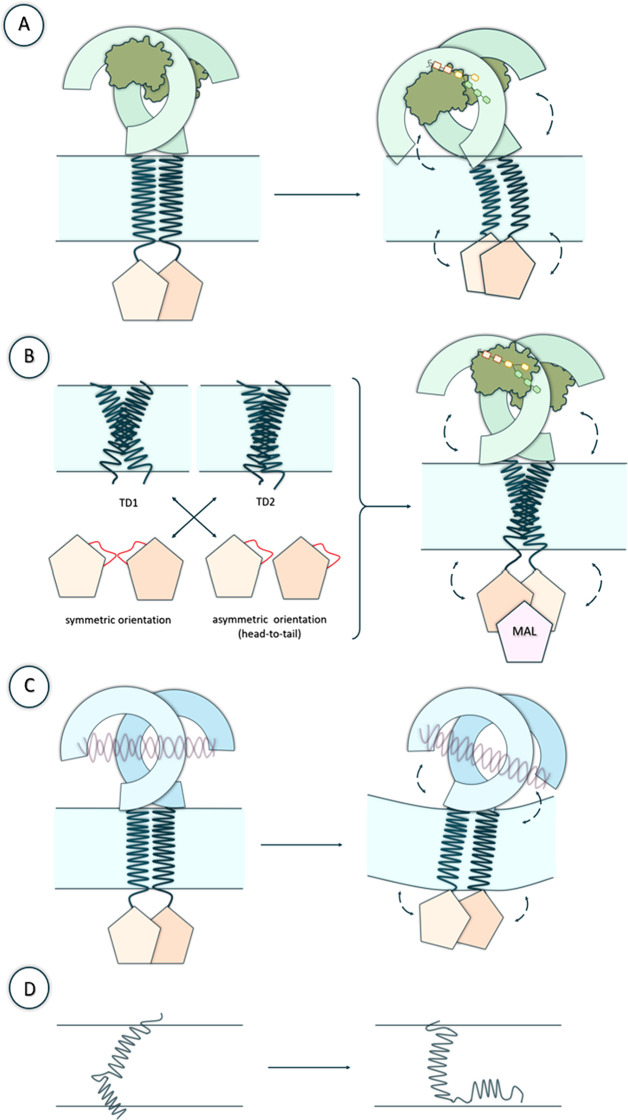
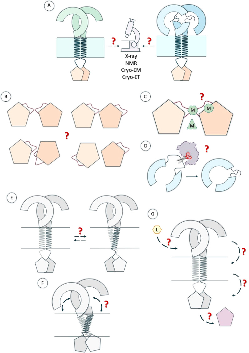
Verification
Claims the paper makes
The claims and statistics of this paper haven’t been checked yet.
Run the check — free, no account needed. It reads the open-access text and checks the sources it cites.
Self-check
Percentages, sums and reported differences re-computed from the paper’s own numbers.
The self-check needs the paper’s full text.
Citations
Cites
Highly accurate protein structure prediction with AlphaFold
Jumper, J., Evans, R., Pritzel, A., Green, T., Figurnov, M., Ronneberger, O., Tunyasuvunakool, K., Bates, R., Žídek, A., Potapenko, A., et al. (2021) · Nature · doi:10.1038/s41586-021-03819-2 · Cited by 43,764
Varadi, M., Anyango, S., Deshpande, M., Nair, S., Natassia, C., Yordanova, G., Yuan, D., Stroe, O., Wood, G., Laydon, A., et al. (2022) · Nucleic Acids Research · doi:10.1093/nar/gkab1061 · Cited by 7,850
Accurate prediction of protein structures and interactions using a 3-track network
Baek, M., DiMaio, F., Anishchenko, I., Dauparas, J., Ovchinnikov, S., Lee, G. R., Wang, J., Cong, Q., Kinch, L. N., Schaeffer, R. D., et al. (2021) · doi:10.1101/2021.06.14.448402 · Cited by 5,549
ClinVar: improving access to variant interpretations and supporting evidence
Landrum, M. J., Lee, J. M., Benson, M., Brown, G. R., Chao, C., Chitipiralla, S., Gu, B., Hart, J., Hoffman, D., Jang, W., et al. (2018) · Nucleic Acids Research · doi:10.1093/nar/gkx1153 · Cited by 4,196
Research works printed but not linked: 115
- 10.1016/j.immuni.2011.05.006 · DOI · Paper page
- 10.1016/j.intimp.2018.03.002 · DOI
- 10.3390/v3060920 · DOI
- 10.3389/fimmu.2018.02379 · DOI
- 10.1038/s41392-021-00687-0 · DOI
- 10.1016/j.cell.2010.01.022 · DOI · Paper page
- 10.1155/2021/9914854 · DOI
- 10.1038/ni.1863 · DOI · Paper page
- 10.1016/S1471-4906(03)00242-4 · DOI
- 10.1186/1471-2164-8-124 · DOI
- 10.1002/pro.4043 · DOI
- 10.1146/annurev.cellbio.21.122303.115827 · DOI
- 10.1111/j.1582-4934.2010.01127.x · DOI
- 10.1002/prot.25179 · DOI
- 10.3389/fphys.2011.00041 · DOI
- 10.3389/fimmu.2014.00461 · DOI
- 10.1038/nri2079 · DOI · Paper page
- 10.4161/cc.21572 · DOI
- 10.1186/s42269-019-0227-2 · DOI
- 10.1038/nrd3203 · DOI
- 10.1002/med.21553 · DOI
- 10.1126/science.abj8754 · DOI · Paper page
- 10.1038/s41541-021-00391-8 · DOI
- 10.1016/j.tips.2016.10.007 · DOI
- 10.3390/ijms17091508 · DOI
- 10.3390/molecules21080994 · DOI
- 10.1038/35040600 · DOI
- 10.1126/science.1115253 · DOI
- 10.1073/pnas.0505077102 · DOI
- 10.1016/j.febslet.2014.08.031 · DOI
- 10.1093/nar/28.1.235 · DOI · Paper page
- 10.1038/s41594-020-00542-w · DOI
- 10.1038/s42003-021-02532-0 · DOI
- 10.1038/s41467-023-37042-6 · DOI
- 10.1021/acs.accounts.9b00631 · DOI
- 10.1016/j.bbagen.2017.07.011 · DOI
- 10.1074/jbc.M110.116046 · DOI
- 10.1074/jbc.M111.227934 · DOI
- 10.1111/febs.14124 · DOI
- 10.1016/j.cell.2007.09.008 · DOI
- 10.1039/C8CC06618C · DOI
- 10.1016/j.ejmech.2019.02.067 · DOI
- 10.1016/j.bcp.2018.04.018 · DOI
- 10.1002/cmdc.201300445 · DOI
- 10.1002/anie.201204910 · DOI
- 10.1016/j.bcp.2020.113957 · DOI
- 10.1002/cmdc.202000060 · DOI
- 10.1021/jacs.8b09223 · DOI
- 10.1021/acs.jmedchem.8b01583 · DOI
- 10.1080/07391102.2018.1474804 · DOI
- 10.1021/acs.jmedchem.7b01803 · DOI
- 10.1038/s41598-018-37421-w · DOI
- 10.1021/acschemneuro.9b00225 · DOI
- 10.1038/mp.2014.177 · DOI
- 10.1016/j.bbi.2009.08.004 · DOI
- 10.1111/bph.13394 · DOI
- 10.1021/acs.jmedchem.5b00426 · DOI
- 10.1021/acs.jcim.7b00717 · DOI
- 10.1096/fj.201900173RRR · DOI
- 10.3390/biomedicines10092326 · DOI
- 10.1016/j.tet.2014.07.026 · DOI
- 10.1016/j.phrs.2016.09.015 · DOI
- 10.1021/acs.jmedchem.1c00300 · DOI
- 10.1080/07391102.2018.1559098 · DOI
- 10.1016/j.ejmech.2019.06.084 · DOI
- 10.1371/journal.pone.0260565 · DOI
- 10.3390/pharmaceutics13020142 · DOI
- 10.1038/s41573-021-00163-y · DOI
- 10.1093/nar/gkaa1100 · DOI · Paper page
- 10.1186/1471-2105-8-4 · DOI
- 10.1385/1-59259-890-0:571 · DOI
- 10.1186/1471-2105-8-424 · DOI
- 10.1111/imm.12889 · DOI
- 10.1093/nar/gky1006 · DOI
- 10.1093/nar/gkx346 · DOI
- 10.1002/jmr.893 · DOI
- 10.1186/1471-2105-14-S6-S4 · DOI
- 10.1093/nar/gkl343 · DOI
- 10.1093/bib/bbaa294 · DOI
- 10.1371/journal.pone.0073957 · DOI
- 10.1093/bib/bbac174 · DOI
- 10.1093/bioinformatics/11.6.681 · DOI
- 10.1038/nmeth.3213 · DOI · Paper page
- 10.1016/j.bpj.2011.10.024 · DOI
- 10.1093/nar/gkt458 · DOI
- 10.1038/nprot.2016.169 · DOI
- 10.1371/journal.pone.0009862 · DOI
- 10.1016/j.imu.2020.100438 · DOI
- 10.1039/D1RA06532G · DOI
- 10.34172/bi.2022.23769 · DOI
- 10.2147/AABC.S366431 · DOI
- 10.2147/IDR.S175114 · DOI
- 10.1038/s41598-018-34254-5 · DOI
- 10.1016/j.vaccine.2018.03.042 · DOI
- 10.3389/fimmu.2022.956776 · DOI
- 10.1016/j.ejps.2020.105279 · DOI
- 10.1038/s41598-019-39299-8 · DOI
- 10.1038/s41598-017-09199-w · DOI
- 10.1016/j.meegid.2018.03.007 · DOI
- 10.1080/14760584.2019.1711057 · DOI
- 10.1007/s10989-021-10283-z · DOI
- 10.1016/j.vaccine.2021.09.025 · DOI
- 10.1016/j.intimp.2022.109238 · DOI
- 10.3389/fmicb.2022.947852 · DOI
- 10.3390/v13112302 · DOI
- 10.3390/pharmaceutics14020423 · DOI
- 10.1038/srep43807 · DOI
- 10.1038/s41598-019-39984-8 · DOI
- 10.1074/jbc.M510442200 · DOI
- 10.1021/acs.jcim.1c00818 · DOI
- 10.1002/prot.25467 · DOI
- 10.1080/07391102.2020.1786457 · DOI
- 10.3389/fimmu.2018.00489 · DOI
- 10.1002/chem.202102995 · DOI
- 10.3390/ijms21082857 · DOI
Metadata
- Authors
- Bzówka, M., Bagrowska, W., & Góra, A.
- Affiliations
- Silesian University of Technology (PL)
- Corresponding author
- Bzówka, Maria
- Venue
- Journal of Chemical Information and Modeling
- Publisher
- American Chemical Society (ACS)
- Published
- 2023-06-07
- Volume
- 63
- Issue
- 12
- Pages
- 3669-3687
- ISSN
- 1549-9596 (ISSN-L)
- 1549-960X
- PMC
- PMC10302489
- OpenAlex
- W4379600654
- Cited by
- 34
- References
- 4
- Citations by year
- 2026: 13 · 2025: 11 · 2024: 9 · 2023: 1
- FWCI
- 0.95
- Type
- journal article
- Language
- en
- Open access
- hybrid
- doi.org
- Licence
- Locations
- Journal of Chemical Information and Modeling (publishedVersion, cc-by)
- ChemRxiv (acceptedVersion, cc-by)
- PubMed Central (publishedVersion, cc-by)
- PubMed (publishedVersion)
- Funders
- Ministerstwo Edukacji i Nauki (DI2018 014148 / 0141/DIA/2019/48)
- Topics
- Life Sciences › Immunology and Microbiology › Immunology › Immune Response and Inflammation
- Life Sciences › Pharmacology, Toxicology and Pharmaceutics › Drug Discovery › Computational Drug Discovery Methods
- Life Sciences › Biochemistry, Genetics and Molecular Biology › Molecular Biology › vaccines and immunoinformatics approaches
- Keywords
- Toll-like receptors, computational methods, Toll-like receptor signaling, pattern recognition, small molecule modulators, vaccine design, innate immunity
- MeSH terms
- Signal Transduction, Toll-Like Receptors
About these checks
Registry, retraction, Cites and statistics results are computed, not judged, with one exception: a printed reference that no index lists is read once by a model before it counts as a citation error. Claims are extracted by a model and kept only when their quote appears verbatim in the text; each cited source is then checked, with a model’s help, for whether it says what the claim relies on. Those verdicts can be wrong — read the claim’s quote and the cited source before relying on one.
Statistics and arithmetic are recomputed, not judged.
Confidence bands are shown only for fields calibrated on our annotated test set. None is calibrated yet, so no band is shown.
What counts as a claim ›How we measure confidence ›How we check this ›
Are you an author, or do you see a problem with this page? Request a review
