Cited by 84

The DOI matches its Crossref record; 1 of the 1 references it cites were found in a registry or an index.

Increased expression of RRM2 by human papillomavirus E7 oncoprotein promotes angiogenesis in cervical cancer

Wang, N, Zhan, T, Ke, T, Huang, X, Ke, D, Wang, Q, Li, H·2014·British Journal of Cancer

Overview

The claims and statistics of this paper haven’t been checked yet.

Run the check — free, no account needed. It reads the open-access text and checks the sources it cites.

A few free checks a day · papers already verified open instantly · results are public

Integrity signals

Registry

Crossref · 10.1038/bjc.2013.817

Cites

1 of 1 references verified0 citation errors · 0 not checkable

Statistics

Figures

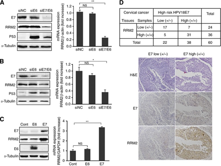
Figure 1 HPVE7 upregulates expression of RRM2. (A) Western blot (left panel) and real-time RT-PCR analyses of RRM2 in HeLa cells with knockdown of E6 or E6/E7 expression. siNC, negative control siRNA; siE6, siRNA against HPV18 E6; siE7/E6, siRNA against E6 and E7; p53, positive control for siRNA against E6; α-tubulin, loading control for western blot analysis. GAPDH was used as an internal control for quantitative RT-PCR analysis. (B) Western blot (left panel) and quantitative real time RT-PCR analyses of RRM2 in CaSki cells with knockdown of E6 or E6/E7 expression. (C) Western blot (left panel) and quantitative real-time RT-PCR analyses of RRM2 in C33A cells with overexpression of E6 or E7. Cont, transfection with a pcDNA3.1 empty vector as control. (D) The expression level of RRM2 positively correlates with the level of the HPVE7 protein in human cervical cancer (upper panel). The expression levels of RRM2 and HPVE7 in 60 human primary cervical cancer specimens were measured by immunostaining with antibodies specific to RRM2 and HPVE7, and correlated. Positive correlation between RRM2 and HPVE7 levels were determined using a Pearson Chi-Square analysis (P<0.01). The lower panel shows immunostaining images of two representative specimens, one with low (+/−) and the other with high (+/+) expression of RRM2 and HPVE7. H&E staining served as control. The data are shown as mean± s.d. of three separate experiments. NS, no significant difference; *P<0.05; **P<0.01.
Figure 1 HPVE7 upregulates expression of RRM2. (A) Western blot (left panel) and real-time RT-PCR analyses of RRM2 in HeLa cells with knockdown of E6 or E6/E7 expression. siNC, negative control siRNA; siE6, siRNA against HPV18 E6; siE7/E6, siRNA against E6 and E7; p53, positive control for siRNA against E6; α-tubulin, loading control for western blot analysis. GAPDH was used as an internal control for quantitative RT-PCR analysis. (B) Western blot (left panel) and quantitative real time RT-PCR analyses of RRM2 in CaSki cells with knockdown of E6 or E6/E7 expression. (C) Western blot (left panel) and quantitative real-time RT-PCR analyses of RRM2 in C33A cells with overexpression of E6 or E7. Cont, transfection with a pcDNA3.1 empty vector as control. (D) The expression level of RRM2 positively correlates with the level of the HPVE7 protein in human cervical cancer (upper panel). The expression levels of RRM2 and HPVE7 in 60 human primary cervical cancer specimens were measured by immunostaining with antibodies specific to RRM2 and HPVE7, and correlated. Positive correlation between RRM2 and HPVE7 levels were determined using a Pearson Chi-Square analysis (P<0.01). The lower panel shows immunostaining images of two representative specimens, one with low (+/−) and the other with high (+/+) expression of RRM2 and HPVE7. H&E staining served as control. The data are shown as mean± s.d. of three separate experiments. NS, no significant difference; *P<0.05; **P<0.01.CC BY-NC-SA · PubMed Central
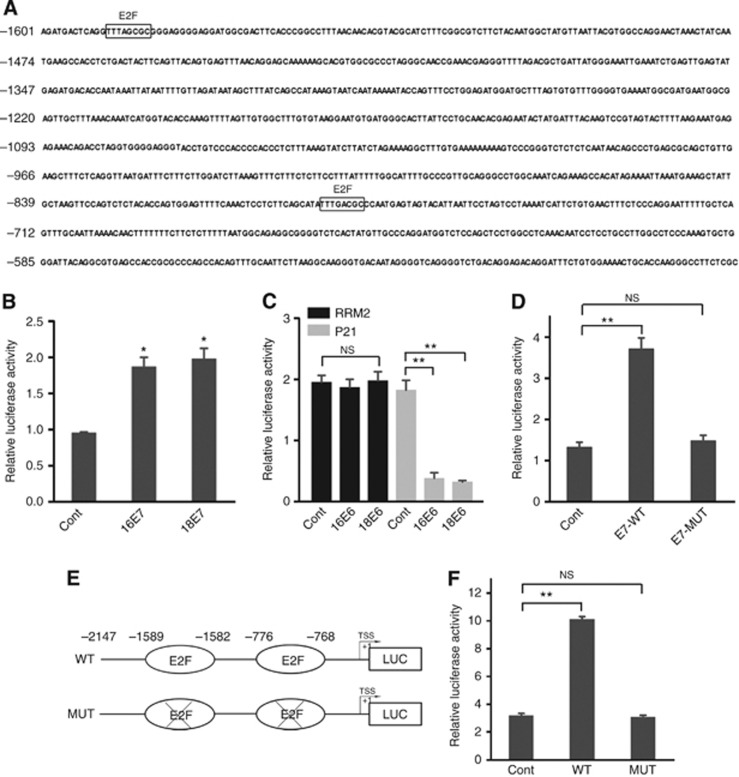
Figure 2 HPVE7 induces increased transactivation of the RRM2 promoter. (A) Sequence of the human RRM2 promoter region. (B) MCF-7 cells were co-transfected with a RRM2 promoter luciferase reporter, an expression plasmid for either HPV-16 E7 or an HPV-18 E7 (an empty vector as control, Cont), and a Renilla luciferase plasmid as an internal control. Twenty-four hours after transfection, cells were collected and used for measuring luciferase activities. The data are shown as mean±s.d. from triplicate wells. The experiment was repeated three times. (C) Luciferase assays for the RRM2 promoter (black columns) and the p21 promoter (white columns) in MCF-7 cells with overexpression of HPV16 E6, HPV18 E6 or with empty vector. (D) Luciferase assays for the RRM2 promoter in MCF-7 cells with overexpression of wild-type HPV16 E7 or mutant HPV16 E7 (E7-MUT), which lose the ability to bind to pRb. Cont, empty vector control. (E) Schematic diagram of the RRM2 promoter luciferase reporter genes. WT, wild type RRM2-Luc reporter; MUT, mutant RRM2-Luc with two consensus E2F-binding sites deleted. (F) Luciferase assays for either the wild-type RRM2 promoter (WT) or a mutant RRM2 promoter with a deletion of the E2F-binding site (MUT) in MCF-7 cells with overexpression of HPV-16 E7. Cont, empty vector control. NS, no significant difference; *P<0.05, **P<0.01.
Figure 2 HPVE7 induces increased transactivation of the RRM2 promoter. (A) Sequence of the human RRM2 promoter region. (B) MCF-7 cells were co-transfected with a RRM2 promoter luciferase reporter, an expression plasmid for either HPV-16 E7 or an HPV-18 E7 (an empty vector as control, Cont), and a Renilla luciferase plasmid as an internal control. Twenty-four hours after transfection, cells were collected and used for measuring luciferase activities. The data are shown as mean±s.d. from triplicate wells. The experiment was repeated three times. (C) Luciferase assays for the RRM2 promoter (black columns) and the p21 promoter (white columns) in MCF-7 cells with overexpression of HPV16 E6, HPV18 E6 or with empty vector. (D) Luciferase assays for the RRM2 promoter in MCF-7 cells with overexpression of wild-type HPV16 E7 or mutant HPV16 E7 (E7-MUT), which lose the ability to bind to pRb. Cont, empty vector control. (E) Schematic diagram of the RRM2 promoter luciferase reporter genes. WT, wild type RRM2-Luc reporter; MUT, mutant RRM2-Luc with two consensus E2F-binding sites deleted. (F) Luciferase assays for either the wild-type RRM2 promoter (WT) or a mutant RRM2 promoter with a deletion of the E2F-binding site (MUT) in MCF-7 cells with overexpression of HPV-16 E7. Cont, empty vector control. NS, no significant difference; *P<0.05, **P<0.01.CC BY-NC-SA · PubMed Central
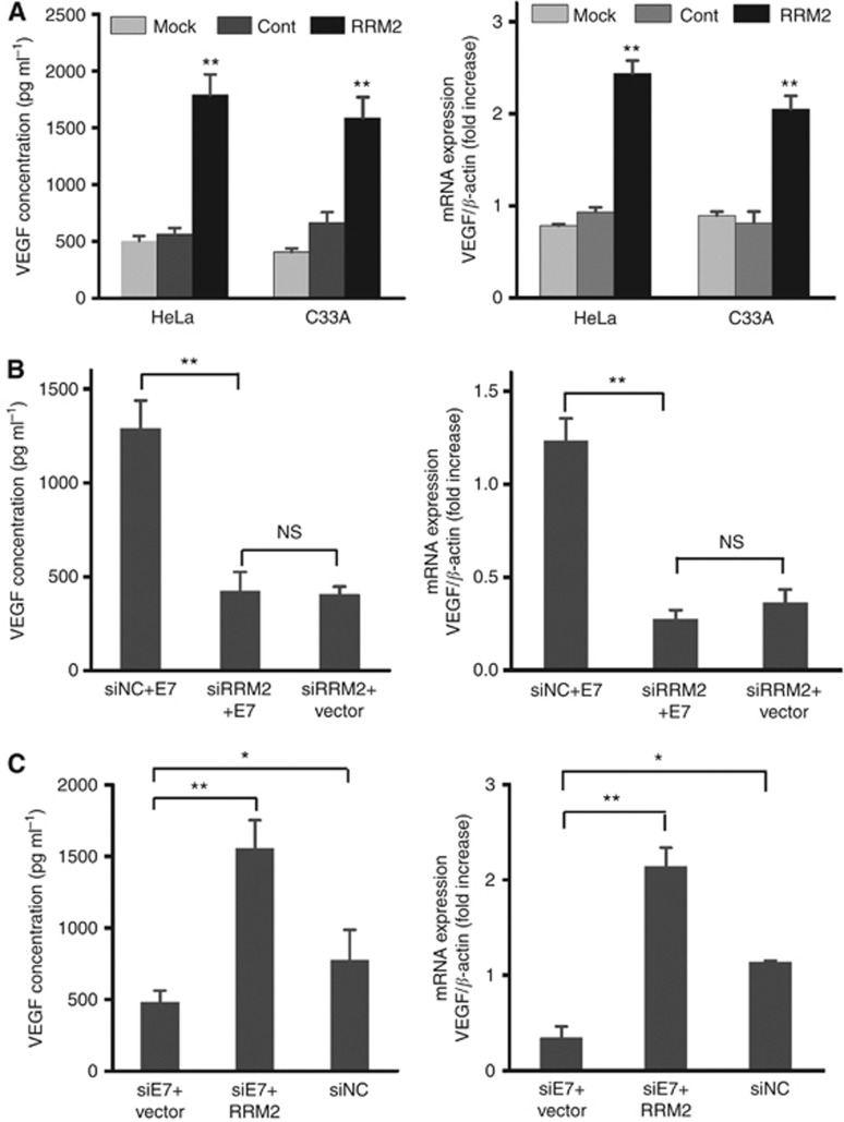
Figure 3 RRM2 mediates HPVE7-induced VEGF expression in cervical cancer cells. (A) C33A and HeLa cells were transiently transfected with a RRM2 expression plasmid. Cells with mock transfection or transfected with the pcDNA3.1 empty vector served as controls. Left panel, the level of secreted VEGF in the conditioned media derived from transfected C33A or HeLa cells was determined by ELISA. Right panel, the expression level of VEGF mRNA in transfected cells was determined by quantitative real time RT-PCR. The data are shown as mean±s.d. from triplicate wells. The experiment was repeated three times. (B) C33A cells were transfected with a HPV16 E7 expression plasmid, and a RRM2 siRNA or a control siRNA. Left panel, ELISA data for the level of secreted VEGF in the conditional media from transfected cells. Right panel, the expression level of VEGF mRNA was determined by quantitative real time RT-PCR. (C) HeLa cells were transfected with a HPV18 E7 siRNA or a control siRNA followed by co-transfection with a RRM2 expression plasmid or the control empty vector. Left panel, ELISA data for the level of secreted VEGF in the conditional media from transfected cells; right panel, quantitative real time RT-PCR data for the expression level of VEGF mRNA. NS, no significant difference; *P<0.05, **P<0.01.
Figure 3 RRM2 mediates HPVE7-induced VEGF expression in cervical cancer cells. (A) C33A and HeLa cells were transiently transfected with a RRM2 expression plasmid. Cells with mock transfection or transfected with the pcDNA3.1 empty vector served as controls. Left panel, the level of secreted VEGF in the conditioned media derived from transfected C33A or HeLa cells was determined by ELISA. Right panel, the expression level of VEGF mRNA in transfected cells was determined by quantitative real time RT-PCR. The data are shown as mean±s.d. from triplicate wells. The experiment was repeated three times. (B) C33A cells were transfected with a HPV16 E7 expression plasmid, and a RRM2 siRNA or a control siRNA. Left panel, ELISA data for the level of secreted VEGF in the conditional media from transfected cells. Right panel, the expression level of VEGF mRNA was determined by quantitative real time RT-PCR. (C) HeLa cells were transfected with a HPV18 E7 siRNA or a control siRNA followed by co-transfection with a RRM2 expression plasmid or the control empty vector. Left panel, ELISA data for the level of secreted VEGF in the conditional media from transfected cells; right panel, quantitative real time RT-PCR data for the expression level of VEGF mRNA. NS, no significant difference; *P<0.05, **P<0.01.CC BY-NC-SA · PubMed Central
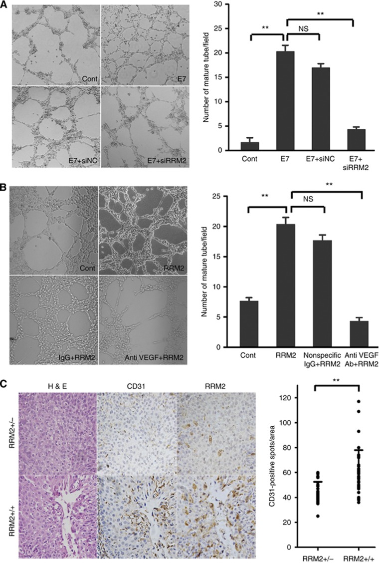
Figure 4 RRM2 is involved in cervical cancer angiogenesis in vitro and in vivo. (A) Results from in vitro matrigel-based angiogenesis assays by HUVECs with conditioned media from C33A cells transfected with pcDNA-HPV18 E7 with or without RRM2 siRNA. Representative images (left panel) and mean numbers of mature tube-forming structures (right panel) at × 100 magnification. HUVECs (1 × 10⁴ cells per well) were seeded onto the surface of 96-well cell culture plates precoated with matrigel and then incubated at 37 °C for 8 h in the conditioned media derived from transfected C33A cells. The data presented are representative of three separate experiments. (B) Application of a VEGF antibody (400 ng ml−1) to neutralise VEGF in three conditioned media abrogated RRM2-enhanced capillary tube formation. (C) Immunohistochemical analysis of 60 cervical cancer samples stained with anti-RRM2 and anti-CD31 antibodies (left panel). Representative images of weak RRM2 expression samples (+/− n=24) and strong RRM2 expression samples (+/+ n=36) are shown. H&E staining served as control. Original magnification, × 100. The data on CD31-positive microvessel densities were plotted in the right panel. Microvessel densities were significantly higher in cancer samples with strong RRM2 expression than that with weak expression. Bars indicate the mean number of vessels. **P<0.01.
Figure 4 RRM2 is involved in cervical cancer angiogenesis in vitro and in vivo. (A) Results from in vitro matrigel-based angiogenesis assays by HUVECs with conditioned media from C33A cells transfected with pcDNA-HPV18 E7 with or without RRM2 siRNA. Representative images (left panel) and mean numbers of mature tube-forming structures (right panel) at × 100 magnification. HUVECs (1 × 10⁴ cells per well) were seeded onto the surface of 96-well cell culture plates precoated with matrigel and then incubated at 37 °C for 8 h in the conditioned media derived from transfected C33A cells. The data presented are representative of three separate experiments. (B) Application of a VEGF antibody (400 ng ml−1) to neutralise VEGF in three conditioned media abrogated RRM2-enhanced capillary tube formation. (C) Immunohistochemical analysis of 60 cervical cancer samples stained with anti-RRM2 and anti-CD31 antibodies (left panel). Representative images of weak RRM2 expression samples (+/− n=24) and strong RRM2 expression samples (+/+ n=36) are shown. H&E staining served as control. Original magnification, × 100. The data on CD31-positive microvessel densities were plotted in the right panel. Microvessel densities were significantly higher in cancer samples with strong RRM2 expression than that with weak expression. Bars indicate the mean number of vessels. **P<0.01.CC BY-NC-SA · PubMed Central
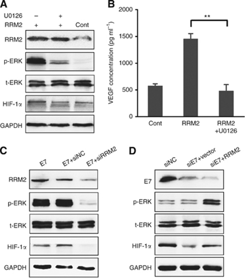
Figure 5 RRM2 mediates HPVE7-induced activation of the ERK1/2 signalling pathway and increases of HIF-1α and VEGF expression. (A) RRM2-induced HIF-1α protein expression was abolished by inhibition of the ERK1/2 signalling pathway by U0126. C33A cells transfected with a RRM2 expression plasmid or a control empty vector were treated with 10 μM U0126 or vehicle control for 16 h. The expression levels of phosphorylated ERK1/2 and HIF-1α were examined by western blot analysis. t-ERK, total ERK levels used as loading control; GAPDH, another loading control. (B) RRM2-induced VEGF expression was abolished by inhibition of the ERK1/2 signalling pathway by U0126. The level of secreted VEGF in the conditioned media was measured by an ELISA assay. The data presented are representative of three independent experiments. (C) C33A cells were transfected with pcDNA-HPV16E7, followed by co-transfection with a RRM2 siRNA (siRRM2) or a control siRNA (siNC) for another 24 h. Cell lysates were used for western blot analysis for phosphorylated ERK1/2 and HIF-1α protein levels. (D) HeLa cells were transfected with a HPV18 E7 siRNA (siE7) or a control siRNA (siNC), followed by co-transfection with pcDNA-RRM2 for another 24 h. Cell lysates were used for western blot analysis for phosphorylated ERK1/2 and HIF-1α protein levels. **P<0.01.
Figure 5 RRM2 mediates HPVE7-induced activation of the ERK1/2 signalling pathway and increases of HIF-1α and VEGF expression. (A) RRM2-induced HIF-1α protein expression was abolished by inhibition of the ERK1/2 signalling pathway by U0126. C33A cells transfected with a RRM2 expression plasmid or a control empty vector were treated with 10 μM U0126 or vehicle control for 16 h. The expression levels of phosphorylated ERK1/2 and HIF-1α were examined by western blot analysis. t-ERK, total ERK levels used as loading control; GAPDH, another loading control. (B) RRM2-induced VEGF expression was abolished by inhibition of the ERK1/2 signalling pathway by U0126. The level of secreted VEGF in the conditioned media was measured by an ELISA assay. The data presented are representative of three independent experiments. (C) C33A cells were transfected with pcDNA-HPV16E7, followed by co-transfection with a RRM2 siRNA (siRRM2) or a control siRNA (siNC) for another 24 h. Cell lysates were used for western blot analysis for phosphorylated ERK1/2 and HIF-1α protein levels. (D) HeLa cells were transfected with a HPV18 E7 siRNA (siE7) or a control siRNA (siNC), followed by co-transfection with pcDNA-RRM2 for another 24 h. Cell lysates were used for western blot analysis for phosphorylated ERK1/2 and HIF-1α protein levels. **P<0.01.CC BY-NC-SA · PubMed Central
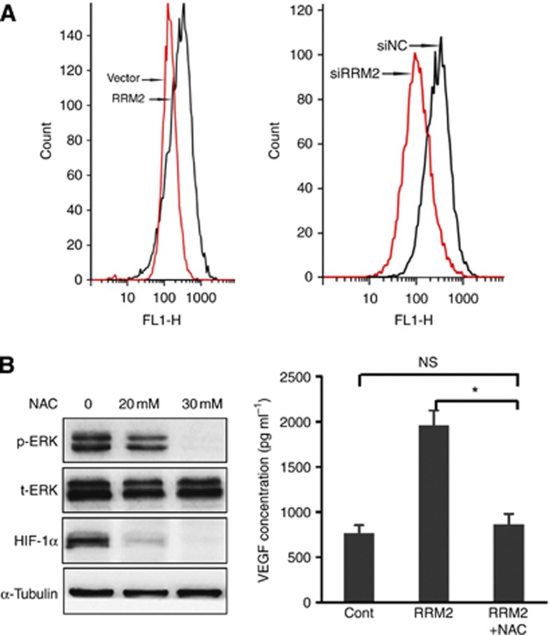
Figure 6 RRM2 elicited production of ROS to activate the ERK1/2 pathway, and enhance HIF-1α and VEGF expression. (A) RRM2 increases generation of ROS in cervical cancer cells. C33A or HeLa cells were transfected with a RRM2 expression plasmid (empty vector as control, left panel) or a RRM2 siRNA (siNC, negative control siRNA, right panel) for 48 h. Cells were then incubated with 20 μM DCFHDA in the dark for 30 min at 37 °C, washed in PBS, resuspended in PBS and analysed by flow cytometry. (B) NAC prevents RRM2 induced activation of ERK1/2 (p-ERK) and increases of HIF-1α and VEGF expression. C33A cells were transfected with a RRM2 expression plasmid for 24 h, followed by treatment with an indicated amount of NAC for 24h. Cells were lysed and used for western blot analysis with antibodies against pERK1/2 and HIF-1α. t-ERK, total ERK levels used as loading control; α-tubulin, loading control. The expression level of VEGF in conditioned media was measured by an ELISA assay. *P<0.05.
Figure 6 RRM2 elicited production of ROS to activate the ERK1/2 pathway, and enhance HIF-1α and VEGF expression. (A) RRM2 increases generation of ROS in cervical cancer cells. C33A or HeLa cells were transfected with a RRM2 expression plasmid (empty vector as control, left panel) or a RRM2 siRNA (siNC, negative control siRNA, right panel) for 48 h. Cells were then incubated with 20 μM DCFHDA in the dark for 30 min at 37 °C, washed in PBS, resuspended in PBS and analysed by flow cytometry. (B) NAC prevents RRM2 induced activation of ERK1/2 (p-ERK) and increases of HIF-1α and VEGF expression. C33A cells were transfected with a RRM2 expression plasmid for 24 h, followed by treatment with an indicated amount of NAC for 24h. Cells were lysed and used for western blot analysis with antibodies against pERK1/2 and HIF-1α. t-ERK, total ERK levels used as loading control; α-tubulin, loading control. The expression level of VEGF in conditioned media was measured by an ELISA assay. *P<0.05.CC BY-NC-SA · PubMed Central
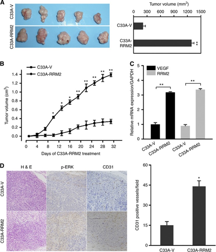
Figure 7 RRM2 overexpression promotes the growth and angiogenesis of cervical cancer xenografts in nude mice. (A) C33A-RRM2 and C33A-vector cells were subcutaneously inoculated into the flank of nude mice. Thirty-two days after the inoculation, the mice were killed and tumour tissues were collected. Left panel shows photographs of the tumours. Right panel shows that overexpression of RRM2 increased the tumour size of C33A-RRM2 at the end of experiment. (B) Overexpression of RRM2 significantly enhanced tumour growth as compared with empty vector. The graph shows tumour growth curves in nude mice inoculated with C33A cells with overexpression of RRM2 or cells with the cloning vector. Data are presented as means ±s.d. (n=5). (C) quantitative RT-PCR analysis of RRM2 and VEGF mRNA in C33A-RRM2 and C33A-vector xenografts. Data are shown as mean±s.d. from five mice. (D) Representative images of tumour sections from RRM2-overexpressed cells and control cells immunostained with pERK1/2 and CD31 antibodies (left panel). The level of phosphorylated ERK1/2 was significantly higher in RRM2-overexpression tumours than in control tumours. H&E staining served as control. Right panel, CD31-positive vessel densities were significantly increased in C33A-RRM2 tumour xenografts compared with that from C33A-vector. Data are presented as mean±s.d. (n=5). *P<0.05, **P<0.01.
Figure 7 RRM2 overexpression promotes the growth and angiogenesis of cervical cancer xenografts in nude mice. (A) C33A-RRM2 and C33A-vector cells were subcutaneously inoculated into the flank of nude mice. Thirty-two days after the inoculation, the mice were killed and tumour tissues were collected. Left panel shows photographs of the tumours. Right panel shows that overexpression of RRM2 increased the tumour size of C33A-RRM2 at the end of experiment. (B) Overexpression of RRM2 significantly enhanced tumour growth as compared with empty vector. The graph shows tumour growth curves in nude mice inoculated with C33A cells with overexpression of RRM2 or cells with the cloning vector. Data are presented as means ±s.d. (n=5). (C) quantitative RT-PCR analysis of RRM2 and VEGF mRNA in C33A-RRM2 and C33A-vector xenografts. Data are shown as mean±s.d. from five mice. (D) Representative images of tumour sections from RRM2-overexpressed cells and control cells immunostained with pERK1/2 and CD31 antibodies (left panel). The level of phosphorylated ERK1/2 was significantly higher in RRM2-overexpression tumours than in control tumours. H&E staining served as control. Right panel, CD31-positive vessel densities were significantly increased in C33A-RRM2 tumour xenografts compared with that from C33A-vector. Data are presented as mean±s.d. (n=5). *P<0.05, **P<0.01.CC BY-NC-SA · PubMed Central

Verification

Claims the paper makes

The claims and statistics of this paper haven’t been checked yet.

Run the check — free, no account needed. It reads the open-access text and checks the sources it cites.

A few free checks a day · papers already verified open instantly · results are public

Claims other papers make about this one

Each is checked against this paper’s own text: does it say what the citing paper says it does?

Self-check

Percentages, sums and reported differences re-computed from the paper’s own numbers.

The self-check needs the paper’s full text.

Citations

Cites

Show all 1 references

Research works printed but not linked: 42

  1. Bodily JM, Mehta KP, Laimins LA (2011) Human papillomavirus E7 enhances hypoxia-inducible factor 1-mediated transcription by inhibiting binding of histone deacetylases. Cancer Res 71 (3): 1187–1195. · DOI · Paper page
  2. Bosch FX, de SS (2007) The epidemiology of human papillomavirus infection and cervical cancer. Dis Markers 23: 213–227. · DOI
  3. Boyer SN, Wazer DE, Band V (1996) E7 protein of human papilloma virus-16 induces degradation of retinoblastoma protein through the ubiquitin-proteasome pathway. Cancer Res 56: 4620–4624.
  4. Branca M, Giorgi C, Santini D, Di Bonito L, Ciotti M, Benedetto A, Paba P, Costa S, Bonifacio D, Di Bonito P, Accardi L, Favalli C, Syrjänen K HPV-Pathogen ISS Study Group (2006) Aberrant expression of VEGF-C is related to grade of cervical intraepithelial neoplasia (CIN) and high risk HPV, but does not predict virus clearance after treatment of CIN or prognosis of cervical cancer. J Clin Pathol 59: 40–47. · DOI
  5. Drigotas M, Affolter A, Mann WJ, Brieger J (2013) Reactive oxygen species activation of MAPK pathway results in VEGF upregulation as an undesired irradiation response. J Oral Pathol Med 42 (8): 612–619. · DOI
  6. Duxbury MS, Whang EE (2007) RRM2 induces NF-kappaB-dependent MMP-9 activation and enhances cellular invasiveness. Biochem Biophys Res Commun 354: 190–196. · DOI
  7. el-Deiry WS, Tokino T, Waldman T, Oliner JD, Velculescu VE, Burrell M, Hill DE, Healy E, Rees JL, Hamilton SR, Kinzler KW, Vogelstein B (1995) Topological control of p21WAF1/CIP1 expression in normal and neoplastic tissues. Cancer Res 55: 2910–2919.
  8. Elford HL, Freese M, Passamani E, Morris HP (1970) Ribonucleotide reductase and cell proliferation. I.Variations of ribonucleotide reductase activity with tumor growth rate in a series of rat hepatomas. J Biol Chem 245: 5228–5233. · DOI
  9. Fan H, Villegas C, Huang A, Wright JA (1998) The mammalian ribonucleotide reductase R2 component cooperates with a variety of oncogenes in mechanisms of cellular transformation. Cancer Res 58: 1650–1653.
  10. Faoro V, Barbazza R, Bonin S, Brunetti D, Sulfaro S, Stanta G (2013) Detection of HPV E7 oncoviral protein in cervical lesions by a new antibody. Appl Immunohistochem Mol Morphol 21: 341–350. · DOI
  11. Garner-Hamrick PA, Fostel JM, Chien WM, Banerjee NS, Chow LT, Broker TR, Fisher C (2004) Global effects of human papillomavirus type 18 E6/E7 in an organotypic keratinocyte culture system. J Virol 78: 9041–9050. · DOI
  12. Gautam A, Li ZR, Bepler G (2003) RRM1-induced metastasis suppression through PTEN-regulated pathways. Oncogene 22: 2135–2142. · DOI
  13. Holland D, Hoppe-Seyler K, Schuller B, Lohrey C, Maroldt J, Dürst M, Hoppe-Seyler F (2008) Activation of the enhancer of zeste homologue 2 gene by the human papillomavirus E7 oncoprotein. Cancer Res 68: 9964–9972. · DOI · Paper page
  14. Huh K, Zhou X, Hayakawa H, Cho JY, Libermann TA, Jin J, Harper JW, Munger K (2007) Human papillomavirus type 16 E7 oncoprotein associates with the cullin 2 ubiquitin ligase complex, which contributes to degradation of the retinoblastoma tumor suppressor. J Virol 81: 9737–9747. · DOI · Paper page
  15. Huh KW, DeMasi J, Ogawa H, Nakatani Y, Howley PM, Münger K (2005) Association of the human papillomavirus type 16 E7 oncoprotein with the 600-kDa retinoblastoma protein-associated factor, p600. Proc Natl Acad Sci USA 102: 11492–11497. · DOI · Paper page
  16. Jemal A, Bray F, Center MM, Ferlay J, Ward E, Forman D (2011) Global cancer statistics. CA Cancer J Clin 61: 69–90. · DOI
  17. Kuner R, Vogt M, Sultmann H, Buness A, Dymalla S, Bulkescher J, Fellmann M, Butz K, Poustka A, Hoppe-Seyler F (2007) Identification of cellular targets for the human papillomavirus E6 and E7 oncogenes by RNA interference and transcriptome analyses. J Mol Med 85: 1253–1262. · DOI
  18. Katich SC, Zerfass-Thome K, Hoffmann I (2001) Regulation of the Cdc25A gene by the human papillomavirus Type 16 E7 oncogene. Oncogene 20: 543–550, 31. · DOI · Paper page
  19. Landt S, Mordelt K, Schwidde I, Barinoff J, Korlach S, Stöblen F, Lichtenegger W, Sehouli J, Kümmel S (2011) Prognostic significance of the angiogenic factors angiogenin, endoglin and endostatin in cervical cancer. Anticancer Res 31: 2651–2655.
  20. Lee DJ, Kang SW (2013) Reactive oxygen species and tumor metastasis. Mol Cells 35: 93–98. · DOI
  21. Liu H, Xiao J, Yang Y, Liu Y, Ma R, Li Y, Deng F, Zhang Y (2011) COX-2 expression is correlated with VEGF-C, lymphangiogenesis and lymph node metastasis in human cervical cancer. Microvasc Res 82: 131–140. · DOI
  22. Liu X, Zhang H, Lai L, Wang X, Loera S, Xue L, He H, Zhang K, Hu S, Huang Y, Nelson RA, Zhou B, Zhou L, Chu P, Zhang S, Zheng S, Yen Y (2013) Ribonucleotide reductase small subunit M2 serves as a prognostic biomarker and predicts poor survival of colorectal cancers. Clin Sci 124: 567–578. · DOI
  23. Meng D, Mei A, Liu J, Kang X, Shi X, Qian R, Chen S (2012) NADPH oxidase 4 mediates insulin-stimulated HIF-1α and VEGF expression, and angiogenesis in vitro. PLoS One 7: e48393. · DOI
  24. Morikawa T, Maeda D, Kume H, Homma Y, Fukayama M (2010) Ribonucleotide reductase M2 subunit is a novel diagnostic marker and a potential therapeutic target in bladder cancer. Histopathology 57: 885–892. · DOI
  25. Nordlund P, Reichard P (2006) Ribonucleotide reductases. Annu Rev Biochem 75: 681–706. · DOI
  26. Policht FA, Song M, Sitailo S, O’Hare A, Ashfaq R, Muller CY, Morrison LE, King W, Sokolova IA (2010) Analysis of genetic copy number changes in cervical disease progression. BMC Cancer 10: 432. · DOI
  27. Ressler S, Scheiden R, Dreier K, Laich A, Müller-Holzner E, Pircher H, Morandell D, Stein I, Viertler HP, Santer FR, Widschwendter A, Even J, Jansen-Dürr P, Capesius C, Zwerschke W (2007) High-risk human papillomavirus E7 oncoprotein detection in cervical squamous cell carcinoma. Clin Cancer Res 13: 7067–7072. · DOI
  28. Rosty C, Sheffer M, Tsafrir D, Stransky N, Tsafrir I, Peter M, De Crémoux P, de La Rochefordière A, Salmon R, Dorval T, Thiery JP, Couturier J, Radvanyi F, Domany E, Sastre-Garau X (2005) Identification of a proliferation gene cluster associated with HPV E6/E7 expression level and viral DNA load in invasive cervical carcinoma. Oncogene 24: 7094–7104. · DOI
  29. Smola-Hess S, Pahne J, Mauch C, Zigrino P, Smola H, Pfister HJ (2005) Expression of membrane type 1 matrix metalloproteinase in papillomavirus-positive cells: role of the human papillomavirus (HPV) 16 and HPV8 E7 gene products. J Gen Virol 86 (Pt 5): 1291–1296. · DOI
  30. Tang S, Tao M, McCoy JP Jr, Zheng ZM (2006) The E7 oncoprotein is translated from spliced E6*I transcripts in high-risk human papillomavirus type 16- or type 18-positive cervical cancer cell lines via translation reinitiation. J Virol 80: 4249–4263. · DOI
  31. Tang X, Zhang Q, Nishitani J, Brown J, Shi S, Le AD (2007) Overexpression of human papillomavirus type 16 oncoproteins enhances hypoxia-inducible factor 1 alpha protein accumulation and vascular endothelial growth factor expression in human cervical carcinoma cells. Clin Cancer Res 13: 2568–2576. · DOI
  32. Thierry F, Benotmane MA, Demeret C, Mori M, Teissier S, Desaintes C (2004) A genomic approach reveals a novel mitotic pathway in papillomavirus carcinogenesis. Cancer Res 64: 895–903. · DOI
  33. Tian J, Yan J, Wang W, Zhong N, Tian L, Sun J, Min Z, Ma J, Lu S (2012) T-2 toxin enhances catabolic activity of hypertrophic chondrocytes through ROS-NF-κB-HIF-2α pathway. Toxicol In Vitro 26: 1106–1113. · DOI
  34. Tian XL, Kadaba R, You SA, Liu M, Timur AA, Yang L, Chen Q, Szafranski P, Rao S, Wu L, Housman DE, DiCorleto PE, Driscoll DJ, Borrow J, Wang Q (2004) Identification of an angiogenic factor that when mutated causes susceptibility to Klippel-Trenaunay syndrome. Nature 42: 640–645. · DOI
  35. Tsai HH, Lee WR, Wang PH, Cheng KT, Chen YC, Shen SC (2013) Propionibacterium acnes-induced iNOS and COX-2 protein expression via ROS-dependent NF-κB and AP-1 activation in macrophages. J Dermatol Sci 69: 122–131. · DOI
  36. Wang F, Xu CQ, He Q, Cai JP, Li XC, Wang D, Xiong X, Liao YH, Zeng QT, Yang YZ, Cheng X, Li C, Yang R, Wang CC, Wu G, Lu QL, Bai Y, Huang YF, Yin D, Yang Q, Wang XJ, Dai DP, Zhang RF, Wan J, Ren JH, Li SS, Zhao YY, Fu FF, Huang Y, Li QX, Shi SW, Lin N, Pan ZW, Li Y, Yu B, Wu YX, Ke YH, Lei J, Wang N, Luo CY, Ji LY, Gao LJ, Li L, Liu H, Huang EW, Cui J, Jia N, Ren X, Li H, Ke T, Zhang XQ, Liu JY, Liu MG, Xia H, Yang B, Shi LS, Xia YL, Tu X, Wang QK (2011) Genome-wide association identifies a susceptibility locus for coronary artery disease in the Chinese Han population. Nat Genet 43: 345–349. · DOI
  37. Werness BA, Levine AJ, Howley PM (1990) Association of human papillomavirus types 16 and 18 E6 proteins with p53. Science 248: 76–79. · DOI · Paper page
  38. Xia C, Meng Q, Liu LZ, Rojanasakul Y, Wang XR, Jiang BH (2007) Reactive oxygen species regulate angiogenesis and tumor growth through vascular endothelial growth factor. Cancer Res 67: 10823–10830. · DOI
  39. Xue L, Zhou B, Liu X, Wang T, Shih J, Qi C, Heung Y, Yen Y (2006) Structurally dependent redox property of ribonucleotide reductase subunit p53R2. Cancer Res 66: 1900–1905. · DOI
  40. Zhang K, Hu S, Wu J, Chen L, Lu J, Wang X, Liu X, Zhou B, Yen Y (2009) Overexpression of RRM2 decreases thrombspondin-1 and increases VEGF production in human cancer cells in vitro and in vivo: implication of RRM2 in angiogenesis. Mol Cancer 8: 11–22. · DOI
  41. Zhou B, Ma R, Si W, Li S, Xu Y, Tu X, Wang Q (2013) MicroRNA-503 targets FGF2 and VEGFA and inhibits tumor angiogenesis and growth. Cancer Lett 333: 159–169. · DOI
  42. Zhou BS, Tsai P, Ker R, Tsai J, Ho R, Yu J, Shih J, Yen Y (1998) Overexpression of transfected human ribonucleotide reductase M2 subunit in human cancer cells enhances their invasive potential. Clin Exp Metastasis 16: 43–49. · DOI

Metadata

Authors
Wang, N., Zhan, T., Ke, T., Huang, X., Ke, D., Wang, Q., & Li, H.
Venue
British Journal of Cancer
Published
2014-01-14
Volume
110
Issue
4
Pages
1034-1044
ISSN
  • 0007-0920
  • 1532-1827
Cited by
84
References
1
Language
en

About these checks

Registry, retraction, Cites and statistics results are computed, not judged, with one exception: a printed reference that no index lists is read once by a model before it counts as a citation error. Claims are extracted by a model and kept only when their quote appears verbatim in the text; each cited source is then checked, with a model’s help, for whether it says what the claim relies on. Those verdicts can be wrong — read the claim’s quote and the cited source before relying on one.

Statistics and arithmetic are recomputed, not judged.

Confidence bands are shown only for fields calibrated on our annotated test set. None is calibrated yet, so no band is shown.

What counts as a claim ›How we measure confidence ›How we check this ›

Are you an author, or do you see a problem with this page? Request a review