Cited by 217

The DOI matches its Crossref record; 10 of the 10 references it cites were found in a registry or an index.

Phosphoinositide 3-kinase enables phagocytosis of large particles by terminating actin assembly through Rac/Cdc42 GTPase-activating proteins

Schlam, Daniel, Bagshaw, Richard D., Freeman, Spencer A., Collins, Richard F., Pawson, Tony, Fairn, Gregory D., Grinstein, Sergio·2015·Nature Communications

Abstract

Phagocytosis is responsible for the elimination of particles of widely disparate sizes, from large fungi or effete cells to small bacteria. Though superficially similar, the molecular mechanisms involved differ: engulfment of large targets requires phosphoinositide 3-kinase (PI3K), while that of small ones does not. Here, we report that inactivation of Rac and Cdc42 at phagocytic cups is essential to complete internalization of large particles. Through a screen of 62 RhoGAP-family members, we demonstrate that ARHGAP12, ARHGAP25 and SH3BP1 are responsible for GTPase inactivation. Silencing these RhoGAPs impairs phagocytosis of large targets. The GAPs are recruited to large—but not small—phagocytic cups by products of PI3K, where they synergistically inactivate Rac and Cdc42. Remarkably, the prominent accumulation of phosphatidylinositol 3,4,5-trisphosphate characteristic of large-phagosome formation is less evident during phagocytosis of small targets, accounting for the contrasting RhoGAP distribution and the differential requirement for PI3K during phagocytosis of dissimilarly sized particles.

Overview

The claims and statistics of this paper haven’t been checked yet.

Run the check — free, no account needed. It reads the open-access text and checks the sources it cites.

A few free checks a day · papers already verified open instantly · results are public

Integrity signals

Registry

Crossref · 10.1038/ncomms9623

Cites

10 of 10 references verified0 citation errors · 0 not checkable

Statistics

Figures

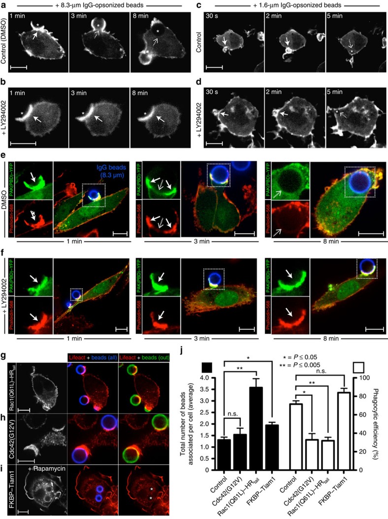
Figure 1 PI3K controls actin disassembly during phagocytosis of large targets. (a–d) Time-lapse confocal micrographs of RAW 264.7 macrophages transiently expressing Lifeact–mRFP and challenged with 8.3-μm (a,b) or 1.6-μm (c,d) IgG-opsonized beads (signalled with a star). Cells were treated with vehicle (DMSO) or the PI3K inhibitor LY294002 for 10 min before initiating phagocytosis. Actin dynamics were followed throughout the course of engulfment, with the 0-min time point corresponding to the initial engagement of the beads. Solid and dashed arrows point to sites of F-actin accumulation and clearance, respectively. (e,f) Confocal micrographs of primary human macrophages transfected with PAK(PBD)–YFP, a biosensor for active Rac/Cdc42, during phagocytosis of 8.3 μm IgG beads. Before phagocytosis, cells were treated with DMSO (e) or LY294002 (f) for 10 min. Phagocytosis was allowed to proceed for the indicated times, before fixing and staining F-actin with phalloidin. Insets (boxed regions) show magnified views of the phagocytic cup. (g–i) Confocal micrographs of RAW 264.7 macrophages transiently co-expressing Lifeact–mRFP in combination with constitutively active Rac1 (g), constitutively active Cdc42 (h) or a recruitable form of the Rac1 GEF Tiam1 (i). Addition of rapamycin triggers translocation of Tiam1 to the plasmalemma, where its rapamycin-binding domain (FKBP) interacts with a second, complementary rapamycin-binding moiety. Transfectants were challenged with 8.3-μm IgG beads, and phagocytosis allowed to proceed for 10 min before fixation. All phagocytic targets were stained with Cy5-conjugated secondary antibody (shown in blue) before phagocytosis. Extracellular beads were identified by staining fixed (non-permeabilized) cells with an Alexa Fluor 488-conjugated secondary antibody (shown in green). Internalized beads are indicated with a star. Scale bar, 10 μm. (j) Quantification of phagocytic indices (total number of beads associated per cell; black bars) and phagocytic efficiencies (ratio of internalized-to-total number of beads per cell; white bars) for the experiments described in (g–i). Values represent the means of three independent replicates±s.e.m. At least 25 cells were assessed per replicate. *P≤0.05, **P≤0.005 or n.s. (not significant) using Student's two-tailed unpaired t-tests.
Figure 1 PI3K controls actin disassembly during phagocytosis of large targets. (a–d) Time-lapse confocal micrographs of RAW 264.7 macrophages transiently expressing Lifeact–mRFP and challenged with 8.3-μm (a,b) or 1.6-μm (c,d) IgG-opsonized beads (signalled with a star). Cells were treated with vehicle (DMSO) or the PI3K inhibitor LY294002 for 10 min before initiating phagocytosis. Actin dynamics were followed throughout the course of engulfment, with the 0-min time point corresponding to the initial engagement of the beads. Solid and dashed arrows point to sites of F-actin accumulation and clearance, respectively. (e,f) Confocal micrographs of primary human macrophages transfected with PAK(PBD)–YFP, a biosensor for active Rac/Cdc42, during phagocytosis of 8.3 μm IgG beads. Before phagocytosis, cells were treated with DMSO (e) or LY294002 (f) for 10 min. Phagocytosis was allowed to proceed for the indicated times, before fixing and staining F-actin with phalloidin. Insets (boxed regions) show magnified views of the phagocytic cup. (g–i) Confocal micrographs of RAW 264.7 macrophages transiently co-expressing Lifeact–mRFP in combination with constitutively active Rac1 (g), constitutively active Cdc42 (h) or a recruitable form of the Rac1 GEF Tiam1 (i). Addition of rapamycin triggers translocation of Tiam1 to the plasmalemma, where its rapamycin-binding domain (FKBP) interacts with a second, complementary rapamycin-binding moiety. Transfectants were challenged with 8.3-μm IgG beads, and phagocytosis allowed to proceed for 10 min before fixation. All phagocytic targets were stained with Cy5-conjugated secondary antibody (shown in blue) before phagocytosis. Extracellular beads were identified by staining fixed (non-permeabilized) cells with an Alexa Fluor 488-conjugated secondary antibody (shown in green). Internalized beads are indicated with a star. Scale bar, 10 μm. (j) Quantification of phagocytic indices (total number of beads associated per cell; black bars) and phagocytic efficiencies (ratio of internalized-to-total number of beads per cell; white bars) for the experiments described in (g–i). Values represent the means of three independent replicates±s.e.m. At least 25 cells were assessed per replicate. *P≤0.05, **P≤0.005 or n.s. (not significant) using Student's two-tailed unpaired t-tests.CC BY · PubMed Central
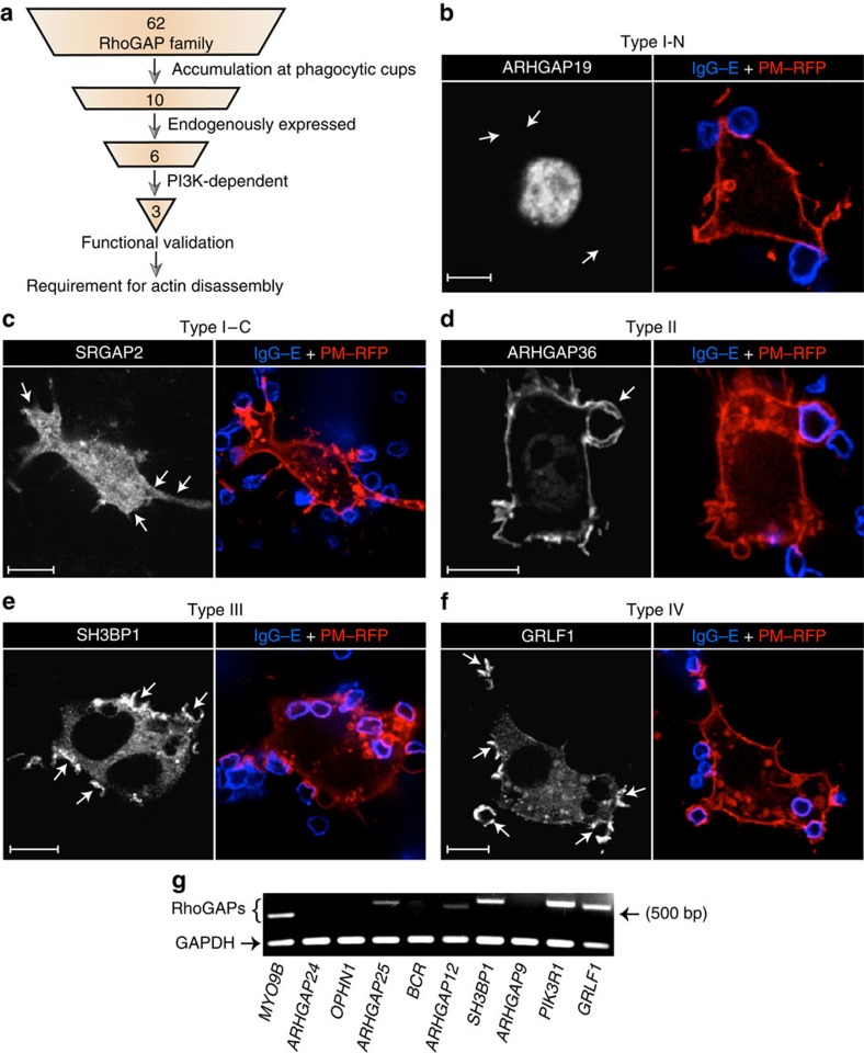
Figure 2 Screening for RhoGAPs responsible for orchestrating actin breakdown. (a) Schematic outlining the experimental approach used in this study. We generated a collection of constructs comprising 62 members of the RhoGAP family conjugated to mCitrine. Each plasmid was independently transfected into RAW 264.7 macrophages, and the subcellular distribution of the encoded RhoGAP followed by confocal microscopy during phagocytosis. The extent of accumulation at phagocytic cups was assessed by ratiometric analysis relative to PM–RFP, a marker for bulk plasma membrane. The 10 RhoGAPs that most significantly accumulated at phagocytic cups were selected for further screening, consisting of assessing endogenous expression by RT–PCR and dependency of recruitment on PI3K. The ability of the remaining candidates to function as RhoGAPs during phagocytosis was validated by overexpressing these proteins and quantifying phagocytic capacity. Lastly, gene silencing was used to determine whether the identified RhoGAPs were necessary for coordinating actin remodelling during phagocytosis. (b–f) Subcellular distribution of a selective subset of RhoGAPs during phagocytosis of IgG-coated erythrocytes (IgG–E). RAW 264.7 macrophages were co-transfected with constructs encoding mCitrine-tagged RhoGAPs plus the plasmalemmal marker PM–RFP. Each panel corresponds to a representative member of four identifiable RhoGAP types, as determined by their recruitment (or lack thereof) to sites of phagocytosis. RhoGAPs that failed to localize to sites of particle engagement and remained nuclear (b) or cytosolic (c) were assigned a type I–N and type I–C nomenclature, respectively. RhoGAPs that constitutively localized to the plasmalemma but did not accumulate at phagocytic cups (d) were designated as type II. In contrast, RhoGAPs that were initially cytosolic but translocated to phagocytic cups (e) were labelled as type III. Lastly, type IV RhoGAPs showed both a cytosolic and plasmalemmal distribution at rest but accumulated at phagocytic cups (f). Arrows point to sites of particle engagement. Scale bar, 10 μm. At least 20 cells were assessed for each RhoGAP in the collection. (g) Representative image of three independent RT–PCR assays, used to ascertain the endogenous expression levels of the 10 RhoGAPs that were most markedly recruited to phagocytic cups. GAPDH was used as a positive control.
Figure 2 Screening for RhoGAPs responsible for orchestrating actin breakdown. (a) Schematic outlining the experimental approach used in this study. We generated a collection of constructs comprising 62 members of the RhoGAP family conjugated to mCitrine. Each plasmid was independently transfected into RAW 264.7 macrophages, and the subcellular distribution of the encoded RhoGAP followed by confocal microscopy during phagocytosis. The extent of accumulation at phagocytic cups was assessed by ratiometric analysis relative to PM–RFP, a marker for bulk plasma membrane. The 10 RhoGAPs that most significantly accumulated at phagocytic cups were selected for further screening, consisting of assessing endogenous expression by RT–PCR and dependency of recruitment on PI3K. The ability of the remaining candidates to function as RhoGAPs during phagocytosis was validated by overexpressing these proteins and quantifying phagocytic capacity. Lastly, gene silencing was used to determine whether the identified RhoGAPs were necessary for coordinating actin remodelling during phagocytosis. (b–f) Subcellular distribution of a selective subset of RhoGAPs during phagocytosis of IgG-coated erythrocytes (IgG–E). RAW 264.7 macrophages were co-transfected with constructs encoding mCitrine-tagged RhoGAPs plus the plasmalemmal marker PM–RFP. Each panel corresponds to a representative member of four identifiable RhoGAP types, as determined by their recruitment (or lack thereof) to sites of phagocytosis. RhoGAPs that failed to localize to sites of particle engagement and remained nuclear (b) or cytosolic (c) were assigned a type I–N and type I–C nomenclature, respectively. RhoGAPs that constitutively localized to the plasmalemma but did not accumulate at phagocytic cups (d) were designated as type II. In contrast, RhoGAPs that were initially cytosolic but translocated to phagocytic cups (e) were labelled as type III. Lastly, type IV RhoGAPs showed both a cytosolic and plasmalemmal distribution at rest but accumulated at phagocytic cups (f). Arrows point to sites of particle engagement. Scale bar, 10 μm. At least 20 cells were assessed for each RhoGAP in the collection. (g) Representative image of three independent RT–PCR assays, used to ascertain the endogenous expression levels of the 10 RhoGAPs that were most markedly recruited to phagocytic cups. GAPDH was used as a positive control.CC BY · PubMed Central
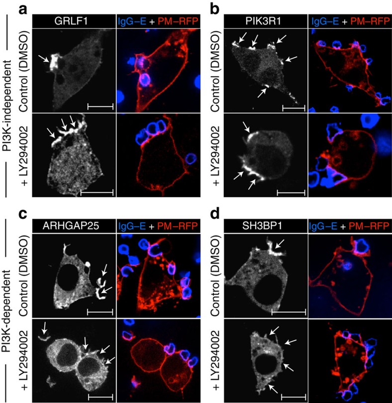
Figure 3 PI3K dependency of RhoGAP recruitment to phagocytic cups. (a–d) RAW 264.7 macrophages were co-transfected with constructs encoding each of the 10 RhoGAPs that most markedly accumulated at phagocytic cups as well as with the plasmalemmal marker PM–RFP. Transfectants were exposed to IgG-opsonized erythrocytes (IgG–E; shown in blue) for 3 min before being fixed and imaged by confocal microscopy. Where indicated, cells were treated with either vehicle (DMSO) or the PI3K inhibitor LY294002 for 10 min before the addition of phagocytic targets. Only a subset of the 10 RhoGAPs that were investigated is shown, namely two PI3K-independent (a,b) and two PI3K-dependent (c,d) proteins. (see Supplementary Fig. 3 for the remaining RhoGAPs that were investigated.) Arrows point to sites of particle engagement. Scale bar, 10 μm. Micrographs are representative of three independent experiments. At least 20 cells were assessed per replicate for each of the RhoGAPs examined.
Figure 3 PI3K dependency of RhoGAP recruitment to phagocytic cups. (a–d) RAW 264.7 macrophages were co-transfected with constructs encoding each of the 10 RhoGAPs that most markedly accumulated at phagocytic cups as well as with the plasmalemmal marker PM–RFP. Transfectants were exposed to IgG-opsonized erythrocytes (IgG–E; shown in blue) for 3 min before being fixed and imaged by confocal microscopy. Where indicated, cells were treated with either vehicle (DMSO) or the PI3K inhibitor LY294002 for 10 min before the addition of phagocytic targets. Only a subset of the 10 RhoGAPs that were investigated is shown, namely two PI3K-independent (a,b) and two PI3K-dependent (c,d) proteins. (see Supplementary Fig. 3 for the remaining RhoGAPs that were investigated.) Arrows point to sites of particle engagement. Scale bar, 10 μm. Micrographs are representative of three independent experiments. At least 20 cells were assessed per replicate for each of the RhoGAPs examined.CC BY · PubMed Central
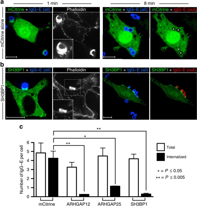
Figure 4 Functional validation of candidate RhoGAPs. (a,b) Constructs encoding mCitrine alone (a) or SH3BP1 (b) were electroporated into RAW 264.7 macrophages to yield high levels of expression. To determine whether these RhoGAPs could indeed inactivate GTPases that are instrumental for phagocytosis, electroporated cells were challenged with IgG-opsonized erythrocytes and allowed to interact with the targets for 1 min (left panels) or 8 min (right panels) before fixation. All phagocytic targets (shown in blue) were stained with a Cy5-conjugated anti-IgG secondary antibody before addition to phagocytes. To determine the number of IgG-coated erythrocytes that were not internalized, fixed (non-permeabilized) cells were stained with Cy3-conjugated anti-IgG (shown in red). Cells were then washed, permeabilized and stained for F-actin with phalloidin. Insets show magnified views of nascent phagocytic cups (boxed region) at the 1 min time point. Phagosomes that had already been formed before addition of the Cy3-conjugated secondary antibody are indicated with a star. Scale bar, 10 μm. Micrographs are representative of three independent experiments. (c) Quantification of the total number of IgG-coated erythrocytes that were engaged (white bars) or internalized (black bars) per phagocyte overexpressing mCitrine alone or mCitrine-tagged ARHGAP12, ARHGAP25 or SH3BP1. Values in (c) represent the means of three independent replicates±s.e.m. *P≤0.05 and **P≤0.005 using Student's two-tailed unpaired t-tests. At least 20 cells were assessed per replicate for each of the RhoGAPs examined.
Figure 4 Functional validation of candidate RhoGAPs. (a,b) Constructs encoding mCitrine alone (a) or SH3BP1 (b) were electroporated into RAW 264.7 macrophages to yield high levels of expression. To determine whether these RhoGAPs could indeed inactivate GTPases that are instrumental for phagocytosis, electroporated cells were challenged with IgG-opsonized erythrocytes and allowed to interact with the targets for 1 min (left panels) or 8 min (right panels) before fixation. All phagocytic targets (shown in blue) were stained with a Cy5-conjugated anti-IgG secondary antibody before addition to phagocytes. To determine the number of IgG-coated erythrocytes that were not internalized, fixed (non-permeabilized) cells were stained with Cy3-conjugated anti-IgG (shown in red). Cells were then washed, permeabilized and stained for F-actin with phalloidin. Insets show magnified views of nascent phagocytic cups (boxed region) at the 1 min time point. Phagosomes that had already been formed before addition of the Cy3-conjugated secondary antibody are indicated with a star. Scale bar, 10 μm. Micrographs are representative of three independent experiments. (c) Quantification of the total number of IgG-coated erythrocytes that were engaged (white bars) or internalized (black bars) per phagocyte overexpressing mCitrine alone or mCitrine-tagged ARHGAP12, ARHGAP25 or SH3BP1. Values in (c) represent the means of three independent replicates±s.e.m. *P≤0.05 and **P≤0.005 using Student's two-tailed unpaired t-tests. At least 20 cells were assessed per replicate for each of the RhoGAPs examined.CC BY · PubMed Central
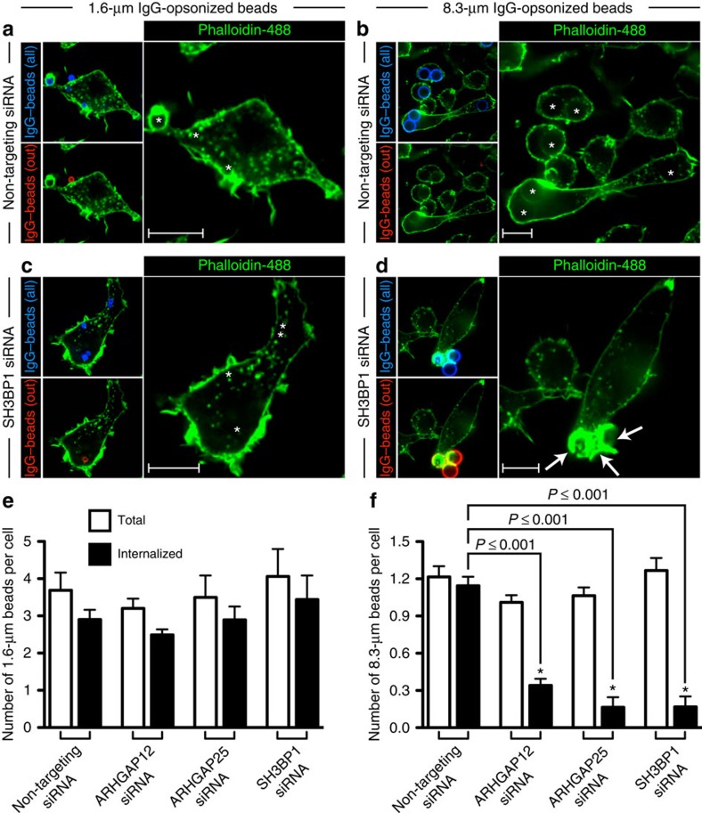
Figure 5 RhoGAPs are necessary for actin breakdown and uptake of large targets. (a–d) RAW 264.7 macrophages were electroporated twice (sequentially within a 48-h interval) with non-targeting control siRNA (a,b) or with siRNA directed against SH3BP1 (c,d), ARHGAP12 or ARHGAP25 and challenged with small (left panel) or large (right panel) IgG-coated beads 96 h after the initial electroporation. Cells were allowed to phagocytose for 10 min before fixation. All phagocytic targets (shown in blue) were stained with Cy5-conjugated anti-IgG secondary antibody before being added to phagocytes, and particles that remained extracellular (shown in red) were identified as in Fig. 4. Cells were then washed, permeabilized and stained for F-actin with phalloidin. Formed phagosomes are indicated with a star. Arrows point to abortive, actin-rich phagocytic cups that develop as a result of RhoGAP silencing in cells attempting to engulf large targets. Scale bar, 10 μm. (e,f) Quantification of the experiment described in (a–d). The total number of IgG-coated beads that were engaged (white bars) or internalized (black bars) per phagocyte is plotted. Values in (e,f) represent the means of three independent replicates±s.e.m. *P≤0.001 using Student's two-tailed unpaired t-tests. At least 20 cells were assessed per replicate for each of the siRNAs examined.
Figure 5 RhoGAPs are necessary for actin breakdown and uptake of large targets. (a–d) RAW 264.7 macrophages were electroporated twice (sequentially within a 48-h interval) with non-targeting control siRNA (a,b) or with siRNA directed against SH3BP1 (c,d), ARHGAP12 or ARHGAP25 and challenged with small (left panel) or large (right panel) IgG-coated beads 96 h after the initial electroporation. Cells were allowed to phagocytose for 10 min before fixation. All phagocytic targets (shown in blue) were stained with Cy5-conjugated anti-IgG secondary antibody before being added to phagocytes, and particles that remained extracellular (shown in red) were identified as in Fig. 4. Cells were then washed, permeabilized and stained for F-actin with phalloidin. Formed phagosomes are indicated with a star. Arrows point to abortive, actin-rich phagocytic cups that develop as a result of RhoGAP silencing in cells attempting to engulf large targets. Scale bar, 10 μm. (e,f) Quantification of the experiment described in (a–d). The total number of IgG-coated beads that were engaged (white bars) or internalized (black bars) per phagocyte is plotted. Values in (e,f) represent the means of three independent replicates±s.e.m. *P≤0.001 using Student's two-tailed unpaired t-tests. At least 20 cells were assessed per replicate for each of the siRNAs examined.CC BY · PubMed Central
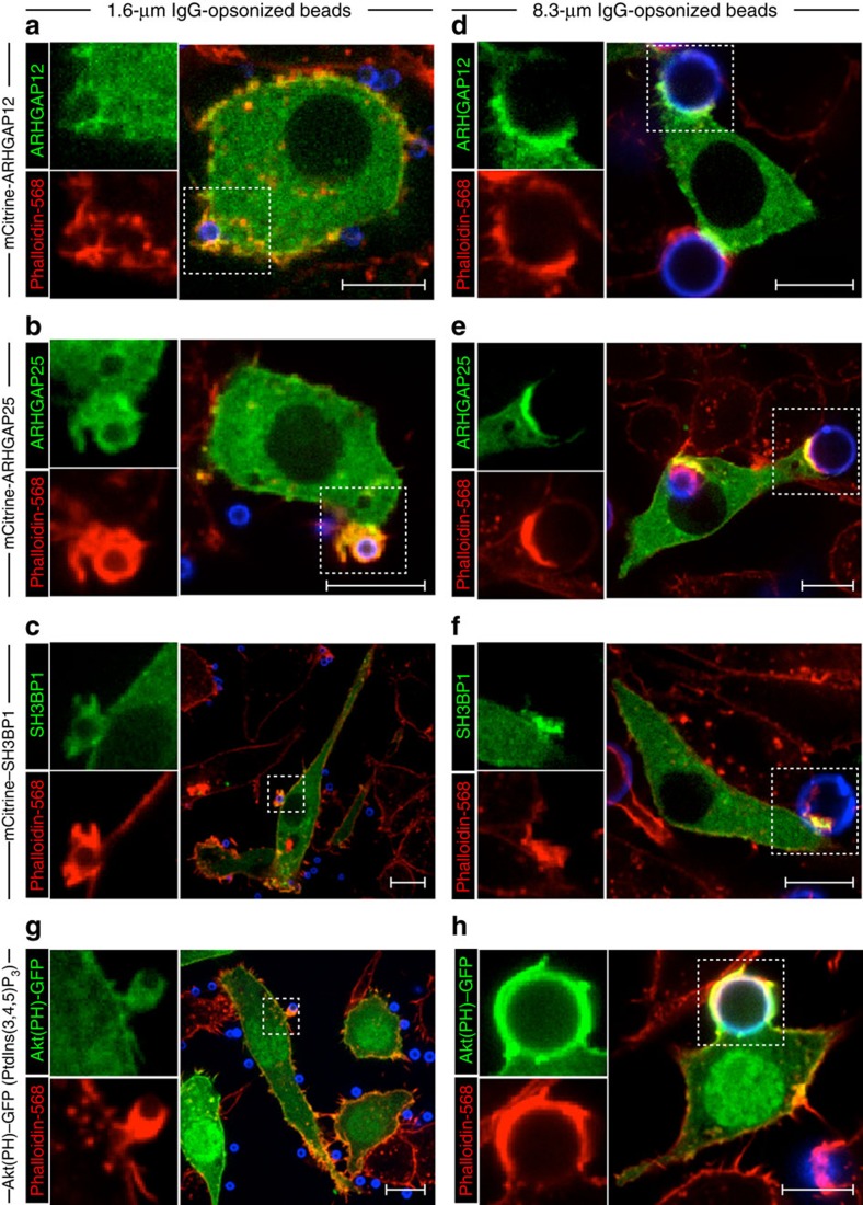
Figure 6 Role of PtdIns(3,4,5)P3 and RhoGAPs during uptake of small particles. Representative confocal micrographs of RAW 264.7 macrophages transfected with constructs encoding mCitrine-tagged ARHGAP12 (a,d), ARHGAP25 (b,e), SH3BP1 (c,f) or with GFP-tagged Akt(PH), a probe for PtdIns(3,4,5)P3 (g,h). The distribution of these fluorescent proteins is shown in green. Cells were challenged with either small (1.6 μm; left panels) or large (8.3 μm; right panels) IgG-coated beads to initiate phagocytosis. All beads were stained with a Cy5-conjugated secondary antibody (shown in blue) before being sedimented onto macrophage surfaces. Phagocytosis was allowed to continue for 8 min before fixation and staining of F-actin with phalloidin-568 (shown in red). Insets (boxed regions) show magnified views of the phagocytic cup. Scale bar, 10 μm. Micrographs are representative of three independent experiments; at least 20 cells were assessed per replicate for each condition.
Figure 6 Role of PtdIns(3,4,5)P3 and RhoGAPs during uptake of small particles. Representative confocal micrographs of RAW 264.7 macrophages transfected with constructs encoding mCitrine-tagged ARHGAP12 (a,d), ARHGAP25 (b,e), SH3BP1 (c,f) or with GFP-tagged Akt(PH), a probe for PtdIns(3,4,5)P3 (g,h). The distribution of these fluorescent proteins is shown in green. Cells were challenged with either small (1.6 μm; left panels) or large (8.3 μm; right panels) IgG-coated beads to initiate phagocytosis. All beads were stained with a Cy5-conjugated secondary antibody (shown in blue) before being sedimented onto macrophage surfaces. Phagocytosis was allowed to continue for 8 min before fixation and staining of F-actin with phalloidin-568 (shown in red). Insets (boxed regions) show magnified views of the phagocytic cup. Scale bar, 10 μm. Micrographs are representative of three independent experiments; at least 20 cells were assessed per replicate for each condition.CC BY · PubMed Central

Verification

Claims the paper makes

The claims and statistics of this paper haven’t been checked yet.

Run the check — free, no account needed. It reads the open-access text and checks the sources it cites.

A few free checks a day · papers already verified open instantly · results are public

Claims other papers make about this one

Each is checked against this paper’s own text: does it say what the citing paper says it does?

Self-check

Percentages, sums and reported differences re-computed from the paper’s own numbers.

The self-check needs the paper’s full text.

Citations

Cites

Show all 10 references

Research works printed but not linked: 35

  1. Flannagan, R. S., Jaumouillé, V. & Grinstein, S. The cell biology of phagocytosis. Annu. Rev. Pathol. 7, 61–98 (2012). · DOI
  2. Cox, D., Tseng, C.-C., Bjekic, G. & Greenberg, S. A requirement for phosphoinositide 3-kinase in pseudopod extension. J. Biol. Chem. 274, 1240–1247 (1999). · DOI · Paper page
  3. Freeman, S. A. & Grinstein, S. Phagocytosis: receptors, signal integration, and the cytoskeleton. Immunol. Rev. 262, 193–215 (2014). · DOI · Paper page
  4. Park, H. & Cox, D. Cdc42 regulates Fc gamma receptor-mediated phagocytosis through the activation and phosphorylation of Wiskott-Aldrich syndrome protein (WASP) and neural-WASP. Mol. Biol. Cell 20, 4500–4508 (2009). · DOI
  5. Cox, D. et al. Requirements for both Rac1 and Cdc42 in membrane ruffling and phagocytosis in leukocytes. J. Exp. Med. 186, 1487–1494 (1997). · DOI · Paper page
  6. Niedergang, F. & Chavrier, P. Regulation of phagocytosis by Rho GTPases. Curr. Top. Microbiol. Immunol. 291, 43–60 (2005).
  7. Chimini, G. & Chavrier, P. Function of Rho family proteins in actin dynamics during phagocytosis and engulfment. Nat. Cell Biol. 2, E191–E196 (2000). · DOI
  8. Riedl, J. et al. Lifeact: a versatile marker to visualize F-actin. Nat. Methods 5, 605–607 (2008). · DOI · Paper page
  9. Hall, A. B. et al. Requirements for Vav guanine nucleotide exchange factors and Rho GTPases in FcgammaR- and complement-mediated phagocytosis. Immunity 24, 305–316 (2006). · DOI
  10. Castellano, F., Montcourrier, P. & Chavrier, P. Membrane recruitment of Rac1 triggers phagocytosis. J. Cell Sci. 113, (Pt 17): 2955–2961 (2000). · DOI
  11. Massol, P., Montcourrier, P., Guillemot, J. C. & Chavrier, P. Fc receptor-mediated phagocytosis requires CDC42 and Rac1. EMBO J. 17, 6219–6229 (1998). · DOI
  12. Srinivasan, S. et al. Rac and Cdc42 play distinct roles in regulating PI(3,4,5)P3 and polarity during neutrophil chemotaxis. J. Cell Biol. 160, 375–385 (2003). · DOI
  13. Beemiller, P. et al. A Cdc42 activation cycle coordinated by PI 3-kinase during Fc receptor-mediated phagocytosis. Mol. Biol. Cell 21, 470–480 (2010). · DOI
  14. Moon, S. Y. & Zheng, Y. Rho GTPase-activating proteins in cell regulation. Trends Cell Biol. 13, 13–22 (2003). · DOI
  15. Cicchetti, P., Ridley, A. J., Zheng, Y., Cerione, R. A. & Baltimore, D. 3BP-1, an SH3 domain binding protein, has GAP activity for Rac and inhibits growth factor-induced membrane ruffling in fibroblasts. EMBO J. 14, 3127–3135 (1995). · DOI
  16. Gentile, A. et al. Met-driven invasive growth involves transcriptional regulation of Arhgap12. Oncogene 27, 5590–5598 (2008). · DOI
  17. Csépányi-Kömi, R., Sirokmány, G., Geiszt, M. & Ligeti, E. ARHGAP25, a novel Rac GTPase-activating protein, regulates phagocytosis in human neutrophilic granulocytes. Blood 119, 573–582 (2012). · DOI
  18. Ferrari, G., Langen, H., Naito, M. & Pieters, J. A coat protein on phagosomes involved in the intracellular survival of mycobacteria. Cell 97, 435–447 (1999). · DOI
  19. Braun, V. et al. TI-VAMP/VAMP7 is required for optimal phagocytosis of opsonised particles in macrophages. EMBO J. 23, 4166–4176 (2004). · DOI
  20. Mohammadi, S. & Isberg, R. R. Cdc42 interacts with the exocyst complex to promote phagocytosis. J. Cell Biol. 200, 81–93 (2013). · DOI
  21. Samie, M. et al. A TRP channel in the lysosome regulates large particle phagocytosis via focal exocytosis. Dev. Cell 26, 511–524 (2013). · DOI
  22. Parrini, M. C. et al. SH3BP1, an exocyst-associated RhoGAP, inactivates Rac1 at the front to drive cell motility. Mol. Cell 42, 650–661 (2011). · DOI
  23. Wiedemann, A. et al. Two distinct cytoplasmic regions of the beta2 integrin chain regulate RhoA function during phagocytosis. J. Cell Biol. 172, 1069–1079 (2006). · DOI
  24. Scott, C. C. et al. Phosphatidylinositol-4,5-bisphosphate hydrolysis directs actin remodeling during phagocytosis. J. Cell Biol. 169, 139–149 (2005). · DOI
  25. Saarikangas, J., Zhao, H. & Lappalainen, P. Regulation of the actin cytoskeleton-plasma membrane interplay by phosphoinositides. Physiol. Rev. 90, 259–289 (2010). · DOI
  26. Levin, R., Grinstein, S. & Schlam, D. Phosphoinositides in phagocytosis and macropinocytosis. Biochim. Biophys. Acta 1851, 805–823 (2014). · DOI
  27. Yeung, T. & Grinstein, S. Lipid signaling and the modulation of surface charge during phagocytosis. Immunol. Rev. 219, 17–36 (2007). · DOI
  28. Sarantis, H. et al. Yersinia entry into host cells requires Rab5-dependent dephosphorylation of PI(4,5)P2 and membrane scission. Cell Host Microbe 11, 117–128 (2012). · DOI
  29. Bohdanowicz, M., Balkin, D. M., De Camilli, P. & Grinstein, S. Recruitment of OCRL and Inpp5B to phagosomes by Rab5 and APPL1 depletes phosphoinositides and attenuates Akt signaling. Mol. Biol. Cell 23, 176–187 (2012). · DOI
  30. Kühbacher, A., Dambournet, D., Echard, A., Cossart, P. & Pizarro-Cerdá, J. Phosphatidylinositol 5-phosphatase oculocerebrorenal syndrome of Lowe protein (OCRL) controls actin dynamics during early steps of Listeria monocytogenes infection. J. Biol. Chem. 287, 13128–13136 (2012). · DOI
  31. Teruel, M. N., Blanpied, T. A., Shen, K., Augustine, G. J. & Meyer, T. A versatile microporation technique for the transfection of cultured CNS neurons. J. Neurosci. Methods 93, 37–48 (1999). · DOI
  32. Chang-Ileto, B. et al. Synaptojanin 1-mediated PI(4,5)P2 hydrolysis is modulated by membrane curvature and facilitates membrane fission. Dev. Cell 20, 206–218 (2011). · DOI
  33. Marshall, J. G. et al. Restricted accumulation of phosphatidylinositol 3-kinase products in a plasmalemmal subdomain during Fc gamma receptor-mediated phagocytosis. J. Cell Biol. 153, 1369–1380 (2001). · DOI
  34. Bohdanowicz, M. & Fairn, G. D. Rapamycin-based inducible translocation systems for studying phagocytosis. Methods Mol. Biol. 748, 183–193 (2011). · DOI
  35. Bohdanowicz, M. et al. Phosphatidic acid is required for the constitutive ruffling and macropinocytosis of phagocytes. Mol. Biol. Cell 24, 1700–1712 (2013). · DOI

Cited by

Metadata

Authors
Schlam, D., Bagshaw, R. D., Freeman, S. A., Collins, R. F., Pawson, T., Fairn, G. D., & Grinstein, S.
Venue
Nature Communications
Published
2015-10-14
Volume
6
Issue
1
ISSN
2041-1723
Cited by
217
References
10
Language
en

About these checks

Registry, retraction, Cites and statistics results are computed, not judged, with one exception: a printed reference that no index lists is read once by a model before it counts as a citation error. Claims are extracted by a model and kept only when their quote appears verbatim in the text; each cited source is then checked, with a model’s help, for whether it says what the claim relies on. Those verdicts can be wrong — read the claim’s quote and the cited source before relying on one.

Statistics and arithmetic are recomputed, not judged.

Confidence bands are shown only for fields calibrated on our annotated test set. None is calibrated yet, so no band is shown.

What counts as a claim ›How we measure confidence ›How we check this ›

Are you an author, or do you see a problem with this page? Request a review