

The DOI matches its Crossref record; 139 of the 140 references it cites were found in a registry or an index.
Hypoxia and aging
Open accessAbstract
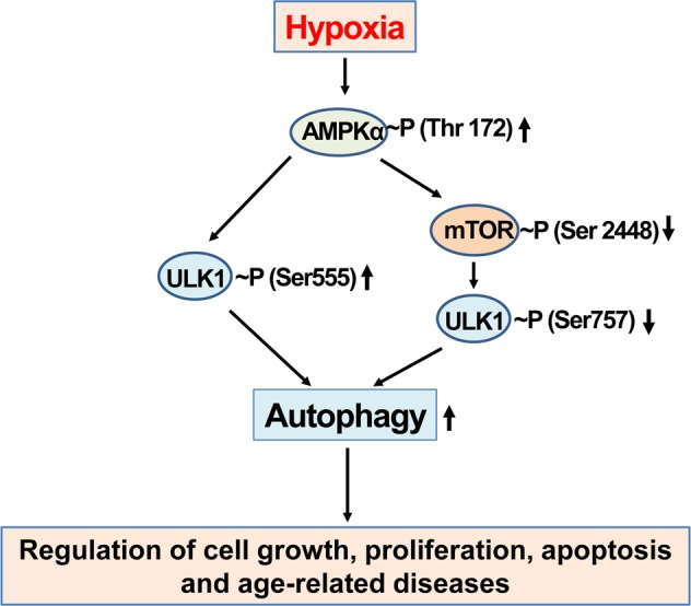
Eukaryotic cells require sufficient oxygen (O2) for biological activity and survival. When the oxygen demand exceeds its supply, the oxygen levels in local tissues or the whole body decrease (termed hypoxia), leading to a metabolic crisis, threatening physiological functions and viability. Therefore, eukaryotes have developed an efficient and rapid oxygen sensing system: hypoxia-inducible factors (HIFs). The hypoxic responses are controlled by HIFs, which induce the expression of several adaptive genes to increase the oxygen supply and support anaerobic ATP generation in eukaryotic cells. Hypoxia also contributes to a functional decline during the aging process. In this review, we focus on the molecular mechanisms regulating HIF-1α and aging-associated signaling proteins, such as sirtuins, AMP-activated protein kinase, mechanistic target of rapamycin complex 1, UNC-51-like kinase 1, and nuclear factor κB, and their roles in aging and aging-related diseases. In addition, the effects of prenatal hypoxia and obstructive sleep apnea (OSA)-induced intermittent hypoxia have been reviewed due to their involvement in the progression and severity of many diseases, including cancer and other aging-related diseases. The pathophysiological consequences and clinical manifestations of prenatal hypoxia and OSA-induced chronic intermittent hypoxia are discussed in detail. Deficient oxygen supplies to the body’s tissues and a reduced ability to deal with the cellular stress of low oxygen availability both contribute to the ageing process. Eui-Ju Yeo from Gachon University in Incheon, South Korea, reviews the links between insufficient oxygen, a condition known as hypoxia, and the functional decline associated with getting old. Yeo highlights the role that the oxygen-sensing proteins known as hypoxia-inducible factors (HIFs) play in facilitating oxygen supply and regulating energy production under limited oxygen conditions. HIFs also interact with a series of signaling proteins that, when abnormally activated, contribute to many aging-associated diseases. Yeo singles out two hypoxia-related health problems, prenatal hypoxia during early brain development and obstructive sleep apnea, and discusses how they can accelerate aging in various ways.
Overview
The claims and statistics of this paper haven’t been checked yet.
Free: it reads the open-access text and checks the first 5 claims that cite a source we can check. Create a free account to check every claim.
Integrity signals
Registry
Cites
Statistics
Figures




Verification
Claims the paper makes
The claims and statistics of this paper haven’t been checked yet.
Free: it reads the open-access text and checks the first 5 claims that cite a source we can check. Create a free account to check every claim.
Claims other papers make about this one
Each is checked against this paper’s own text: does it say what the citing paper says it does?
The source backs part of the claim, not all of it — or backs it for a different population, condition or comparison.
In the citing paper
“Hypoxia blocks the ubiquitination and proteasome degradation of HIF1, which is shuttled to the nucleus and induces the transcription of more than 100 genes involved in adaptive processes that increase oxygen supply and support anaerobic ATP generation, such as angiogenesis, erythropoiesis, anaerobic glycolytic metabolism, pH regulation, cell proliferation, cell survival, inflammation, and immunity”
From Proteomics in aging research: A roadmap to clinical, translational research (2021) · cites this paper as Yeo, 2019
Experimental — accuracy not yet measuredThe source backs part of the claim, not all of it — or backs it for a different population, condition or comparison.
In the citing paper
“Interestingly, HIF1α downregulates mitochondrial biogenesis, thereby reducing ATP production, and possibly further contributing to the oxidative stress that triggers the nuclear factor κB (NFkB)‐mediated production of cytokines (Yeo, 2019).”
From Proteomics in aging research: A roadmap to clinical, translational research (2021) · cites this paper as Yeo, 2019
Experimental — accuracy not yet measuredThe cited source says what the claim attributes to it.
In the citing paper
“Finally, the HIF1 pathway cross‐talks with other pathways that are relevant for aging, including sirtuins, AMP‐activated protein kinase (AMPK), mechanistic target of rapamycin complex 1 (mTORC1), UNC‐51‐like kinase 1 (ULK1), NFκB pathways, suggesting that chronic or episodic hypoxia may be connected to biological aging (Beek et al., 2019; Yeo, 2019).”
From Proteomics in aging research: A roadmap to clinical, translational research (2021) · cites this paper as Yeo, 2019
In the citing paper
“There is extensive evidence that dysregulation of HIF1 occurs with aging and is involved in the pathogenesis of many age‐related chronic conditions, including cancer growth and metastasis, cardiac disease, pulmonary disease, liver disease, and kidney disease (Yeo, 2019).”
From Proteomics in aging research: A roadmap to clinical, translational research (2021) · cites this paper as Yeo, 2019
Self-check
Percentages, sums and reported differences re-computed from the paper’s own numbers.
The self-check needs the paper’s full text.
Citations
Cites
AMPK and mTOR regulate autophagy through direct phosphorylation of Ulk1
Kim, J., Kundu, M., Viollet, B., & Guan, K.-L. (2011) · Nature Cell Biology · doi:10.1038/ncb2152 · Cited by 7,227
Targeting HIF-1 for cancer therapy
Semenza, G. L. (2003) · Nature Reviews Cancer · doi:10.1038/nrc1187 · Cited by 6,717
Wang, G. L., Jiang, B. H., Rue, E. A., & Semenza, G. L. (1995) · Proceedings of the National Academy of Sciences · doi:10.1073/pnas.92.12.5510 · Cited by 6,212
Jaakkola, P., Mole, D. R., Tian, Y.-M., Wilson, M. I., Gielbert, J., Gaskell, S. J., Kriegsheim, A. V., Hebestreit, H. F., Mukherji, M., Schofield, C. J., et al. (2001) · Science · doi:10.1126/science.1059796 · Cited by 5,535
HIFα Targeted for VHL-Mediated Destruction by Proline Hydroxylation: Implications for O 2 Sensing
Ivan, M., Kondo, K., Yang, H., Kim, W., Valiando, J., Ohh, M., Salic, A., Asara, J. M., Lane, W. S., & Kaelin Jr., W. G. (2001) · Science · doi:10.1126/science.1059817 · Cited by 4,735
AMPK: a nutrient and energy sensor that maintains energy homeostasis
Hardie, D. G., Ross, F. A., & Hawley, S. A. (2012) · Nature Reviews Molecular Cell Biology · doi:10.1038/nrm3311 · Cited by 4,602
mTOR: from growth signal integration to cancer, diabetes and ageing
Zoncu, R., Efeyan, A., & Sabatini, D. M. (2011) · Nature Reviews Molecular Cell Biology · doi:10.1038/nrm3025 · Cited by 3,941
TSC2 Mediates Cellular Energy Response to Control Cell Growth and Survival
Inoki, K., Zhu, T., & Guan, K.-L. (2003) · Cell · doi:10.1016/s0092-8674(03)00929-2 · Cited by 3,914
AMPK Phosphorylation of Raptor Mediates a Metabolic Checkpoint
Gwinn, D. M., Shackelford, D. B., Egan, D. F., Mihaylova, M. M., Mery, A., Vasquez, D. S., Turk, B. E., & Shaw, R. J. (2008) · Molecular Cell · doi:10.1016/j.molcel.2008.03.003 · Cited by 3,849
Comprehensive molecular characterization of clear cell renal cell carcinoma
The Cancer Genome Atlas Research Network (2013) · Nature · doi:10.1038/nature12222 · Cited by 3,485
Cited by
Pathophysiological mechanisms and therapeutic approaches in obstructive sleep apnea syndrome
Lv, R., Liu, X., Zhang, Y., Dong, N., Wang, X., He, Y., Yue, H., & Yin, Q. (2023) · Signal Transduction and Targeted Therapy · doi:10.1038/s41392-023-01496-3 · Cited by 477
HIF-1α promotes SARS-CoV-2 infection and aggravates inflammatory responses to COVID-19
Tian, M., Liu, W., Li, X., Zhao, P., Shereen, M. A., Zhu, C., Huang, S., Liu, S., Yu, X., Yue, M., et al. (2021) · Signal Transduction and Targeted Therapy · doi:10.1038/s41392-021-00726-w · Cited by 253
Hadanny, A., & Efrati, S. (2020) · Biomolecules · doi:10.3390/biom10060958 · Cited by 204
“The decreased level of SIRT1, in both transcriptional and posttranscriptional stages during aging is considered to be a major metabolic pathway that attenuates mitochondrial biogenesis and causes aging-related diseases [14].”
BackgroundCell Senescence, DNA Damage, and Metabolism
Shmulevich, R., & Krizhanovsky, V. (2021) · Antioxidants & Redox Signaling · doi:10.1089/ars.2020.8043 · Cited by 157
“2) senescence (25, 45, 95).”
Proteomics in aging research: A roadmap to clinical, translational research
Moaddel, R., Ubaida‐Mohien, C., Tanaka, T., Lyashkov, A., Basisty, N., Schilling, B., Semba, R. D., Franceschi, C., Gorospe, M., & Ferrucci, L. (2021) · Aging Cell · doi:10.1111/acel.13325 · Cited by 139
“…that are relevant for aging, including sirtuins, AMP- activated protein kinase (AMPK), mechanistic target of rapamycin complex 1 (mTORC1), UNC- 51- like kinase 1 (ULK1), NFκB pathways, suggesting that chronic or episodic hypoxia may be connected to biological aging (Beek et al., 2019; Yeo, 2019).”
BackgroundInfluential
Metadata
- Authors
- Yeo, E.-J.
- Affiliations
- Gachon University (KR)
- Corresponding author
- Yeo, Eui-Ju
- ORCID
- Yeo, Eui-Ju
- Venue
- Experimental & Molecular Medicine
- Published
- 2019-06
- Volume
- 51
- Issue
- 6
- Pages
- 1-15
- ISSN
- 1226-3613 (ISSN-L)
- 2092-6413
- PMC
- PMC6586788
- OpenAlex
- W2953255246
- Cited by
- 175
- References
- 140
- Citations by year
- 2026: 8 · 2025: 27 · 2024: 27 · 2023: 24 · 2022: 30 · 2021: 36 · 2020: 21 · 2019: 1
- FWCI
- 2.11
- Type
- journal article
- Language
- en
- Open access
- gold
- doi.org
- Licence
- cc-by
- creativecommons.org/licenses/by/4.0 (tdm)
- creativecommons.org/licenses/by/4.0 (vor, 19 d)
- Locations
- Experimental & Molecular Medicine (publishedVersion, cc-by)
- PubMed Central (publishedVersion, cc-by)
- PubMed (publishedVersion)
- DOAJ (DOAJ: Directory of Open Access Journals) (submittedVersion, cc-by-sa)
- Funders
- National Research Foundation of Korea (2017R1D1A1B03033499)
- Topics
- Life Sciences › Biochemistry, Genetics and Molecular Biology › Cancer Research › Cancer, Hypoxia, and Metabolism
- Life Sciences › Biochemistry, Genetics and Molecular Biology › Aging › Genetics, Aging, and Longevity in Model Organisms
- Life Sciences › Biochemistry, Genetics and Molecular Biology › Cell Biology › Autophagy in Disease and Therapy
- Keywords
- hypoxia, aging, hypoxia-inducible factor, HIF-1α, oxygen sensing, sirtuins, AMPK, mTORC1, NF-κB, age-related diseases, prenatal hypoxia, obstructive sleep apnea, intermittent hypoxia, cancer, brain development
- MeSH terms
- Mechanistic Target of Rapamycin Complex 1, Aging, Animals, Hypoxia, Humans, Signal Transduction, Cell Hypoxia, Sirtuins, Hypoxia-Inducible Factor 1, alpha Subunit, AMP-Activated Protein Kinases
About these checks
Registry, retraction, Cites and statistics results are computed, not judged, with one exception: a printed reference that no index lists is read once by a model before it counts as a citation error. Claims are extracted by a model and kept only when their quote appears verbatim in the text; each cited source is then checked, with a model’s help, for whether it says what the claim relies on. Those verdicts can be wrong — read the claim’s quote and the cited source before relying on one.
Statistics and arithmetic are recomputed, not judged.
Confidence bands are shown only for fields calibrated on our annotated test set. None is calibrated yet, so no band is shown.
What counts as a claim ›How we measure confidence ›How we check this ›
Are you an author, or do you see a problem with this page? Request a review
How to cite
Cite CoChat for the checks on this page, and the paper for its own findings.
- This page
- CoChat. (2026). Citation check: Hypoxia and aging. https://app.cochat.ai/papers/10.1038/s12276-019-0233-3
- The paper
- Yeo, E.-J. (2019). Hypoxia and aging. Experimental & Molecular Medicine. https://doi.org/10.1038/s12276-019-0233-3
Data sources
Bibliographic data: Semantic Scholar, OpenAlex, Crossref.
Contains information from the Semantic Scholar Academic Graph Datasets, made available under the ODC Attribution License (ODC-BY 1.0).
Semantic Scholar’s data is described in: Kinney, R., et al. (2023). The Semantic Scholar Open Data Platform. arXiv:2301.10140.
